

Radiation Exposure in Computed Tomography
- PMID: 36633449
- PMCID: PMC10198168
- DOI: 10.3238/arztebl.m2022.0395
Radiation Exposure in Computed Tomography
Abstract
Background: Computed tomography (CT) studies are requested by specialists from most medical disciplines and play a vital role in the diagnosis and treatment of patients. It follows that physicians of all specialties should possess basic knowledge of computed tomography, its proper use, and the radiation exposure associated with it.
Methods: This review is based on publications retrieved by a selective search of the literature.
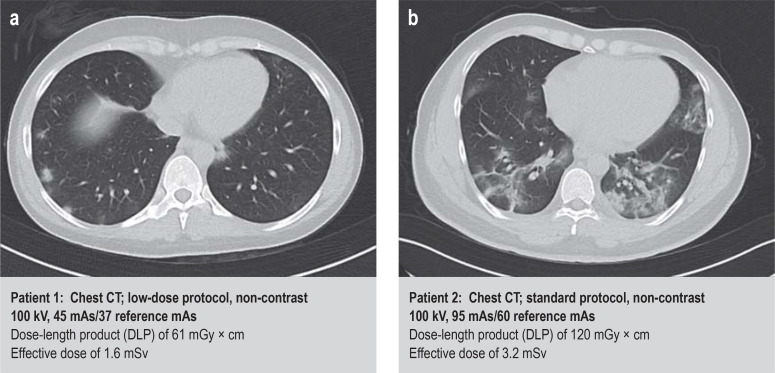
Results: Approximately 12 million CT studies are carried out in Germany each year, and the trend is rising. Approximately 9% of all diagnostic studies involving ionizing radiation are CT studies. On average, more than 60% of the collective effective dose due to medical radiation exposure is attributable to CT. There are two types of radiation effects caused by ionizing radiation: sto - chastic and deterministic. The additional, individual relative lifetime cancer mortality risk due to ionizing radiation with wholebody exposure at a low single dose is estimated at 5% per sievert. Radiation exposure from CT studies of the head and trunk, e.g. of a patient with polytrauma, corresponds to an additional lifetime cancer mortality risk of approximately 0.1% at an effective dose of approximately 20 millisievert.
Conclusion: The radiation exposure due to CT, and the risks to which patients are subjected by it, have become more important with greater use of CT. Technical advances, targeted dose monitoring, and analyses of dose data can help identify areas where improvement is necessary, in furtherance of the overriding goal of lowering patients' radiation exposure while preserving adequate image quality.
Figures
References
-
- Hounsfield GN. Computerized transverse axial scanning (tomography): part 1 Description of system. Br J Radiol. 1973;46:1016–1022. - PubMed
-
- Kalender WA, Seissler W, Klotz E, Vock P. Spiral volumetric CT with single-breath-hold technique, continuous transport, and continuous scanner rotation. Radiology. 1990;176:181–183. - PubMed
-
- Huda W, Mettler FA. Volume CT dose index and dose-length product displayed during CT: what good are they? Radiology. 2011;258:236–242. - PubMed
-
- International Commission on Radiological Protection. The 2007 recommendations of the International Commission on Radiological Protection. ICRP publication 103. Ann ICRP. 2007;37:1–332. - PubMed
Publication types
MeSH terms
LinkOut - more resources
Full Text Sources