Review
. 2023 Mar 1;134(3):529-548.
doi: 10.1152/japplphysiol.00497.2022. Epub 2023 Jan 12.

Lactate as a myokine and exerkine: drivers and signals of physiology and metabolism

Affiliations
Review

Lactate as a myokine and exerkine: drivers and signals of physiology and metabolism

George A Brooks et al. J Appl Physiol (1985). .

Abstract

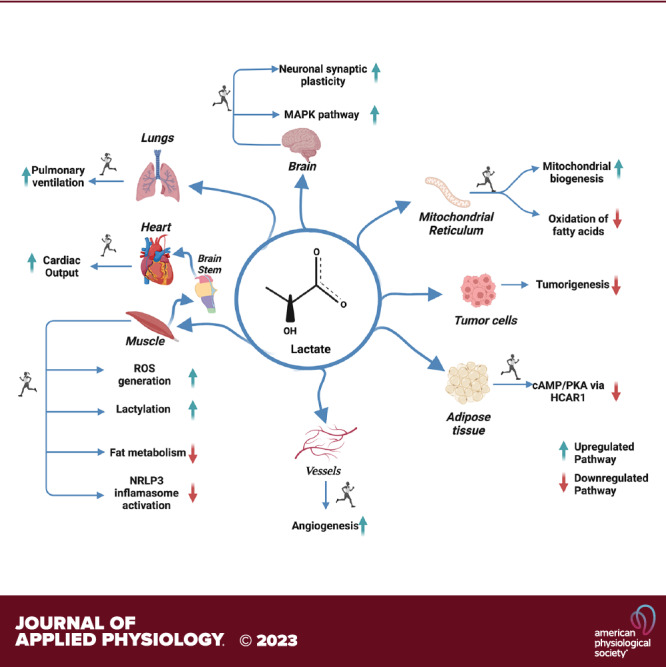
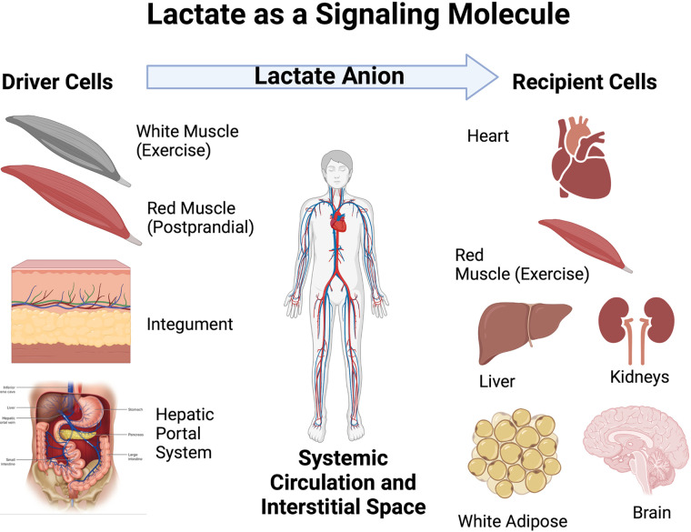
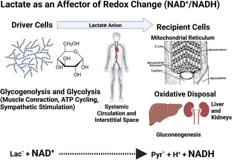
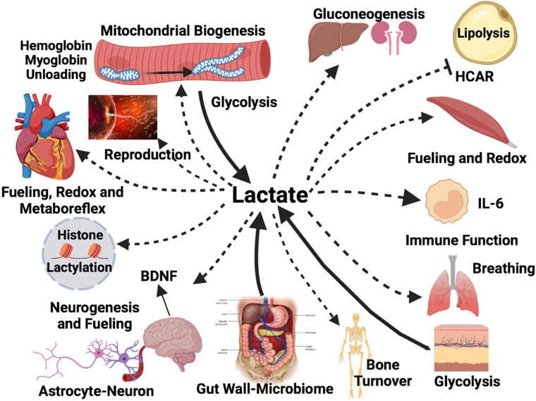
No longer viewed as a metabolic waste product and cause of muscle fatigue, a contemporary view incorporates the roles of lactate in metabolism, sensing and signaling in normal as well as pathophysiological conditions. Lactate exists in millimolar concentrations in muscle, blood, and other tissues and can rise more than an order of magnitude as the result of increased production and clearance limitations. Lactate exerts its powerful driver-like influence by mass action, redox change, allosteric binding, and other mechanisms described in this article. Depending on the condition, such as during rest and exercise, following carbohydrate nutrition, injury, or pathology, lactate can serve as a myokine or exerkine with autocrine-, paracrine-, and endocrine-like functions that have important basic and translational implications. For instance, lactate signaling is: involved in reproductive biology, fueling the heart, muscle adaptation, and brain executive function, growth and development, and a treatment for inflammatory conditions. Lactate also works with many other mechanisms and factors in controlling cardiac output and pulmonary ventilation during exercise. Ironically, lactate can be disruptive of normal processes such as insulin secretion when insertion of lactate transporters into pancreatic β-cell membranes is not suppressed, and in carcinogenesis when factors that suppress carcinogenesis are inhibited, whereas factors that promote carcinogenesis are upregulated. Lactate signaling is important in areas of intermediary metabolism, redox biology, mitochondrial biogenesis, neurobiology, gut physiology, appetite regulation, nutrition, and overall health and vigor. The various roles of lactate as a myokine and exerkine are reviewed.NEW & NOTEWORTHY Lactate sensing and signaling is a relatively new and rapidly changing field. As a physiological signal lactate works both independently and in concert with other signals. Lactate operates via covalent binding and canonical signaling, redox change, and lactylation of DNA. Lactate can also serve as an element of feedback loops in cardiopulmonary regulation. From conception through aging lactate is not the only a myokine or exerkine, but it certainly deserves consideration as a physiological signal.

Keywords: cardiopulmonary regulation; glucose paradox; lactate shuttle; lactylation; metabolic signaling.


Conflict of interest statement

No conflicts of interest, financial or otherwise, are declared by the authors.

Figures

None
Graphical abstract
Figure 1.
Figure 1.
Illustration of the roles of driver and recipient cells in lactate shuttle signaling. Lactate fluxes from sites of production and high concentration in driver cell compartments and tissues to sites of lower concentration in recipient disposal sites. Depending on metabolic conditions some sites can switch from driver to recipient cells. Examples of switching are several and include initial lactate release from muscle beds at the onset of exercise to uptake by the same muscle bed as blood flow and oxygenation increase to meet metabolic demands. At that time, other tissues such as the integument become lactate shuttle drivers. Another example occurs after carbohydrate nutrition when red skeletal muscle takes up glucose and releases lactate as part of the “postprandial lactate shuttle.” Seen from the perspective of Fig. 1, lactate shuttling provides for fuel energy carbon exchange and metabolic signaling. Figure modified from Ref. . Recreated with BioRender.com.
Figure 2.
Figure 2.
Illustration of the cellular redox exchange caused by lactate shuttling. At driver sites, lactate production results for reduction of pyruvate to lactate. However, at recipient sites oxidation of lactate to pyruvate occurs. Pyruvate reduction to lactate and subsequent oxidation of lactate to pyruvate result in millimolar changes in cellular NADH/NAD+ ratios. Among other forms of lactate signaling described in text or Fig. 3, changes in cell redox caused by lactate shuttling are most profound. Figure is a pictorial representation of data in Ref. , created with BioRender.com.
Figure 3.
Figure 3.
Illustration of diverse forms of intracellular lactate shuttling. Lactate producer (Driver) cells and tissues (broad solid lines and arrow heads) contributing to circulating lactate include contributions from the integument, gut, fast-glycolytic skeletal muscle, postprandial red skeletal muscle, and mixed skeletal muscle at the onset of exercise. Lactate consumer (Recipient) sites disposing of lactate (dashed lines and lesser arrow heads) include mitochondrial lactate oxidation in red and mixed skeletal muscle, the heart and brain during steady rate exercise. Also included are (dashed lines and lesser arrow heads) for lactate disposal via gluconeogenesis in the liver and kidneys, and for brain neurons (as part of the ANLS). Lactate-stimulated IL-6 release from monocytes and working muscle is an example of lactate-stimulated cytokine release. Whether drivers or recipients, all cells experience redox signaling effects. Signaling sites not involving carbon exchange or transformation include white adipose where lactate inhibits lipolysis via HCAR and CREB signaling, the heart when peripheral muscle lactate accumulation stimulates the metaboreflex with afferent signaling to the medullary cardiovascular center via Types III- and -IV sensory fibers which increases cardiac output, pulmonary ventilation via the carotid body olfactory receptor (Olfr78), the skeletal muscle where stimulates mitochondrial biogenesis via peroxisome proliferator-activated receptor gamma coactivator-1 alpha (PGC-1α), reactive oxygen species (ROS) and sirtuin activation. Further, lactate has the following actions: In working muscle lactate dissociates oxymyoglobin and blood oxyhemoglobin; in the brain lactate from the arterial circulation of glycolysis in astrocytes fuels neurons and participates in glutamatergic signaling as well as stimulates neurogenesis in the hippocampus and brain-derived neurotropic factor (BDNF) secretion. Moreover, lactatemia and tissue lactate accumulation have an epigenetic effect via lactylation of histones, and lactate has anti-inflammatory effects. Tissues involved starting top left and looking clockwise: skeletal muscle fibers, gluconeogenic organs the liver and kidneys, white adipose tissue, working red skeletal muscle, monocytes, the lungs, integument, skeleton, gut wall and microbiome, the brain, all nucleated cells containing DNA, the heart, ova, and sperm. Created with BioRender.com. Solid and dashed lines indicate flux directions, but not rates because typically lactate Ra = Rd in a steady state.

Similar articles

Cited by

References

    1. Brooks GA. Lactate: glycolytic end product and oxidative substrate during sustained exercise in mammals—the “lactate shuttle”. In: Circulation, Respiration, and Metabolism. Proceedings in Life Sciences, edited by Gilles R. Berlin, Heidelberg: Springer, 1985, p. 208–218.
    1. Brooks GA. Lactate shuttles in nature. Biochem Soc Trans 30: 258–264, 2002. doi:10.1042/bst0300258. - DOI - PubMed
    1. Brooks GA. The science and translation of lactate shuttle theory. Cell Metab 27: 757–785, 2018. doi:10.1016/j.cmet.2018.03.008. - DOI - PubMed
    1. Brooks GA, Arevalo JA, Osmond AD, Leija RG, Curl CC, Tovar AP. Lactate in contemporary biology: a phoenix risen. J Physiol 600: 1229–1251, 2022. doi:10.1113/JP280955. - DOI - PMC - PubMed
    1. Hui S, Ghergurovich JM, Morscher RJ, Jang C, Teng X, Lu W, Esparza LA, Reya T, Le Z, Yanxiang Guo J, White E, Rabinowitz JD. Glucose feeds the TCA cycle via circulating lactate. Nature 551: 115–118, 2017. doi:10.1038/nature24057. - DOI - PMC - PubMed

Publication types