

Type II taste cells participate in mucosal immune surveillance
- PMID: 36634039
- PMCID: PMC9836272
- DOI: 10.1371/journal.pbio.3001647
Type II taste cells participate in mucosal immune surveillance
Abstract
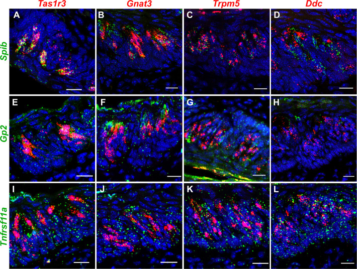
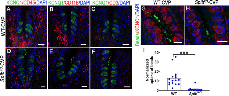
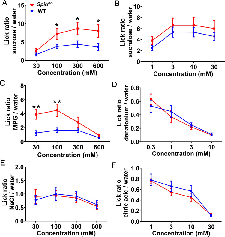
The oral microbiome is second only to its intestinal counterpart in diversity and abundance, but its effects on taste cells remains largely unexplored. Using single-cell RNASeq, we found that mouse taste cells, in particular, sweet and umami receptor cells that express taste 1 receptor member 3 (Tas1r3), have a gene expression signature reminiscent of Microfold (M) cells, a central player in immune surveillance in the mucosa-associated lymphoid tissue (MALT) such as those in the Peyer's patch and tonsils. Administration of tumor necrosis factor ligand superfamily member 11 (TNFSF11; also known as RANKL), a growth factor required for differentiation of M cells, dramatically increased M cell proliferation and marker gene expression in the taste papillae and in cultured taste organoids from wild-type (WT) mice. Taste papillae and organoids from knockout mice lacking Spib (SpibKO), a RANKL-regulated transcription factor required for M cell development and regeneration on the other hand, failed to respond to RANKL. Taste papillae from SpibKO mice also showed reduced expression of NF-κB signaling pathway components and proinflammatory cytokines and attracted fewer immune cells. However, lipopolysaccharide-induced expression of cytokines was strongly up-regulated in SpibKO mice compared to their WT counterparts. Like M cells, taste cells from WT but not SpibKO mice readily took up fluorescently labeled microbeads, a proxy for microbial transcytosis. The proportion of taste cell subtypes are unaltered in SpibKO mice; however, they displayed increased attraction to sweet and umami taste stimuli. We propose that taste cells are involved in immune surveillance and may tune their taste responses to microbial signaling and infection.
Copyright: © 2023 Qin et al. This is an open access article distributed under the terms of the Creative Commons Attribution License, which permits unrestricted use, distribution, and reproduction in any medium, provided the original author and source are credited.
Conflict of interest statement
The authors have declared that no competing interests exist.
Figures

Comment in
-
A possible role for taste receptor cells in surveying the oral microbiome.PLoS Biol. 2023 Jan 13;21(1):e3001953. doi: 10.1371/journal.pbio.3001953. eCollection 2023 Jan. PLoS Biol. 2023. PMID: 36638078 Free PMC article.
Similar articles
-
Aggravated gut inflammation in mice lacking the taste signaling protein α-gustducin.Brain Behav Immun. 2018 Jul;71:23-27. doi: 10.1016/j.bbi.2018.04.010. Epub 2018 Apr 17. Brain Behav Immun. 2018. PMID: 29678794 Free PMC article.
-
A subset of broadly responsive Type III taste cells contribute to the detection of bitter, sweet and umami stimuli.PLoS Genet. 2020 Aug 13;16(8):e1008925. doi: 10.1371/journal.pgen.1008925. eCollection 2020 Aug. PLoS Genet. 2020. PMID: 32790785 Free PMC article.
-
c-Rel is dispensable for the differentiation and functional maturation of M cells in the follicle-associated epithelium.Immunobiology. 2017 Feb;222(2):316-326. doi: 10.1016/j.imbio.2016.09.008. Epub 2016 Sep 18. Immunobiology. 2017. PMID: 27663963 Free PMC article.
-
Development of Peyer's patches, follicle-associated epithelium and M cell: lessons from immunodeficient and knockout mice.Semin Immunol. 1999 Jun;11(3):183-91. doi: 10.1006/smim.1999.0174. Semin Immunol. 1999. PMID: 10381864 Review.
-
An alternative pathway for sweet sensation: possible mechanisms and physiological relevance.Pflugers Arch. 2020 Dec;472(12):1667-1691. doi: 10.1007/s00424-020-02467-1. Epub 2020 Oct 8. Pflugers Arch. 2020. PMID: 33030576 Review.
Cited by
-
Altered peripheral taste function in a mouse model of inflammatory bowel disease.Sci Rep. 2023 Nov 2;13(1):18895. doi: 10.1038/s41598-023-46244-3. Sci Rep. 2023. PMID: 37919307 Free PMC article.
-
Interkingdom Detection of Bacterial Quorum-Sensing Molecules by Mammalian Taste Receptors.Microorganisms. 2023 May 16;11(5):1295. doi: 10.3390/microorganisms11051295. Microorganisms. 2023. PMID: 37317269 Free PMC article. Review.
-
Organoids in gastrointestinal diseases: from bench to clinic.MedComm (2020). 2024 Jun 29;5(7):e574. doi: 10.1002/mco2.574. eCollection 2024 Jul. MedComm (2020). 2024. PMID: 38948115 Free PMC article. Review.
-
A possible role for taste receptor cells in surveying the oral microbiome.PLoS Biol. 2023 Jan 13;21(1):e3001953. doi: 10.1371/journal.pbio.3001953. eCollection 2023 Jan. PLoS Biol. 2023. PMID: 36638078 Free PMC article.
-
Deciphering the M-cell niche: insights from mouse models on how microfold cells "know" where they are needed.Front Immunol. 2024 May 28;15:1400739. doi: 10.3389/fimmu.2024.1400739. eCollection 2024. Front Immunol. 2024. PMID: 38863701 Free PMC article. Review.
References
Publication types
MeSH terms
Substances
Grants and funding
LinkOut - more resources
Full Text Sources
Molecular Biology Databases
