Differential effects of anti-CD20 therapy on CD4 and CD8 T cells and implication of CD20-expressing CD8 T cells in MS disease activity
- PMID: 36634138
- PMCID: PMC9934304
- DOI: 10.1073/pnas.2207291120
Differential effects of anti-CD20 therapy on CD4 and CD8 T cells and implication of CD20-expressing CD8 T cells in MS disease activity
Abstract
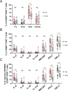
A small proportion of multiple sclerosis (MS) patients develop new disease activity soon after starting anti-CD20 therapy. This activity does not recur with further dosing, possibly reflecting deeper depletion of CD20-expressing cells with repeat infusions. We assessed cellular immune profiles and their association with transient disease activity following anti-CD20 initiation as a window into relapsing disease biology. Peripheral blood mononuclear cells from independent discovery and validation cohorts of MS patients initiating ocrelizumab were assessed for phenotypic and functional profiles using multiparametric flow cytometry. Pretreatment CD20-expressing T cells, especially CD20dimCD8+ T cells with a highly inflammatory and central nervous system (CNS)-homing phenotype, were significantly inversely correlated with pretreatment MRI gadolinium-lesion counts, and also predictive of early disease activity observed after anti-CD20 initiation. Direct removal of pretreatment proinflammatory CD20dimCD8+ T cells had a greater contribution to treatment-associated changes in the CD8+ T cell pool than was the case for CD4+ T cells. Early disease activity following anti-CD20 initiation was not associated with reconstituting CD20dimCD8+ T cells, which were less proinflammatory compared with pretreatment. Similarly, this disease activity did not correlate with early reconstituting B cells, which were predominantly transitional CD19+CD24highCD38high with a more anti-inflammatory profile. We provide insights into the mode-of-action of anti-CD20 and highlight a potential role for CD20dimCD8+ T cells in MS relapse biology; their strong inverse correlation with both pretreatment and early posttreatment disease activity suggests that CD20-expressing CD8+ T cells leaving the circulation (possibly to the CNS) play a particularly early role in the immune cascades involved in relapse development.
Keywords: CD20-expressing T cells; CD20dim T cells; CD20dimCD8+ T cells; anti-CD20 therapy; ocrelizumab.
Conflict of interest statement
The authors have organizational affiliations to disclose. J.L.B. has received personal fees and nonfinancial support from Chugai Pharmaceutical, Viela Bio/Horizon Therapeutics, Equillium, Frequency Therapeutics, Mitsubishi-Tanabe, Reistone Bio, Abbvie, Clene Neuroscience, Alexion, Genentech, and Roche; and grants from Mallinckrodt and Novartis. H.-C.v.B. is an employee of F. Hoffmann-La Roche Ltd. R.C. is Site Investigator for studies funded by Roche, Novartis, MedImmune, and EMD Serono and has received honoraria from Roche, EMD 75 Serono, Sanofi, Biogen, Novartis, and Teva. K.R.E. has received research/grant support from Biogen, Sanofi Genzyme, F. Hoffmann-La Roche Ltd., Genentech, Inc., and Novartis. R.F. has nothing to declare. P.S.G. has received honoraria for speaking, advisory boards or consultation fees from Actelion, Alexion, Biogen, Bristol Myers Squibb-Celgene, EMD Serono, Sanofi Genzyme, Innodem Neurosciences, McKesson, Novartis, Pendopharm, F. Hoffmann-La Roche Ltd., and Teva Neuroscience. He also serves as a scientific advisor for Innodem Neurosciences. B.M.G. has received consulting fees from Alexion, Novartis, EMD Serono, Viela Bio, Genentech/Roche, Greenwhich Biosciences, Axon Advisors, Rubin Anders, ABCAM, Signant, IQVIA, Sandoz, DruggabilityTechnologies, Genzyme, Immunovant, and PRIME Education. He has received grant funding from PCORI, NIH, NMSS, The Siegel Rare Neuroimmune Association, Clene Nanomedicine, and the Guthy Jackson Charitable Foundation for NMO. He serves as an unpaid member of the board of the Siegel Rare Neuroimmune Association. He receives royalties from UpToDate. D.A.H. has in the past 10 y consulted for the following companies: Bayer, Biohaven Pharmaceuticals, Bristol-Myers Squibb, Compass Therapeutics, Eisai, EMD Serono, Genentech, Inc., Juno Therapeutics, McKinsey & Co., MedImmune/AstraZeneca, Mylan Pharmaceuticals, Neurophage Pharmaceuticals, NKT Therapeutics, Novartis, Proclara Biosciences, Questcor Pharmaceuticals, F. Hoffmann-La Roche Ltd., Sage Therapeutics, Sanofi Genzyme, Toray Industries, and Versant Venture. C.I. has received consulting fees from EMD Serono and Sanofi Genzyme. C.B.L. has served on scientific advisory boards or as a speaker for Biogen, Sanofi, EMD Serono, Alexion, and Bristol-Myers Squibb and has done consulting for InterX, Inc. and Diagnose Early. E.E.L. has received honoraria for consulting from Genentech, Inc., Genzyme, EMD Serono, Bristol Myers Squibb, TG Therapeutics, NGM Bio, Janssen, and Biogen. G.P. has served on advisory boards and/or speaker bureaus for Biogen, Celgene/Bristol Myers Squibb, EMD Serono, Genentech, Inc., F. Hoffmann-La Roche Ltd., Novartis, Sanofi Genzyme, Greenwich Biosciences, Teva, and VielaBio/Horizon. F.P. has received fees for serving on DMC in clinical trials with Chugai, Lundbeck and Roche, and preparation of expert witness statement for Novartis. M.S.W. is serving as an editor for PLoS One and has received travel funding and/or speaker honoraria from Biogen, Merck Serono, Novartis, F. Hoffmann-La Roche Ltd., Teva, Bayer, and Genzyme. T.Z. received personal compensation from Almirall, Biogen, Bayer, Celgene, Novartis, Roche, Sanofi, and Teva for the consulting services. D.J. received consulting fees and/or research support from: Biogen, Genentech, Novartis, EMD Serono, Banner Life Sciences, Bristol Myers Squibb, Horizon, and Sanofi Genzyme. J.M.G. has received consulting fees from Biogen. A.H.C. has received fees or honoraria for consulting for Biogen, Celgene/Bristol Myers Squibb, EMD Serono, F. Hoffmann-La Roche Ltd., Genentech, Inc., and Novartis, and TG Therapeutics and received fees for serving on scientific advisory boards and reviewing grants for the Conrad N. Hilton Foundation and Race to Erase MS. B.C., B.M., R.C.W., X.J., C.T.H., and A.H. are employees of Genentech, Inc. A.B.-O. has participated as a speaker in meetings sponsored by and received consulting fees from Janssen Pharmaceuticals/Actelion, Atara Biotherapeutics, Biogen Idec, Celgene/Receptos, Roche/Genentech, Mapi Pharma, MedImmune, Merck/EMD Serono, Novartis, and Sanofi Genzyme. Yes, the authors have stock ownership to disclose. B.C., B.M., R.C.W., X.J., C.T.H., and A.H. are shareholders of F. Hoffmann-La Roche Ltd. R. Yes, the authors have research support to disclose. J.L.B. has received personal fees and nonfinancial support from Chugai Pharmaceutical, Viela Bio/Horizon Therapeutics, Equillium, Frequency Therapeutics, Mitsubishi-Tanabe, Reistone Bio, Abbvie, Clene Neuroscience, Alexion, Genentech, and Roche and grants from Mallinckrodt and Novartis. R.C. receives research support from Teva Innovation Canada, Roche Canada, and Vancouver Coastal Health Research Institute. K.R.E. has received research/grant support from Biogen, Sanofi Genzyme, F. Hoffmann-La Roche Ltd., Genentech, Inc., and Novartis. P.S.G. has received research or educational grant from Biogen, EMD Serono, Genzyme-Sanofi, Hoffmann-La Roche, and Teva Innovation Canada. He has received honoraria for speaking and advisory board participation from Actelion, Allergan, Biogen Idec, EMD Serono, Sanofi Genzyme, Merz, Novartis, Pendopharm, F. Hoffmann-La Roche Ltd., and Teva Neuroscience. D.A.H. has received generous support by grants from the National Institutes of Health (U10 AI089992, R25 NS079193, P01 AI073748, U24 AI11867, R01 AI22220, UM 1HG009390, P01 AI039671, P50 CA121974, and R01 CA227473) and the National Multiple Sclerosis Society (CA 1061-A-18, RG-1802-30153); is also supported by grants from the National Institute of Neurological Disorders and Stroke and the Nancy Taylor Foundation for Chronic Diseases; and has received funding for his laboratory from Bristol-Myers Squibb, Genentech, Inc., Novartis Questcor, Sanofi Genzyme, and Race to Erase MS. C.I. has received search support from F. Hoffmann-La Roche Ltd, Genentech, Inc., Biogen, and Sanofi Genzyme. E.E.L. has received research funding from Genentech, Inc. and Biogen. F.P. has received research grants from Janssen, Merck KGaA, and UCB. M.S.W. receives research support from the Deutsche Forschungsgemeinschaft (WE 3547/5-1), Novartis, Teva, Biogen Idec, F. Hoffmann-La Roche Ltd., Merck, and the ProFutura Programm of the Universitätsmedizin Göttingen. T. Ziemssen received additional financial support for the research activities from Biogen, Novartis, Teva, and Sanofi. D.J. received consulting fees and/or research support from Biogen, Genentech, Novartis, EMD Serono, Banner Life Sciences, Bristol Myers Squibb, Horizon, and Sanofi Genzyme. J.M.G. has received research support to UCSF from Roche/Genentech and Vigil Neurosciences for clinical trials. A.B.-O. has received grant support from Biogen Idec, Roche/Genentech, Merck/EMD Serono, Novartis, and Sanofi-Genzyme.
Figures


Comment in
-
Learning multiple sclerosis immunopathogenesis from anti-CD20 therapy.Proc Natl Acad Sci U S A. 2023 Feb 7;120(6):e2221544120. doi: 10.1073/pnas.2221544120. Epub 2023 Jan 31. Proc Natl Acad Sci U S A. 2023. PMID: 36719925 Free PMC article. No abstract available.
References
-
- Li R., Patterson K. R., Bar-Or A., Reassessing B cell contributions in multiple sclerosis. Nat. Immunol. 19, 696–707 (2018). - PubMed
-
- Bar-Or A., et al. , Rituximab in relapsing-remitting multiple sclerosis: A 72-week, open-label, phase I trial. Ann. Neurol. 63, 395–400 (2008). - PubMed
-
- Hauser S. L., et al. , B-cell depletion with rituximab in relapsing-remitting multiple sclerosis. N. Engl. J. Med. 358, 676–688 (2008). - PubMed
-
- Kappos L., et al. , Ocrelizumab in relapsing-remitting multiple sclerosis: A phase 2, randomised, placebo-controlled, multicentre trial. Lancet 378, 1779–1787 (2011). - PubMed
Publication types
MeSH terms
Substances
Grants and funding
LinkOut - more resources
Full Text Sources
Medical
Research Materials

