. 2023 Jan 13;379(6628):201-206.
doi: 10.1126/science.add3598. Epub 2023 Jan 12.

Excessive mechanotransduction in sensory neurons causes joint contractures

Affiliations

Excessive mechanotransduction in sensory neurons causes joint contractures

Shang Ma et al. Science. .

Abstract

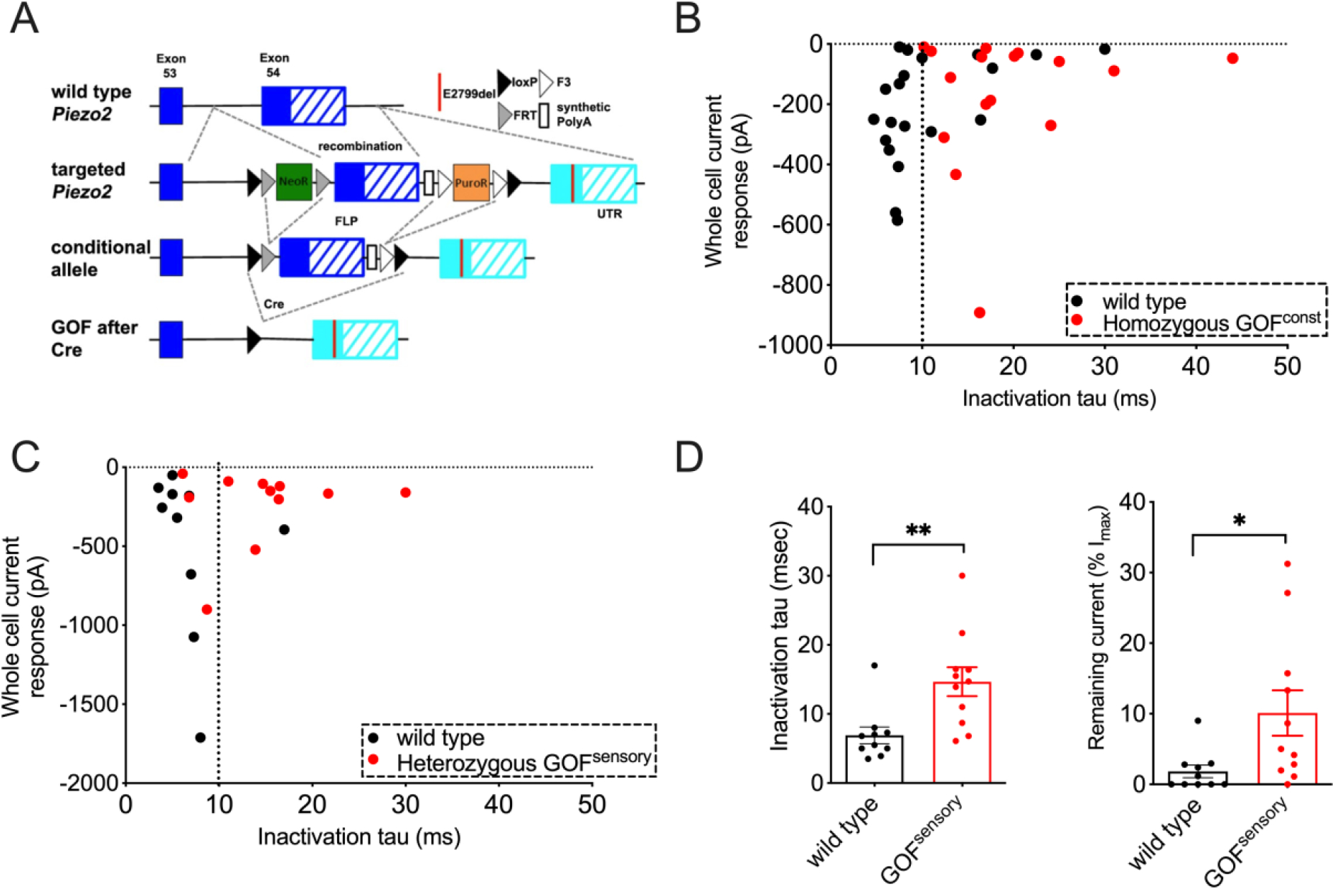
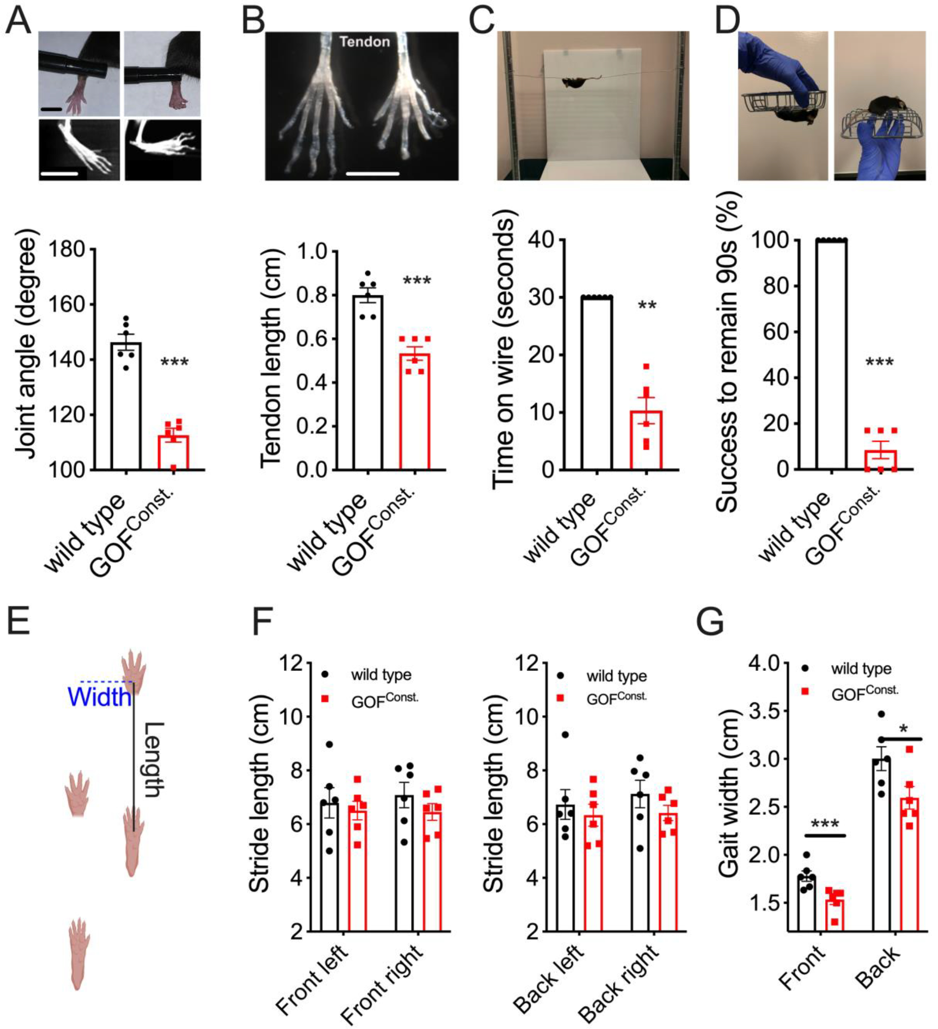
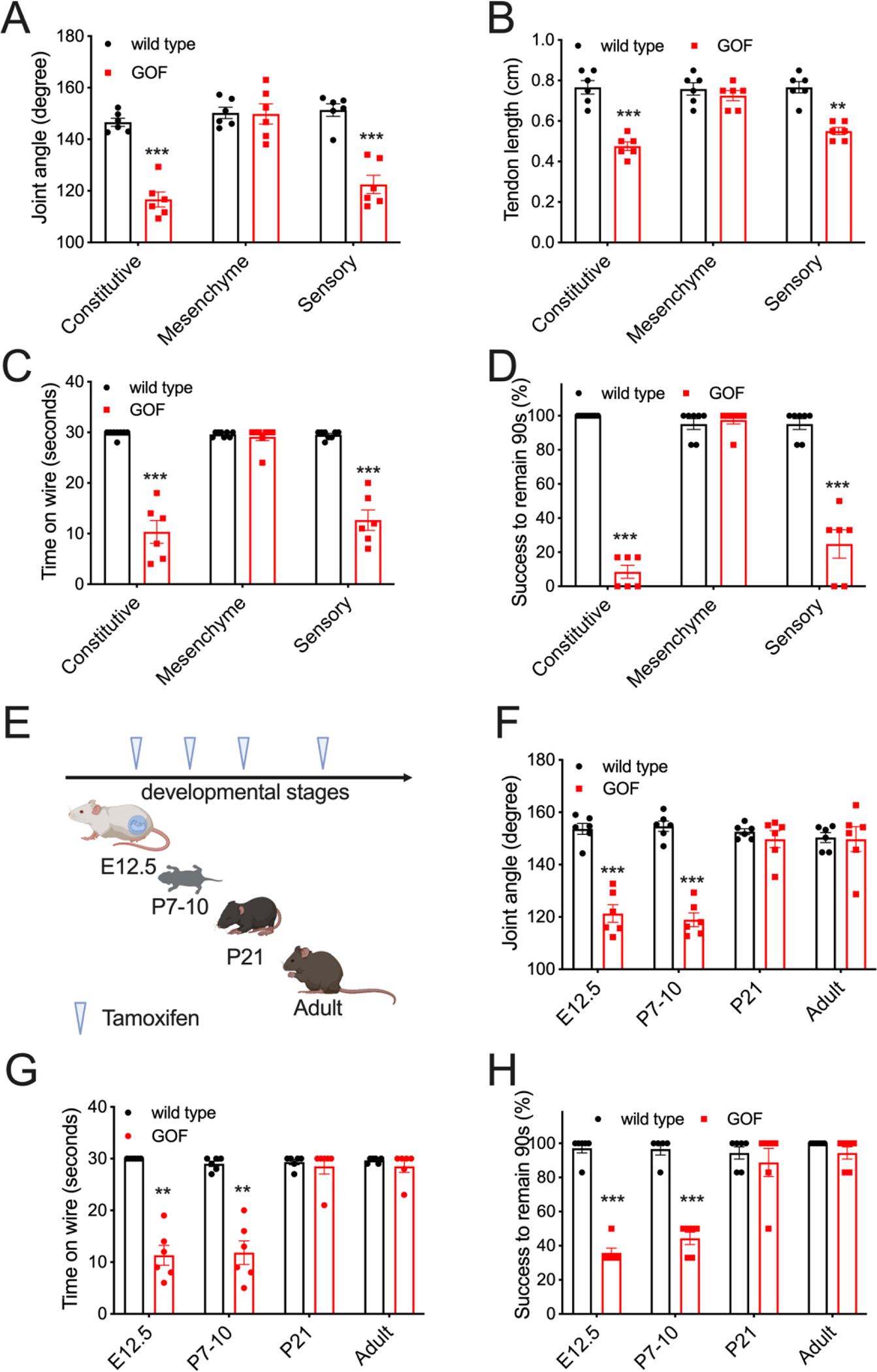
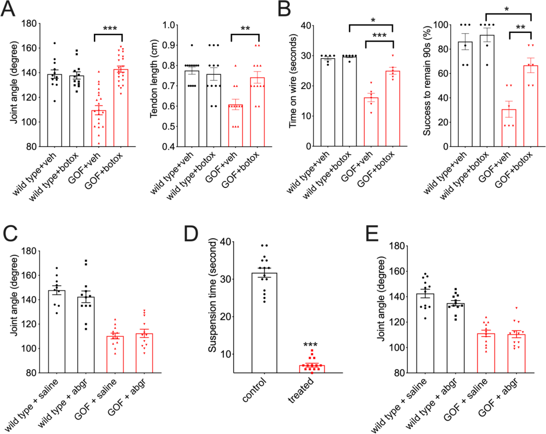
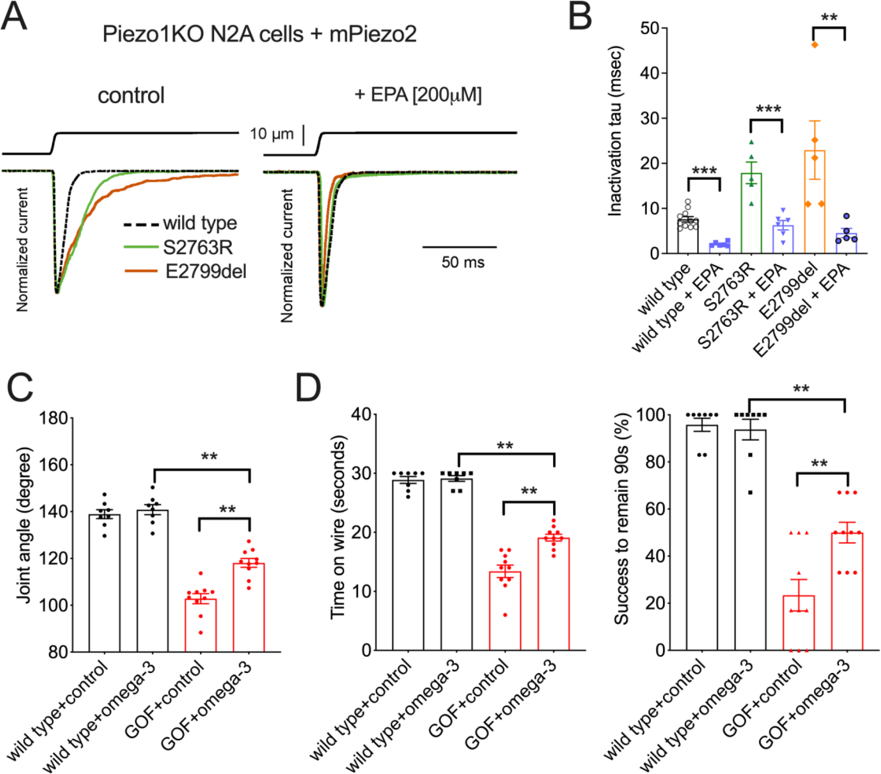
Distal arthrogryposis (DA) is a collection of rare disorders that are characterized by congenital joint contractures. Most DA mutations are in muscle- and joint-related genes, and the anatomical defects originate cell-autonomously within the musculoskeletal system. However, gain-of-function mutations in PIEZO2, a principal mechanosensor in somatosensation, cause DA subtype 5 (DA5) through unknown mechanisms. We show that expression of a gain-of-function PIEZO2 mutation in proprioceptive sensory neurons that mainly innervate muscle spindles and tendons is sufficient to induce DA5-like phenotypes in mice. Overactive PIEZO2 causes anatomical defects through increased activity within the peripheral nervous system during postnatal development. Furthermore, botulinum toxin (Botox) and a dietary fatty acid that modulates PIEZO2 activity reduce DA5-like deficits. This reveals a role for somatosensory neurons: Excessive mechanosensation within these neurons disrupts musculoskeletal development.


Conflict of interest statement

Competing interests: The authors declare no competing financial interests.

Figures

Figure 1.
Figure 1.. Gain-of-function (GOF) PIEZO2 increases mechanosensitivity of sensory neurons.
(A) Conditional GOF Piezo2 mouse model. Cre-dependent replacement of wild type exon (blue) by GOF-carrying exon (cyan). (B) Mechanically activated currents and inactivation time constant (tau) in DRG sensory neurons from wild type (black) and constitutively homozygous GOF Piezo2 mice (red). 87% WT and 70% GOF neurons were responsive to the stimulus. (C) Mechanically activated currents and inactivation time constant in Tdtomato+ neurons from PvalbCre/Ai9 (wild type, black) and GOF Piezo2; PvalbCre/Ai9 mice (red). (D) Bar graphs representing inactivation time constant (ms) and remaining current at the end of the stimulus (% of Imax value) in DRG sensory neurons. *p < 0.05, **p < 0.01, and ***p < 0.001 (Student’s t-test). Each data point represents a single DRG neuron.
Figure 2.
Figure 2.. GOF Piezo2 mice develop limb defects.
(A) Joint morphology of live animals and CT scan (upper images) and phalange-metacarpal joint angles (lower graph) in the hindlimb. Left: wild type; Right: GOF mice. Scale bars: 0.5cm. (B) Intact tendon (upper) and tendon length (lower) from hindlimbs. Scale bar: 0.5cm. (C) Hanging wire test (upper) and Time (sec) that animals remained on the metal wire, with 30 sec as cutoff (lower). (D) Inverted screen test (upper) and quantification as percentage of animals successfully remaining on the rotating screen for 90 seconds (lower). (E) Gait assay: the stride length measures forelimb-hindlimb distance in the first (front) and second (back) stride. Gait width is the distance between forelimbs (front) or between hindlimbs (back) (F-G) Quantifications for E. *p < 0.05, **p < 0.01, and ***p < 0.001 (Student’s t-test). Each data point represents a single animal.
Figure 3.
Figure 3.. GOF PIEZO2 in sensory neurons causes limb defects.
(A) Phalange-metacarpal joint angle of the hindlimbs in wild type and tissue specific GOF mice (black: wild type; red: GOF Piezo2). (B) Tendon length of hindlimbs. (C) Quantification of hanging wire test results. (D) Quantification of inverted screen test results. (E) Tamoxifen induction of GOF PIEZO2 in proprioceptive neurons by AdvillinCre-ERT2 at various developmental stages. (F) Phalange-metacarpal joint angle of the hindlimbs in E. (G) Quantification of hanging wire test results in E. (H) Quantification of inverted screen test in E. *p < 0.05, **p < 0.01, and ***p < 0.001 (One-way ANOVA followed by Tukey’s multiple comparison). Each data point represents a single animal.
Figure 4.
Figure 4.. Pharmacological effects on limb phenotypes in GOF mice.
(A) Phalange-metacarpal joint angle and tendon length of the hindlimbs in young adult wild type and GOF Piezo2; PvalbCre mice that received Botulinum toxin (Botox) at P7–10. (B) Quantification of behavioral tests in mice treated with Botox. (C) Phalange-metacarpal joint angle of the hindlimbs in young adult mice receiving a single dose of alpha-Bungarotoxin (abgr) at P7–10. (D) Quantification for motor defects (in a modified hanging assay) in young adult mice receiving 5-day alpha-Bungarotoxin (abgr) treatment at P7–10. (E) Phalange-metacarpal joint angle of the hindlimbs in young adult mice receiving daily alpha-Bungarotoxin (abgr) treatment from P5–10 in D.
Figure 5.
Figure 5.. EPA diet rescues joint defects in GOF mice.
(A) Mechanically activated currents in N2APiezo1−/− cells heterologously expressing mouse PIEZO2 wild type (stippled) and GOF mutants S2763R (green) and E2799del (brown). (B) Quantification of the inactivation time constant results in A. (C) Phalange-metacarpal joint angle of the hindlimbs in young adult wild type and GOF Piezo2; PvalbCre mice that received EPA-enriched diet. Control: sunflower-oil-based food, which has a similar level of fat content and energy. (D) Quantification of hanging wire and inverted screen test results in C. *p < 0.05, **p < 0.01, and ***p < 0.001 (Two-way ANOVA followed by Tukey honestly significance test).

References

    1. Bamshad M, Van Heest AE, Pleasure D, Arthrogryposis: A Review and Update. Journal of Bone and Joint Surgery. 91, 40–46 (2009). - PMC - PubMed
    1. Kowalczyk B, Feluś J, Arthrogryposis: an update on clinical aspects, etiology, and treatment strategies. aoms. 1, 10–24 (2016). - PMC - PubMed
    1. Tajsharghi H, Kimber E, Kroksmark A-K, Jerre R, Tulinius M, Oldfors A, Embryonic Myosin Heavy-Chain Mutations Cause Distal Arthrogryposis and Developmental Myosin Myopathy That Persists Postnatally. Arch Neurol. 65 (2008). - PubMed
    1. Beals RK, Weleber RG, Distal arthrogryposis 5: A dominant syndrome of peripheral contractures and ophthalmoplegia. Am. J. Med. Genet. 131A, 67–70 (2004). - PubMed
    1. Coste B, Houge G, Murray MF, Stitziel N, Bandell M, Giovanni MA, Philippakis A, Hoischen A, Riemer G, Steen U, Steen VM, Mathur J, Cox J, Lebo M, Rehm H, Weiss ST, Wood JN, Maas RL, Sunyaev SR, Patapoutian A, Gain-of-function mutations in the mechanically activated ion channel PIEZO2 cause a subtype of Distal Arthrogryposis. Proc. Natl. Acad. Sci. U.S.A. 110, 4667–4672 (2013). - PMC - PubMed