. 2023 Feb 8;31(2):305-319.e10.
doi: 10.1016/j.chom.2022.12.011. Epub 2023 Jan 11.

Regulators of male and female sexual development are critical for the transmission of a malaria parasite

Affiliations

Regulators of male and female sexual development are critical for the transmission of a malaria parasite

Andrew J C Russell et al. Cell Host Microbe. .

Abstract

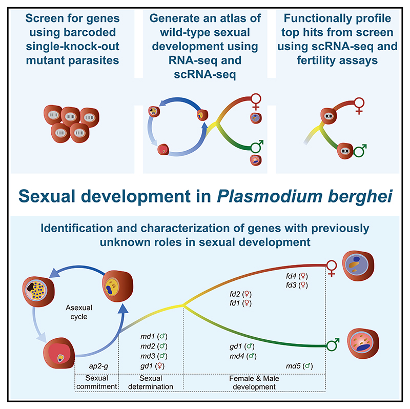
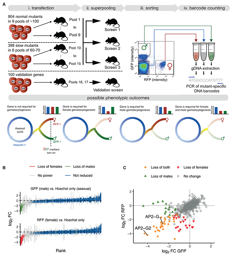
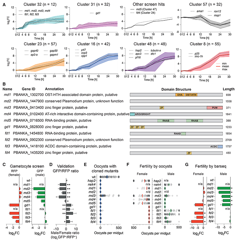
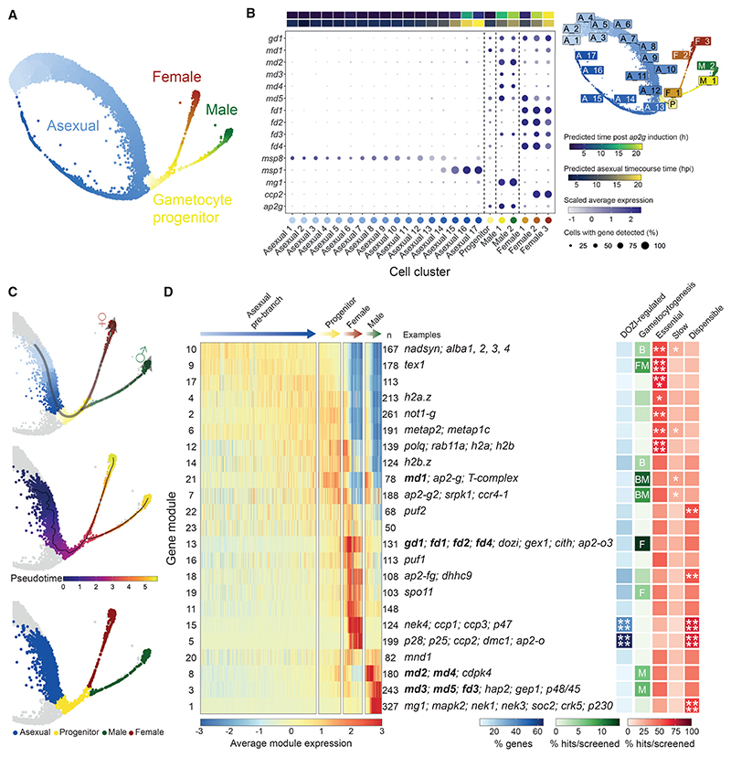
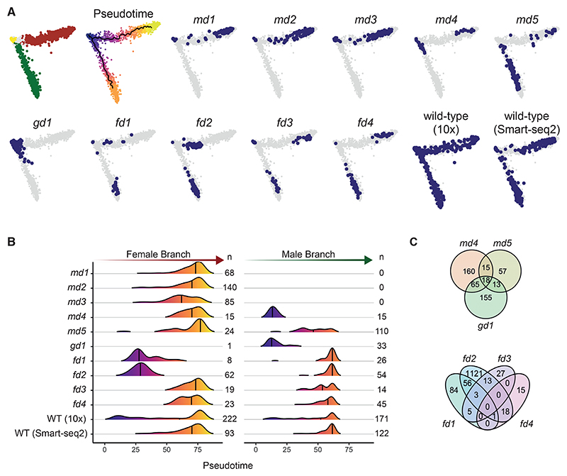
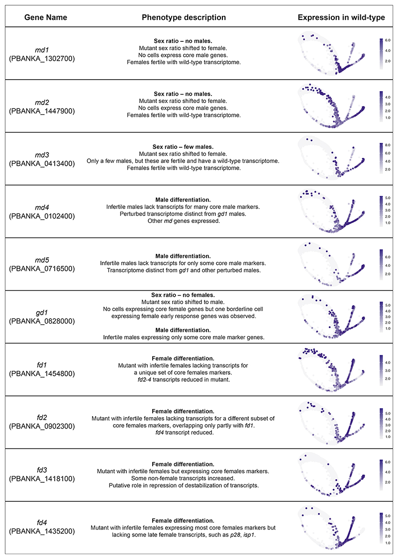
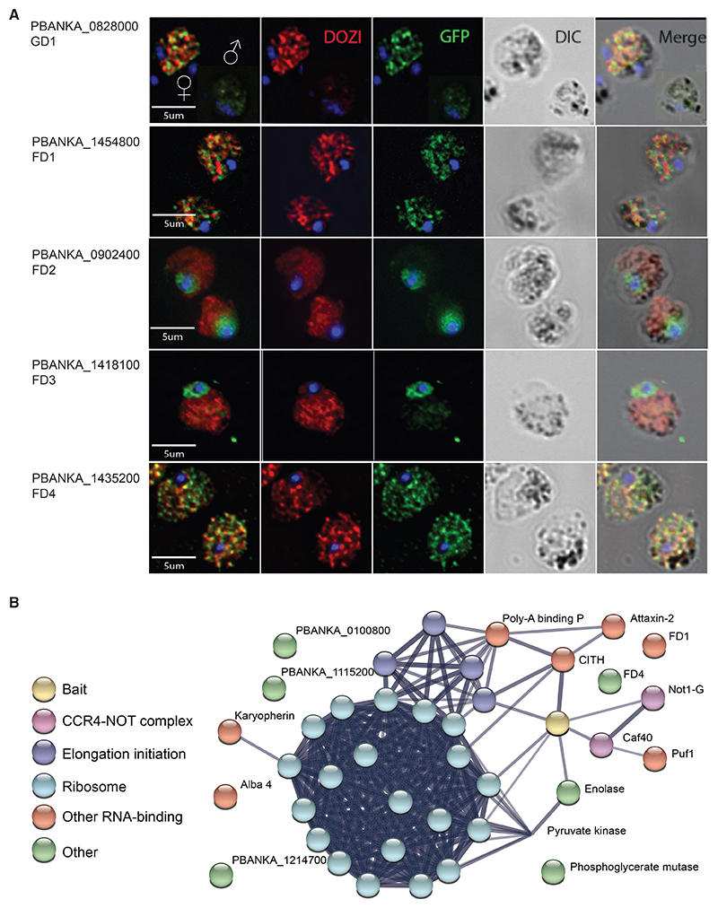
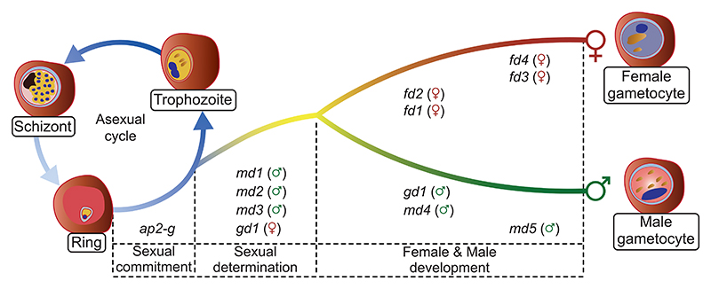
Malaria transmission to mosquitoes requires a developmental switch in asexually dividing blood-stage parasites to sexual reproduction. In Plasmodium berghei, the transcription factor AP2-G is required and sufficient for this switch, but how a particular sex is determined in a haploid parasite remains unknown. Using a global screen of barcoded mutants, we here identify genes essential for the formation of either male or female sexual forms and validate their importance for transmission. High-resolution single-cell transcriptomics of ten mutant parasites portrays the developmental bifurcation and reveals a regulatory cascade of putative gene functions in the determination and subsequent differentiation of each sex. A male-determining gene with a LOTUS/OST-HTH domain as well as the protein interactors of a female-determining zinc-finger protein indicate that germ-granule-like ribonucleoprotein complexes complement transcriptional processes in the regulation of both male and female development of a malaria parasite.

Keywords: Plasmodium; development; differentiation; malaria; sex determination; sex ratio; single cell analysis; transmission.


Conflict of interest statement

Declaration of interests The authors declare no competing interests.

Figures

Graphical abstract
Graphical abstract
Figure 1
Figure 1. A bar-seq screen for sexual reporter expression in P. berghei
(A) Schematic overview showing how PlasmoGEM vectors were allocated to pools for transfection and the resulting mutants combined into superpools for sorting on reporter expression. Duplicate barcode PCRs were performed for each sorted population and converted into sequencing libraries for barcode counting. Possible phenotypic outcomes are illustrated, using sexual marker expression as a proxy for gametocytogenesis genes that affect sexual development after the sex-specific promoters are turned on (dashed line) are unlikely to be identified in the screen. (B) All robustly quantified mutants are ranked by the degree to which cells expressing either the male or the female reporter gene were underrepresented. Error bars show standard deviations from at least 4 independent screening experiments. (C) Combined results from both reporters, showing filled symbols where the underrepresentation was significant for either one or both sexes. Two highlighted mutants in ApiAP2 genes confirm the expected loss of both markers from the population, as published.
Figure 2
Figure 2. Selection and validation of ten P. berghei genes with sex-specific roles in gametocytogenesis
(A) Selected gene expression clusters from a bulk RNA-seq time course of induced sexual development. Ring-stage parasites were reprogramed at t = 0 h by inducing ap2-g. Relative transcript abundances are given as log2-fold change relative to uninduced, asexually developing parasites. Shown are selected clusters (number of genes) with screen hits designated md, fd, or gd. Selected well-characterized marker genes of male, female, and asexual development are also shown. (B) Schematic illustration of genes with validated roles in sexual development. OST-HTH, oskar-TDRD5/TDRD7 winged helix-turn-helix domain; OHA, OST-HTH associated domain; ARID/BRIGHT, AT-rich interaction domain; ZN, C3H1 zinc finger; PUM, Pumilio RNA-binding repeat profile; RNAB, RNA-binding domain; ACDC, apetala 2 domain-coincident C-terminal domain; PH-like, PH domain like. (C) Fold-change (FC) in reporter-positive cells in the bar-seq screen. Error bars show standard deviations. (D) Sex ratio in individual mutants determined by flow cytometry. Error bars show standard deviations from 2 to 4 biological replicates with cloned mutants, except for fd3, where the uncloned population is shown. * p < 0.05; ** p < 0.01 in unpaired t test. (E) Transmission efficiency of mutant clones in vivo determined by counting oocysts on midguts 10 days after an infectious blood meal. (F) Male and female fertility as determined by the ability of mutant clones to give rise to oocysts in mosquitoes when crossed to nek4 and hap2 mutants, which provide fertile male or female gametes, respectively. Oocysts counts show combined data from 25 to 80 dissected mosquitoes from 2 to 3 independent experiments. n/d, not done; n/a, not applicable. (G) Fold-change (FC) in female and male fertility determined by bar-seq of 10-day infected midguts following mutagenesis of female-only or male-only lines, respectively. Error bars show standard deviations from four biological replicates. * p < 0.001 in unpaired t test.
Figure 3
Figure 3. Combined analysis of wild-type 10x and Smart-seq2 transcriptomes from P. berghei-infected mouse red blood cells
(A) UMAP plot of all 6,880 wild-type cells (Smart-seq2 and 10x), colored by their assigned sex designation and progression along development. (B) Dot plot showing expression of known marker genes alongside the candidate genes in 23 cell clusters. The estimated average time point of each cluster is annotated by correlating single-cell data to bulk time course data (this study and another study conducted by Hoo et al.). Cluster locations are shown on the UMAP plot (top right). (C) Wild-type cells were sub-clustered and 10x-only cells (non-gray dots) were selected in order to re-calculate pseudotime for the branches of interest and construct modules of genes that are co-expressed over pseudotime. These selected cells are colored by sex assignments (bottom), recalculated pseudotime values (middle), and a composite of sex assignments and pseudotime (top), superimposed on the UMAP in (A). Non-branch cells are colored gray. (D) Heatmap showing the scaled average expression of gene modules in cells shown in (C). n, the number of genes per module; DOZI-regulated, % of DOZI-regulated genes within each cluster according to Guerreiro et al. M, F, and B represent significant (p ≤ 0.05) enrichment of screen phenotype in males only, females only, or both sexes, respectively. Essential, Slow, Dispensable, % genes per cluster with asexual blood-stage phenotype according to Bushell et al. Significance of enrichment: * p ≤ 0.05; ** p ≤ 0.01; *** p ≤ 0.001; **** p ≤ 0.0001. See Figure 5 for details. Exemplary genes for each cluster are shown.
Figure 4
Figure 4. Smart-seq2 analysis of in vitro-matured wild-type and mutant parasites
(A) Principal-component analysis (PCA) plots of 3,012 transcriptomes from single parasitized red blood cells of the sexual branch that were obtained by merging all wild-type and mutant cells and subsetting the branch of interest (Figure S5). (B) Density plots showing the distribution of assigned female (left) and male (right) cells along each pseudotime trajectory and grouped by genotype. n, the number of cells for each condition as shown in (A). WT, wild type. The line indicates the median. (C) Venn diagrams showing the numbers of genes with differences in transcript abundance in female and male gametocytes, respectively, relative to wild-type. Genes that result in the absence of a sex cannot be evaluated.
Figure 5
Figure 5. Summary data from Smart-seq2 experiments of mutants
A description of the phenotype observed from the single-cell RNA-seq data is given along with a summary of the fertility phenotype (Figures 2E–2G). On the right-hand side, the scaled expression of the gene in wild-type only cells is shown with the 5th and 95th quantiles set as the minimum and maximum expression values, respectively, to eliminate any outliers having a strong influence on visualization.
Figure 6
Figure 6. Functional analysis of GD1 and female development genes FD1-4
(A) Immunofluorescence images of fixed gametocytes expressing C-terminally GFP-tagged proteins from their endogenous promoters. Female gametocytes are shown except where indicated. The stress granule helicase DOZI served as a female marker. Images are representative of ca. 500 inspected cells. (B) STRING association network of selected proteins specifically co-immunoprecipitated with GD1-3xHA with a significance analysis of interactome (SAINT) score probability > 0.9. See Table S6 for a complete list of putative interactors with gene IDs.
Figure 7
Figure 7. Model of how sexual determination and differentiation is affected by a cascade dominated by putative nucleic acid-binding proteins identified in this study
In addition to its role in commitment, ap2g probably has sex-specific roles in differentiation, together withthe female-specific transcription factor AP2-FG, which binds in the promoter regions of fd2 and fd4.

Comment in

References

    1. Brancucci NMB, Bertschi NL, Zhu L, Niederwieser I, Chin WH, Wampfler R, Freymond C, Rottmann M, Felger I, Bozdech Z, Voss TS. Heterochromatin protein 1 secures survival and transmission of malaria parasites. Cell Host Microbe. 2014;16:165–176. - PubMed
    1. Filarsky M, Fraschka SA, Niederwieser I, Brancucci NMB, Carrington E, Carrió E, Moes S, Jenoe P, Bártfai R, Voss TS. GDV1 induces sexual commitment of malaria parasites by antagonizing HP1-dependent gene silencing. Science. 2018;359:1259–1263. - PMC - PubMed
    1. Kafsack BFC, Rovira-Graells N, Clark TG, Bancells C, Crowley VM, Campino SG, Williams AE, Drought LG, Kwiatkowski DP, Baker DA, et al. A transcriptional switch underlies commitment to sexual development in malaria parasites. Nature. 2014;507:248–252. - PMC - PubMed
    1. Sinha A, Hughes KR, Modrzynska KK, Otto TD, Pfander C, Dickens NJ, Religa AA, Bushell E, Graham AL, Cameron R, et al. A cascade of DNA-binding proteins for sexual commitment and development in Plasmodium. Nature. 2014;507:253–257. - PMC - PubMed
    1. Llorà-Batlle O, Michel-Todó L, Witmer K, Toda H, Fernández-Becerra C, Baum J, Cortés A. Conditional expression of PfAP2-G for controlled massive sexual conversion in Plasmodium falciparum. Sci Adv. 2020;6:eaaz5057. - PMC - PubMed

Substances