. 2023 Feb;299(2):102894.
doi: 10.1016/j.jbc.2023.102894. Epub 2023 Jan 10.

Assembly chaperone Nas6 selectively destabilizes 26S proteasomes with defective regulatory particle-core particle interfaces

Affiliations

Assembly chaperone Nas6 selectively destabilizes 26S proteasomes with defective regulatory particle-core particle interfaces

Jennifer L Warnock et al. J Biol Chem. 2023 Feb.

Abstract

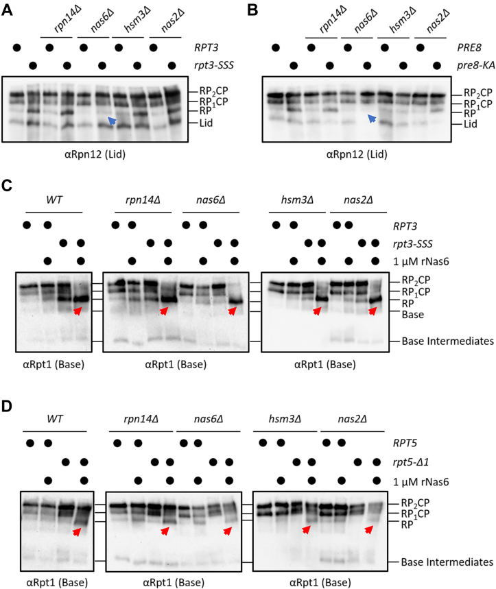
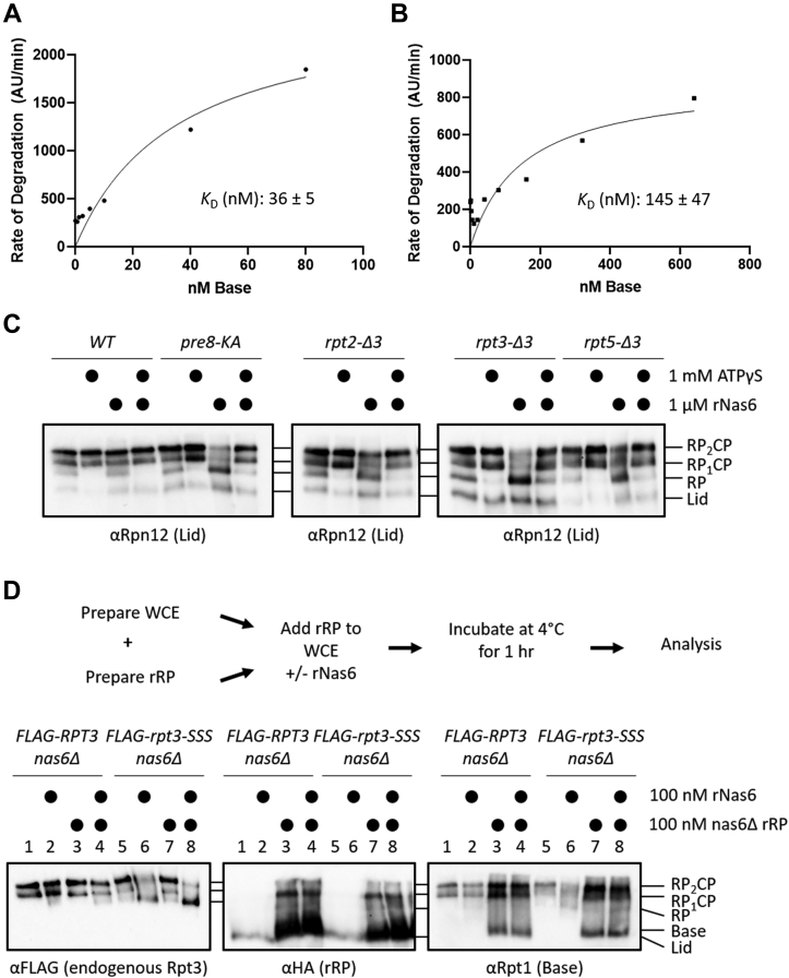
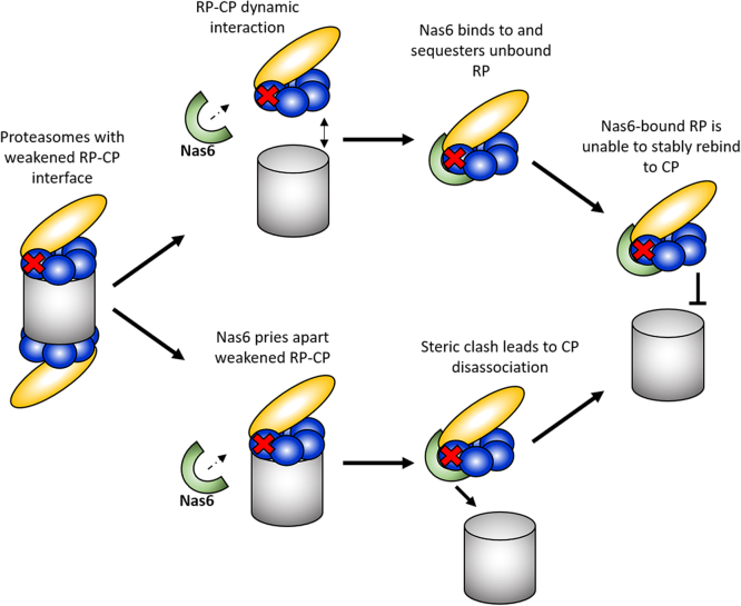
The 26S proteasome is a 66-subunit-chambered protease present in all eukaryotes that maintains organismal health by degrading unneeded or defective proteins. Defects in proteasome function or assembly are known to contribute to the development of various cancers, neurodegeneration, and diabetes. During proteasome biogenesis, a family of evolutionarily conserved chaperones assembles a hexameric ring of AAA+ family ATPase subunits contained within the proteasomal regulatory particle (RP) and guide their docking onto the surface of the proteolytic core particle (CP). This RP-CP interaction couples the substrate capture and unfolding process to proteolysis. We previously reported a mutation in the proteasome that promoted dissociation of the RP and CP by one of these chaperones, Nas6. However, the nature of the signal for Nas6-dependent proteasome disassembly and the generality of this postassembly proteasome quality control function for Nas6 remain unknown. Here, we use structure-guided mutagenesis and in vitro proteasome disassembly assays to demonstrate that Nas6 more broadly destabilizes 26S proteasomes with a defective RP-CP interface. We show that Nas6 can promote dissociation of mature proteasomes into RP and CP in cells harboring defects on either side of the RP-CP interface. This function is unique to Nas6 and independent from other known RP assembly chaperones. Further biochemical experiments suggest that Nas6 may exploit a weakened RP-CP interface to dissociate the RP from the CP. We propose that this postassembly role of Nas6 may fulfill a quality control function in cells by promoting the recycling of functional subcomplexes contained within defective proteasomes.

Keywords: 26S proteasome; assembly; chaperone; macromolecular complex; proteolysis; quality control; ubiquitin.


Conflict of interest statement

Conflict of interest The authors declare that they no conflicts of interest with the contents of this article.

Figures

Figure 1
Figure 1
A frameshift mutant altering the Rpt3 C-terminal tail leads to proteasome structural and functional defects.A, growth assay analysis of Rpt3 frameshift mutant (rpt3-YA,ext), which results in an extension of the Rpt3 C-terminal tail, with deletion of RPN4. Serial dilutions of indicated strains were plated on YPD media and incubated at indicated temperatures for 2 days. Growth of the double mutant is extremely sick at normal growing temperatures and lethal at elevated temperatures. B, native PAGE analysis of the panel of Rpt3 frameshift mutant (rpt3-YA,ext) and genetic separation of rpt3-ext and rpt3-YA mutants. Structural defects characterized by an accumulation of free RP, Lid, and CP observed in the frameshift mutant is due to the extension of the Rpt3 tail. Also shown is an in-gel peptidase assay showing a peptidase defect characterized by severely lower AMC fluorescence. The peptidase defect of the frameshift mutant is also due to extension of the Rpt3 tail. AMC, 7-amino-4-methylcoumarin; CP, core particle; RP, regulatory particle.
Figure 2
Figure 2
Disruption of the Rpt3 tail docking into the CP promotes an RP–CP interaction defect.A, overview of Rpt3 C-terminal tail docking mutants. In red is the conserved HbYX motif. Highlighted are introduced mutations upstream of the HbYX motif. (Right) Cartoon diagram of the Rpt tails of the base docking into the pockets formed between CP α subunits. Shown in red, the Rpt3 tail docking into CP α2 (PRE8) pocket. B, native PAGE immunoblotting of the mutants shown in (A). C, cartoon (left) and atomic structure (right; PDB 6FVY) of the RP base (blue) docking onto the CP (gray). The Rpt3 tail (cyan) docks into the CP in the pocket formed by the α1 (brown) and the α2 (green) subunits. Spheres highlighting close contacts (≥4 Å) formed between the Rpt3 tail and the α1 subunit. HbYX motif shown in red. Other base and CP subunits as well as the N-terminal region of Rpt3 are omitted for clarity. D, in-gel peptidase assay of the panel of Rpt3 tail docking mutants (numbered as in A). Rpt3 tail docking mutants show a decrease in overall peptidase activity, indicated by lower AMC fluorescence. Quantification of proteasomal peptidase activity. Peptidase activity was normalized to total RP2CP and RP1CP abundances (B; α20S), with WT peptidase activity then set to 100%. One-way ANOVA with Tukey’s test for multiple comparisons; error bars represent ± SD; ns, not significant; ∗p < 0.05; ∗∗p < 0.01; ∗∗∗p < 0.001; ∗∗∗∗p < 0.0001; N = 5. AMC, 7-amino-4-methylcoumarin; CP, core particle; RP, regulatory particle.
Figure 3
Figure 3
RP assembly chaperone Nas6 associates with free RP in mutants with weakened RP–CP interfaces.A, native PAGE-immunoblotting of extracts from the mutants shown in Figure 2A. Each strain also expresses 3xFLAG-tagged Nas6 from the chromosomal locus. B, native PAGE-immunoblotting of extracts from rpt3-SSS (top panels) and pre8-KA (bottom panels) cells expressing the indicated 3xFLAG-tagged chaperones. A red arrow indicates accumulation of Nas6-bound RP. C, top, experimental workflow for Nas6-induced dissociation experiment. Bottom, native PAGE-immunoblotting of the indicated extracts pretreated with increasing concentrations of recombinant Nas6 (rNas6; 0, 0.1, or 1 μM). D, deletion of NAS6 rescues the RP-CP association defect in rpt3-SSS and pre8-KA cells. Red and blue arrowheads indicate the changes in RP2CP and free RP, respectively observed via native PAGE. E, NAS6 deletion rescues the structural defect of rpt3-SSS proteasomes, but not the peptidase defect. Native PAGE-separated extracts of the indicated strains were either immunoblotted for the CP or subjected to in-gel peptidase assay as described above. Peptidase activity was quantified as described in Figure 2D. One-way ANOVA with Tukey’s test for multiple comparisons; error bars represent ± SD; ns, not significant; ∗∗∗p < 0.001; N = 4. CP, core particle; RP, regulatory particle.
Figure 4
Figure 4
Nas6 preferentially destabilizes proteasomes with an RP-CP defect.A, native PAGE-immunoblotting of the extracts of the indicated strains treated with or without recombinant Nas6 as shown in Figure 3C. Blue and red arrows indicate the loss of doubly capped proteasomes and accumulation of free RP, respectively. B, extracts of the indicated strains were analyzed and annotated as in (A). C, the strains shown were transformed with plasmids encoding the indicated proteins. The transformants were then spotted as serial dilutions onto selective media and incubated for 2 days at the temperatures shown before imaging. CP, core particle; RP, regulatory particle.
Figure 5
Figure 5
Nas6 has a specific and independent role in destabilizing proteasomes with a defective RP-CP interface.A and B, native PAGE-immunoblotting of the indicated strains. A blue arrowhead indicates the rescue of RP accumulation upon deletion of NAS6. C and D, Nas6 can destabilize proteasomes in the absence of other RP chaperones. The indicated cell extracts were treated with rNas6 before native PAGE-immunoblotting. Red arrows indicate the accumulation of free RP. CP, core particle; RP, regulatory particle.
Figure 6
Figure 6
The ability of Nas6 to dissociate RP and CP correlates with the RP-CP affinity.A and B, Michaelis–Menten analyses of base-CP interaction using WT (A) or rpt3-Δ3 (B) base purified from nas6Δ cells. Note the different scales on the x-axes. The KD for each is shown with the SD (N = 3). C, ATPγS prevents Nas6-dependent destabilization of proteasomes. Extracts of the indicated strains were prepared in the presence of ATP or ATPγS as indicated and then treated or not with rNas6 before separation by native PAGE and immunoblotting. D, top, experimental design for the measurement of recombinant RP (rRP) exchange. Bottom, native PAGE-immunoblots of the indicated strains incubated with increasing concentrations of rRP. CP, core particle; RP, regulatory particle.
Figure 7
Figure 7
Model for Nas6-dependent dissociation of proteasomes. Nas6 may function either by sequestering RPs as they transiently dissociate from proteasomes with defective RP-CP interfaces (top path) and/or by forming a ternary complex with proteasomes, triggering dissociation of RP and CP and preventing rebinding (bottom path). See text for additional discussion. CP, core particle; RP, regulatory particle.

References

    1. Thibaudeau T.A., Smith D.M. A practical review of proteasome pharmacology. Pharmacol. Rev. 2019;71:170–197. - PMC - PubMed
    1. Rousseau A., Bertolotti A. Regulation of proteasome assembly and activity in health and disease. Nat. Rev. Mol. Cell Biol. 2018;19:697–712. - PubMed
    1. Bard J.A.M., Goodall E.A., Greene E.R., Jonsson E., Dong K.C., Martin A. Structure and function of the 26S proteasome. Annu. Rev. Biochem. 2018;87:697–724. - PMC - PubMed
    1. Groll M., Huber R. Substrate access and processing by the 20S proteasome core particle. Int. J. Biochem. Cel. Biol. 2003;35:606–616. - PubMed
    1. Jäger S., Groll M., Huber R., Wolf D.H., Heinemeyer W. Proteasome beta-type subunits: unequal roles of propeptides in core particle maturation and a hierarchy of active site function. J. Mol. Biol. 1999;291:997–1013. - PubMed

Publication types

MeSH terms

Substances

LinkOut - more resources