Review
. 2023 Mar;47(2):153-163.
doi: 10.4093/dmj.2022.0305. Epub 2023 Jan 12.

The Link between Mitochondrial Dysfunction and Sarcopenia: An Update Focusing on the Role of Pyruvate Dehydrogenase Kinase 4

Affiliations
Review

The Link between Mitochondrial Dysfunction and Sarcopenia: An Update Focusing on the Role of Pyruvate Dehydrogenase Kinase 4

Min-Ji Kim et al. Diabetes Metab J. 2023 Mar.

Abstract

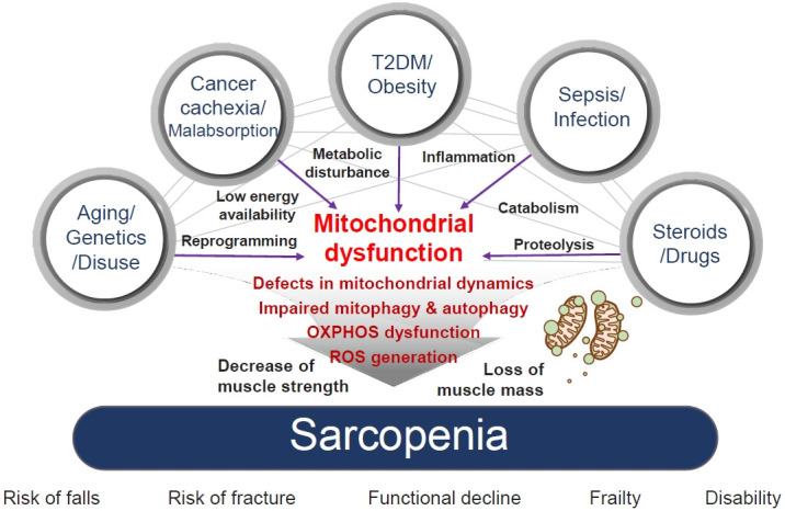
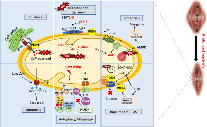
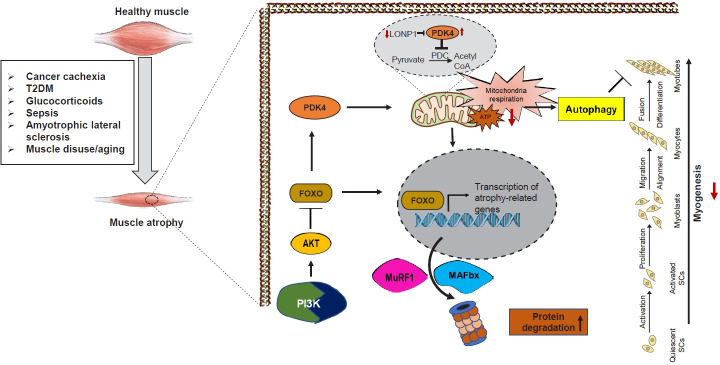
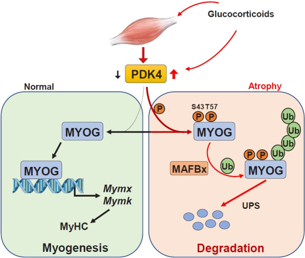
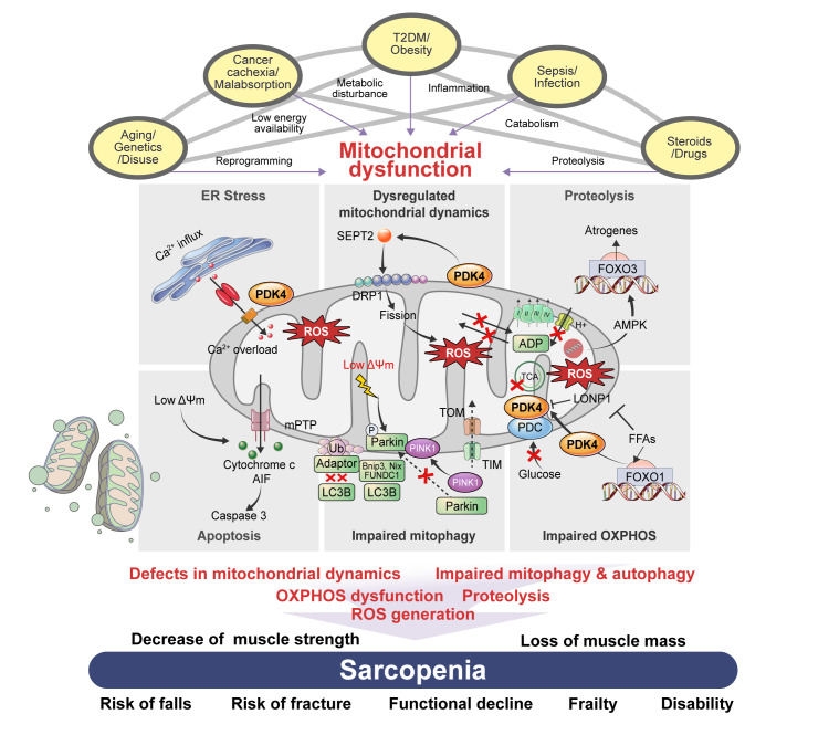
Sarcopenia, defined as a progressive loss of muscle mass and function, is typified by mitochondrial dysfunction and loss of mitochondrial resilience. Sarcopenia is associated not only with aging, but also with various metabolic diseases characterized by mitochondrial dyshomeostasis. Pyruvate dehydrogenase kinases (PDKs) are mitochondrial enzymes that inhibit the pyruvate dehydrogenase complex, which controls pyruvate entry into the tricarboxylic acid cycle and the subsequent adenosine triphosphate production required for normal cellular activities. PDK4 is upregulated in mitochondrial dysfunction-related metabolic diseases, especially pathologic muscle conditions associated with enhanced muscle proteolysis and aberrant myogenesis. Increases in PDK4 are associated with perturbation of mitochondria-associated membranes and mitochondrial quality control, which are emerging as a central mechanism in the pathogenesis of metabolic disease-associated muscle atrophy. Here, we review how mitochondrial dysfunction affects sarcopenia, focusing on the role of PDK4 in mitochondrial homeostasis. We discuss the molecular mechanisms underlying the effects of PDK4 on mitochondrial dysfunction in sarcopenia and show that targeting mitochondria could be a therapeutic target for treating sarcopenia.

Keywords: Metabolic diseases; Mitochondria; Muscular atrophy; Pyruvate dehydrogenase acetyl-transferring kinase; Pyruvate dehydrogenase complex; Sarcopenia.


Conflict of interest statement

CONFLICTS OF INTEREST

No potential conflict of interest relevant to this article was reported.

Figures

Fig. 1.
Fig. 1.
The association between risk factors of sarcopenia and mitochondrial dysfunction. Sarcopenia, generally defined as aging-related loss of muscle mass and function, is closely associated with other genetic or environmental factors. Sarcopenia pathogenesis occurs in multiple ways: genetic predisposition, aging, and disuse of muscle lead to transcriptional reprogramming or epigenetic modification; cancer cachexia or nutrient malabsorption cause low energy availability; metabolic diseases, such as type 2 diabetes mellitus (T2DM) and obesity, lead to metabolic disturbances; infection and sepsis cause inflammation; and steroids and other drugs can lead to catabolism and proteolysis. All these risk factors cause mitochondrial dysfunction, which results in sarcopenia via several mechanisms: defects in mitochondrial dynamics, such as fusion/fission; impaired mitochondrial quality control, including mitochondrial biogenesis; mitophagy and autophagy; dysfunctions in oxidative phosphorylation (OXPHOS), which is important in energy generation; and reactive oxygen species (ROS) production, which originates in dysregulated OXPHOS. Mechanistically, these factors decrease muscle strength and cause loss of muscle mass, which are associated with an increased risk of falls and fractures, functional decline, frailty, disability, and hospitalization with poorer health outcomes.
Fig. 2.
Fig. 2.
Muscle atrophy induces mitochondrial dysfunction through multiple pathways. Prolonged inactivity and other myopathies elevate reactive oxygen species (ROS) levels, which regulate different mitochondrial systems. Increased endoplasmic reticulum (ER) stress caused by calcium (Ca2+) overload and ROS results in Ca2+ influx into mitochondria through pyruvate dehydrogenase kinase 4 (PDK4)-mediated stabilization of inositol 1,4,5 trisphosphate receptor type 1 (IP3R1)–glucose-regulated protein 75 (GRP75)–voltage-dependent anion-selective channel 1 complex at the mitochondria-associated ER membrane. Increased mitochondrial Ca2+ content causes a drop in mitochondrial membrane potential (ΔΨm) leading to the opening of mitochondrial permeability transition pores (mPTP) to release cytochrome c, which activates apoptosis-inducing factor (AIF), leading to the onset of caspase 3-mediated cell death. Muscle atrophy causes increased fission via septin 2 (SEPT2)-mediated dynamin-related protein 1 (DRP1) upregulation. This imbalance between fusion and fission generates ROS and a lower ΔΨm, leading to dysfunctional mitochondrial-mediated autophagy (mitophagy). This occurs via suppressed PTEN-induced kinase 1 (PINK1)-PARKIN interaction, as well as reduced autophagic flux by lysosome adapter accumulation. Fission also causes a drop in ATP synthesis and impaired electron transport chain activity, leading to the activation of 5´ AMP-activated protein kinase (AMPK)–forkhead box protein O3 (FOXO3)-dependent atrogenes, which cause protein degradation (proteolysis). An increase in free-fatty acids (FFAs) caused by myopathologic metabolic alterations results in a switch from glucose oxidation to beta-oxidation. This causes an increase in FOXO1-PDK4 activity leading to the inactivation of the pyruvate dehydrogenase complex (PDC) by PDK4. Synergistically, FFAs also inhibit Lon peptidase 1 (LonP1), which degrades PDK4 in the mitochondria. Overall, this decreases oxidative phosphorylation (OXPHOS), leading to metabolic inflexibility-related myopathies. Acetyl-CoA, acetyl coenzyme A; ADP, adenosine diphosphate; BNIP3, BCL2/adenovirus E1B 19 kDa protein-interacting protein 3; FIS1, mitochondrial fission 1 protein; FUNDC1, FUN14 domain containing 1; H+, proton ion; LC3B, microtubule-associated proteins 1A/1B light chain 3B; MFF, mitochondrial fission factor; NDP52, nuclear dot protein 52; OPTN, optineurin; P62, sequestosome 1 (SQSTM1); TCA, tricarboxylic acid cycle; TIM, translocase of the inner membrane; TOM, translocase of the outer membrane; Ub, ubiquitin.
Fig. 3.
Fig. 3.
Schematic of the involvement of pyruvate dehydrogenase kinase 4 (PDK4) in regulating muscle protein degradation and atrophic gene expression. In the pathological state, PDK4 is activated and can contribute to mitochondrial damage, as well as transcription of atrophy-related genes culminating in muscle atrophy. The transcription factor forkhead box protein O1 (FOXO1) is proposed to upregulate transcription of PDK4. During starvation, a decline in insulin levels activates FOXO1, which enhances PDK4 expression. PDK4 is highly expressed in cancer cachexia, type 2 diabetes mellitus (T2DM), glucocorticoid-induced myopathy, sepsis, amyotrophic lateral sclerosis (ALS), muscle disuse, and aging. Overexpression of PDK4 induces mitochondrial dysfunction and muscle protein degradation, giving PDK4 a central role in mediating metabolic instability-induced skeletal muscle dysfunction. Acetyl-CoA, acetyl coenzyme A; ATP, adenosine triphosphate; LonP1, Lon peptidase 1; MAFbx, muscle atrophy F-box; MuRF1, muscle RING finger 1; PDC, pyruvate dehydrogenase complex; PI3K, phosphatidylinositol-3-kinase; SC, satellite cell.
Fig. 4.
Fig. 4.
Pyruvate dehydrogenase kinase 4 (PDK4) and regulation of myogenesis. The ubiquitin-proteasome pathway serves a pivotal role in the mediation of protein degradation in muscle atrophy. In normal muscle, the transcription factor myogenin (MYOG) activates transcription of the myogenic genes MYMX (myomixer, myoblast fusion factor) and MYMK (myomaker, myoblast fusion factor). By contrast, during dexamethasone-induced muscle atrophy, induction of PDK4 leads to phosphorylation (P) of myogenin (MYOG) and recruitment of muscle atrophy F-box (MAFbx), which polyubiquitinates (Ub) MYOG, leading to its degradation and preventing transcription of myogenic genes. MyHC, myosin heavy chain; UPS, ubiquitin-proteasome system.
None

Similar articles

Cited by

References

    1. Shimokata H, Shimada H, Satake S, Endo N, Shibasaki K, Ogawa S, et al. Chapter 2 Epidemiology of sarcopenia. Geriatr Gerontol Int. 2018;18 Suppl 1:13–22. - PubMed
    1. Fu X, Zhu M, Zhang S, Foretz M, Viollet B, Du M. Obesity impairs skeletal muscle regeneration through inhibition of AMPK. Diabetes. 2016;65:188–200. - PMC - PubMed
    1. Liu X, Qu H, Zheng Y, Liao Q, Zhang L, Liao X, et al. Mitochondrial glycerol 3-phosphate dehydrogenase promotes skeletal muscle regeneration. EMBO Mol Med. 2018;10:e9390. - PMC - PubMed
    1. Hyatt H, Deminice R, Yoshihara T, Powers SK. Mitochondrial dysfunction induces muscle atrophy during prolonged inactivity: a review of the causes and effects. Arch Biochem Biophys. 2019;662:49–60. - PMC - PubMed
    1. Pin F, Novinger LJ, Huot JR, Harris RA, Couch ME, O’Connell TM, et al. PDK4 drives metabolic alterations and muscle atrophy in cancer cachexia. FASEB J. 2019;33:7778–90. - PMC - PubMed

Publication types