Review
. 2023 Jun;24(6):382-400.
doi: 10.1038/s41576-022-00562-w. Epub 2023 Jan 12.

Autophagy genes in biology and disease

Affiliations
Review

Autophagy genes in biology and disease

Hayashi Yamamoto et al. Nat Rev Genet. 2023 Jun.

Abstract

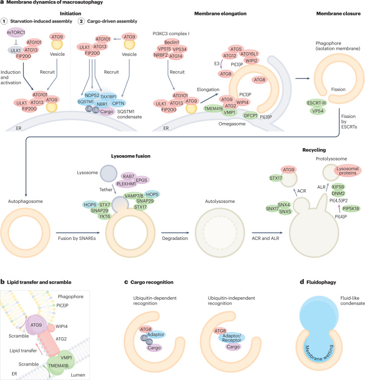
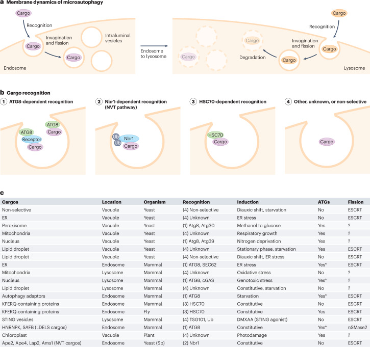
Macroautophagy and microautophagy are highly conserved eukaryotic cellular processes that degrade cytoplasmic material in lysosomes. Both pathways involve characteristic membrane dynamics regulated by autophagy-related proteins and other molecules, some of which are shared between the two pathways. Over the past few years, the application of new technologies, such as cryo-electron microscopy, coevolution-based structural prediction and in vitro reconstitution, has revealed the functions of individual autophagy gene products, especially in autophagy induction, membrane reorganization and cargo recognition. Concomitantly, mutations in autophagy genes have been linked to human disorders, particularly neurodegenerative diseases, emphasizing the potential pathogenic implications of autophagy defects. Accumulating genome data have also illuminated the evolution of autophagy genes within eukaryotes as well as their transition from possible ancestral elements in prokaryotes.


Conflict of interest statement

The authors declare no competing interests.

Figures

Fig. 1
Fig. 1. Membrane dynamics of macroautophagy.
a, (1) At initiation of macroautophagy, the ULK complex assembles near the ER membrane upon starvation and recruits ATG9 vesicles via its interaction with the ATG13–ATG101 subcomplex. (2) Alternatively, cargo adaptors such as p62, NDP52 and TAX1BP1 induce assembly of the ULK complex via interaction with FIP200, whereas ATG9 vesicles are recruited by OPTN. At the membrane-elongation step, the ULK complex recruits the class III phosphatidylinositol 3–kinase complex I (PI3KC3–C1) that produces PI(3)P, which further recruits its effector proteins, DFCP1 to omegasomes and WIPI2 and WIPI4 to phagophores. WIPI4 directs ATG2 to the phagophore membrane, which transfers phospholipids from the ER in concert with ATG9, VMP1 and TMEM41B (see b). WIPI2 recruits the ATG12–ATG5–ATG16L1 complex to promote LC3 lipidation on the phagophore membrane. Autophagosomes are closed by the action of the ESCRT machinery. Subsequently, PLEKHM1, EPG5 and RAB7 tether autophagosomes with lysosomes, and the two SNARE complexes, STX17–SNAP29–VAMP7/8 and YKT6–SNAP29–STX7, trigger fusion. After prolonged starvation, lysosomal membrane proteins on autolysosomes are recycled via autophagic lysosome reformation (ALR), whereas autophagosomal membrane proteins are recycled via autophagosomal components recycling (ACR). b, The lipid transfer protein ATG2 tethers the ER and phagophore membranes and transfers phospholipids from the ER to the phagophore. ATG9 on the phagophore membrane and VMP1 and TMEM41B on the ER membrane scramble phospholipids. c, In selective macroautophagy, cargos are recognized in a ubiquitin (Ub)-dependent manner (through Ub-binding adaptors) or a Ub-independent manner. Cargo adaptors/receptors bind to ATG8 on the autophagic membrane. d, In macro-fluidophagy, the phagophore membrane adheres to fluid-like condensates via membrane wetting.
Fig. 2
Fig. 2. Membrane dynamics of microautophagy.
a, Microautophagy involves the invagination of endosomal membranes (left) or lysosomal membranes (right) to incorporate cytoplasmic material. The resulting intraluminal vesicles are degraded inside lysosomes or the vacuole. b, Cargos are recognized by (1) ATG8, (2) Nbr1, (3) HSC70 or (4) other proteins. Microautophagy can also uptake cytoplasmic materials non-selectively. c, Several types of microautophagy in mammals, yeasts, flies and plants are summarized. The numbers in the ‘Recognition’ column correspond to those in panel b. The ‘ATGs’ column indicates dependency on autophagy-related (ATG) proteins. Asterisks indicate dependence on only ATG8. Sp, Schizosaccharomyces pombe. Ub, ubiquitin.

References

    1. Nakatogawa H. Mechanisms governing autophagosome biogenesis. Nat. Rev. Mol. Cell Biol. 2020;21:439–458. doi: 10.1038/s41580-020-0241-0. - DOI - PubMed
    1. Schuck S. Microautophagy — distinct molecular mechanisms handle cargoes of many sizes. J. Cell Sci. 2020;133:jcs246322. doi: 10.1242/jcs.246322. - DOI - PubMed
    1. Mizushima N, Komatsu M. Autophagy: renovation of cells and tissues. Cell. 2011;147:728–741. doi: 10.1016/j.cell.2011.10.026. - DOI - PubMed
    1. Levine B, Kroemer G. Biological functions of autophagy genes: a disease perspective. Cell. 2019;176:11–42. doi: 10.1016/j.cell.2018.09.048. - DOI - PMC - PubMed
    1. Mejlvang J, et al. Starvation induces rapid degradation of selective autophagy receptors by endosomal microautophagy. J. Cell Biol. 2018;217:3640–3655. doi: 10.1083/jcb.201711002. - DOI - PMC - PubMed

Publication types