. 2023 Mar;48(4):671-682.
doi: 10.1038/s41386-023-01531-5. Epub 2023 Jan 12.

Xanomeline restores endogenous nicotinic acetylcholine receptor signaling in mouse prefrontal cortex

Affiliations

Xanomeline restores endogenous nicotinic acetylcholine receptor signaling in mouse prefrontal cortex

Saige K Power et al. Neuropsychopharmacology. 2023 Mar.

Abstract

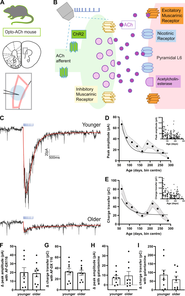
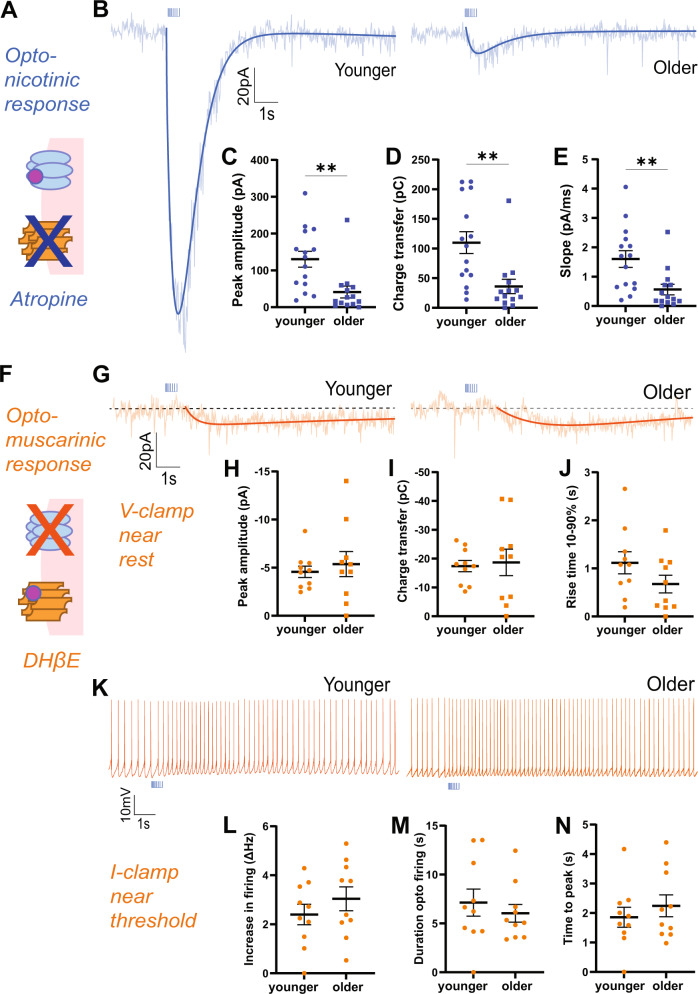
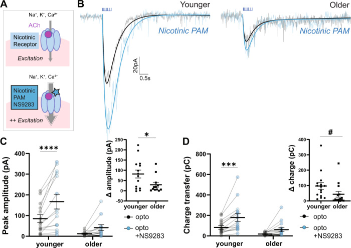
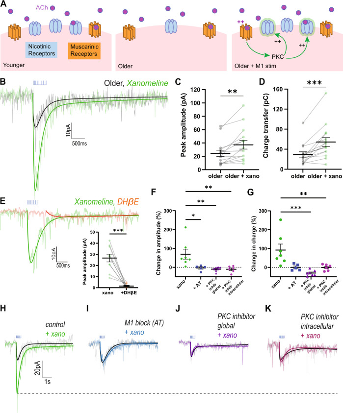
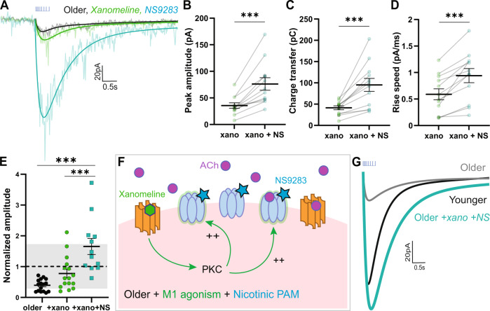
Cholinergic synapses in prefrontal cortex are vital for attention, but this modulatory system undergoes substantial pre- and post-synaptic alterations during adulthood. To examine the integrated impact of these changes, we optophysiologically probe cholinergic synapses ex vivo, revealing a clear decline in neurotransmission in middle adulthood. Pharmacological dissection of synaptic components reveals a selective reduction in postsynaptic nicotinic receptor currents. Other components of cholinergic synapses appear stable, by contrast, including acetylcholine autoinhibition, metabolism, and excitation of postsynaptic muscarinic receptors. Pursuing strategies to strengthen cholinergic neurotransmission, we find that positive allosteric modulation of nicotinic receptors with NS9283 is effective in young adults but wanes with age. To boost nicotinic receptor availability, we harness the second messenger pathways of the preserved excitatory muscarinic receptors with xanomeline. This muscarinic agonist and cognitive-enhancer restores nicotinic signaling in older mice significantly, in a muscarinic- and PKC-dependent manner. The rescued nicotinic component regains youthful sensitivity to allosteric enhancement: treatment with xanomeline and NS9283 restores cholinergic synapses in older mice to the strength, speed, and receptor mechanism of young adults. Our results reveal a new and efficient strategy to rescue age-related nicotinic signaling deficits, demonstrating a novel pathway for xanomeline to restore cognitively-essential endogenous cholinergic neurotransmission.


Conflict of interest statement

The authors declare no competing interests.

Figures

Fig. 1
Fig. 1. Responses to optogenetically-released acetylcholine decrease with age.
A Image depicts coronal brain section taken for whole-cell electrophysiological recordings. The area outlined in pink shows layer 6 of the prefrontal (prelimbic and infralimbic) cortex. The recording electrode is represented in black and the delivered optogenetic stimulus is represented in blue. See caveats section for further discussion of the model of opto-ACh stimulation. B Schematic represents the cholinergic prefrontal synapse receiving optogenetic stimulus (0.5 s pulse train of decreasing frequency). Afferents release endogenous acetylcholine (ACh) which binds to nicotinic and muscarinic receptors, exciting postsynaptic pyramidal neurons. C Example recordings of opto-ACh current from a neuron from younger and older mice, measured in voltage clamp (Vm = −75 mV, stimuli in blue, triple exponential fit in red). D, E Graphs show the decrease with age of the peak amplitude and the charge transfer of the opto-Ach current (n = 185 neurons, 43 mice). F, G Graphs show the increase in opto-ACh current peak and charge transfer with M2 muscarinic antagonist AF-DX 116 are not different between younger (<P150) and older (≥P150) responses (younger: P87 ± 16.1, P50-130; older: P225 ± 32.2, P159-303). H, I Graphs show the change in peak amplitude and charge transfer of the opto-ACh current with galantamine are not different between younger and older responses (younger: P104 ± 18.7, P57-138; older: P202 ± 16.1, P150-281).
Fig. 2
Fig. 2. Nicotinic response is decreased and muscarinic response is preserved through adulthood.
A Schematic illustrates muscarinic receptor blockade to isolate the nicotinic response. B Example optogenetically-elicited nicotinic responses recorded in voltage clamp (Vm = −75 mV) in the presence of muscarinic receptor blocker atropine (AT) from younger and older mice. Graphs show significant age-group decreases in opto-ACh nicotinic responses: (C) peak amplitude, (D) charge transfer and (E) rising slope (**P < 0.01, unpaired t-test, n = 29 neurons, 18 mice) (younger: P92 ± 5.2, P68-114; older: P184 ± 8.5, P150-226). F Schematic illustrates nicotinic receptor blockade to isolate the postsynaptic muscarinic response. G Example responses recorded in voltage clamp (Vm = −75 mV) in the presence of nicotinic receptor blocker DHβE show muscarinic currents in neurons from younger and older mice. Graphs show no significant age group differences in opto-ACh muscarinic currents: (H) peak amplitude, (I) charge transfer, or (J) 10–90% rise time. K Example responses recorded in current clamp with depolarizing current injected to bring the neuron to fire action potentials in the presence of nicotinic receptor blocker DHβE show muscarinic firing response in neurons from younger and older mice. Graphs show no significant age-group difference in (L) peak firing frequency, (M) duration of opto-ACh firing change, or (N) time to peak firing (younger: P86 ± 20.2, P55-143; older: P236 ± 23.1, P167-303).
Fig. 3
Fig. 3. Older opto-ACh responses are less sensitive to nicotinic allosteric potentiation.
A Schematics show acetylcholine activation of nicotinic receptors at baseline and with NS9283 positive allosteric modulation. B Paired recordings in voltage clamp (Vm = −75 mV) show opto-ACh current responses before (black) and after (blue) bath-application of nicotinic positive allosteric modulator (PAM) NS9283 in neurons from younger and older mice. C Graph shows peak amplitude of paired opto-ACh currents before and after NS9283 in neurons from younger and older mice (*P < 0.05, Interaction, two-way ANOVA; ****P < 0.0001, Sidak’s multiple comparisons). Inset graph shows the change in current amplitude imparted by NS9283 is significantly greater in younger than older mice (*P < 0.05, unpaired t-test). D Graph shows charge transfer of paired opto-ACh currents before and after NS9283 in neurons from younger and older mice (****P < 0.0001, NS9283, **P < 0.01, Age, two-way ANOVA; ***P < 0.001, Sidak’s multiple comparisons). Inset graph shows the change in charge transfer imparted by NS9283 trends toward a difference between younger and older mice (t24 = 1.8, #P = 0.08) (younger: P97 ± 12.1, P56-138; older: P227 ± 17.8, P166-295).
Fig. 4
Fig. 4. Older nicotinic responses improved by muscarinic M1 agonist xanomeline.
A Schematic of cholinergic receptor activation in prefrontal layer 6 pyramidal neurons in younger, older, and older with intervention to improve nicotinic receptor availability. B Paired recordings in voltage clamp (Vm = −75 mV) in older mice (mean: P223 ± 20.7 range: P150-358) show opto-ACh response before (black) and after (green) bath-application of muscarinic M1 agonist xanomeline. C, D Graphs show peak amplitude and charge transfer of older responses significantly increases with xanomeline (**P < 0.01, ***P < 0.001, paired t-test). E Paired recordings in voltage clamp (Vm = −75 mV) show opto-ACh response with xanomeline (green) is greatly reduced by nicotinic receptor blocker DHβE (orange). Graph (inset) shows peak amplitude of responses with xanomeline are significantly reduced with application of DHβE (***P < 0.001, paired t-test). F, G Graphs show potentiation with xanomeline is blocked in the presence of muscarinic receptor blocker atropine (AT), global PKC inhibitors (chelerythrine or Go6983), and intracellular PKC inhibitor (PKC 19-31). There is a significant drug effect on the percent change of peak amplitude, (**P < 0.01, one-way ANOVA; *P < 0.05, ** P < 0.01, Dunett’s post hoc tests) and charge transfer (*P < 0.05, one-way ANOVA; ** P < 0.01, Dunett’s post hoc tests). Example responses show xanomeline-potentiation of the opto-ACh response, (H) under control conditions, is blocked by pre-application of (I) muscarinic receptor antagonist atropine (AT), (J) global PKC inhibitor (Go6983), or (K) intracellular PKC inhibitor (PKC 19-31 peptide).
Fig. 5
Fig. 5. Older opto-ACh responses rescued to younger levels by combined treatment of M1 agonist xanomeline and nicotinic PAM NS9283.
A Recordings in voltage clamp (Vm = −75 mV) show the opto-ACh response in a neuron from an older mouse at baseline (black), with xanomeline (green), and with NS9283 (teal). In paired experiments, (B) peak amplitude, (C) charge transfer, and (D) rising slope of older (mean: P333 ± 5.9, range: P325–344) xanomeline-enhanced responses (xano) significantly increase with NS9283 (***P < 0.001, paired t-test). E Graph shows peak amplitude of opto-ACh responses normalized to the mean peak amplitude of the younger opto-ACh responses, with gray denoting one standard deviation of younger opto-ACh responses. There is a significant effect of pharmacological interventions in rescuing older responses (****P < 0.0001, one-way ANOVA; *** P < 0.001, Tukey’s post hoc tests). F Schematic shows model for nicotinic receptor changes with age and the effects of combined xanomeline and NS9283 pharmacological intervention. G Example exponential fits of opto-ACh responses from younger, older, and older neuron treated with xanomeline and NS9283. Combined pharmacological intervention rescues older opto-ACh responses to younger levels.

Similar articles

Cited by

References

    1. Filley CM, Cullum CM. Attention and vigilance functions in normal aging. Appl Neuropsychol. 1994;1:29–32. doi: 10.1207/s15324826an0101&2_6. - DOI - PubMed
    1. Mani TM, Bedwell JS, Miller LS. Age-related decrements in performance on a brief continuous performance test. Arch Clin Neuropsychol. 2005;20:575–86. doi: 10.1016/j.acn.2004.12.008. - DOI - PubMed
    1. Gamboz N, Zamarian S, Cavallero C. Age-related differences in the attention network test (ANT) Exp Aging Res. 2010;36:287–305. doi: 10.1080/0361073X.2010.484729. - DOI - PubMed
    1. Kaufman DAS, Sozda CN, Dotson VM, Perlstein WM. An Event-Related Potential Investigation of the Effects of Age on Alerting, Orienting, and Executive Function. Front Aging Neurosci. 2016;8:99. doi: 10.3389/fnagi.2016.00099. - DOI - PMC - PubMed
    1. Zhou S, Fan J, Lee TMC, Wang C, Wang K. Age-related differences in attentional networks of alerting and executive control in young, middle-aged, and older Chinese adults. Brain Cogn. 2011;75:205–10. doi: 10.1016/j.bandc.2010.12.003. - DOI - PubMed

Publication types