. 2023 Jan 5:18:65-78.
doi: 10.2147/IJN.S385395. eCollection 2023.

Exosomal miR-133a-3p Derived from BMSCs Alleviates Cerebral Ischemia-Reperfusion Injury via Targeting DAPK2

Affiliations

Exosomal miR-133a-3p Derived from BMSCs Alleviates Cerebral Ischemia-Reperfusion Injury via Targeting DAPK2

Xuanyong Yang et al. Int J Nanomedicine. .

Abstract

Background: Cerebral ischemia-reperfusion (CI/R) injury is a subtype of complication after treatment of ischemic stroke. It has been reported that exosomes derived from BMSCs could play an important role in CI/R injury. However, whether BMSCs-derived exosomes could regulate CI/R injury via carrying miRNAs remains to be further explored.

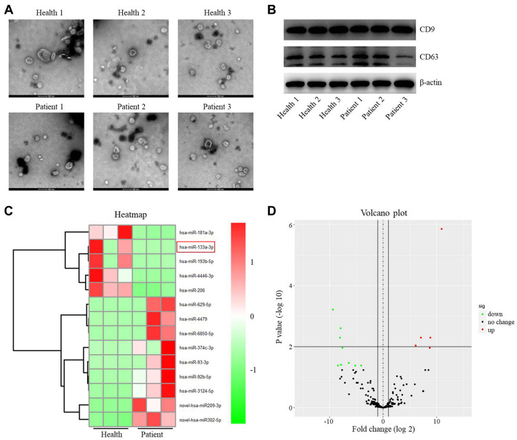
Methods: RNA sequencing was performed to identify the differentially expressed miRNAs. To mimic CI/R in vitro, SH-SY5Y cells were exposed to oxygen glucose deprivation/reoxygenation (OGD/R). The viability of SH-SY5Y cells was tested by CCK8 assay, and TUNEL staining was performed to detect the cell apoptosis.

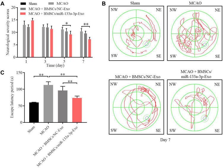
Results: MiR-133a-3p was identified to be reduced in exosomes derived from the plasma of patients with IS. Upregulation of miR-133a-3p significantly reversed OGD/R-induced SH-SY5Y cell growth inhibition. Consistently, BMSCs-derived exosomal miR-133a-3p could restore OGD/R-decreased SH-SY5Y cell proliferation via inhibiting apoptosis. Meanwhile, DAPK2 was a direct target of miR-133a-3p. In addition, OGD/R notably upregulated the level of DAPK2 and weakened the expressions of p-Akt and p-mTOR in SH-SY5Y cells, whereas exosomal miR-133a-3p derived from BMSCs notably reversed these phenomena. Exosomal miR-133a-3p derived from BMSCs could reverse OGD/R-induced cell apoptosis via inhibiting autophagy. Furthermore, exosomal miR-133a-3p derived from BMSCs markedly alleviated the symptom of CI/R injury in vivo.

Conclusion: Exosomal miR-133a-3p derived from BMSCs alleviates CI/R injury via targeting DAPK2/Akt signaling. Thus, our study might shed new light on discovering new strategies against CI/R injury.

Keywords: BMSC; CI/R; DAPK2; autophagy; miR-133a-3p.


Conflict of interest statement

These authors declared no competing interests in this study.

Figures

Figure 1
Figure 1
Differentially expressed miRNAs between exosomes derived from plasma of healthy people and patients with IS. (A) The extracellular vesicles were extracted from the plasma of patients with IS (n=3) or healthy people (n=3). Then, the extracellular vesicles were identified by TEM. (B) The expressions of CD9 and CD63 in extracellular vesicles from healthy people and patients with IS were detected by Western blot. (C) The differentially expressed miRNAs between the exosomes from healthy people and patients with IS were assessed using heatmap. (D) Volcano plots illustrating the differentially expressed miRNAs in IS. Red indicates a higher expression level, while green indicates a lower expression level.
Figure 2
Figure 2
MiR-133a-3p upregulation reversed OGD/R-induced apoptosis in SH-SY5Y cells. (A) The expression of miR-133a-3p in plasma of healthy people or patients with IS was investigated by RT-qPCR. (B) SH-SY5Y cells were treated with OGD/R. The level of miR-133a-3p in SH-SY5Y cells was tested by RT-qPCR. (C) SH-SY5Y cells were transfected with NC or miR-133a-3p mimics. The expression of miR-133a-3p in SH-SY5Y cells was tested by RT-qPCR. (D) SH-SY5Y cells were treated with OGD/R, OGD/R + mimics-ctrl or OGD/R + miR-133a-3p mimics. The viability of SH-SY5Y cells was tested by CCK8 assay. (E) The LDH level in supernatants of SH-SY5Y cells was tested by ELISA. (F) The apoptosis in SH-SY5Y cells was tested by TUNEL staining. *P<0.05, **P< 0.01.
Figure 3
Figure 3
Exosomal miR-133a-3p derived from BMSCs reversed OGD/R-induced SH-SY5Y cell injury. SH-SY5Y cells were treated with OGD/R, OGD/R + BMSCs/NC-Exo or OGD/R + BMSCs/miR-133a-3p-Exo. (A) The level of miR-133a-3p in SH-SY5Y cells was investigated by RT-qPCR. (B) The viability of SH-SY5Y cells was tested by CCK8 assay. (C) The proliferation of SH-SY5Y cells was detected by EdU staining. (D) The LDH level in supernatants of SH-SY5Y cells was tested by ELISA. (E) The apoptosis in SH-SY5Y cells was tested by TUNEL staining. (FH) The levels of MDA, SOD and GSH-Px in supernatants of SH-SY5Y cells were investigated by ELISA. **P< 0.01.
Figure 4
Figure 4
MiR-133a-3p directly targeted DAPK2. (A) Targetscan was used to predict the downstream mRNA of miR-133a-3p. (B) The relative luciferase activity in WT/MT-DAPK2 was detected by dual luciferase assay. (C) SH-SY5Y cells were transfected with mimics-ctrl or miR-133a-3p mimics. The levels of DAPK2 in SH-SY5Y cells were tested by RT-qPCR. (D) SH-SY5Y cells were treated with OGD/R, OGD/R + BMSCs/NC-Exo or OGD/R + BMSCs/miR-133a-3p-Exo. The protein levels of DAPK2, Akt, p-Akt, mTOR and p-mTOR in SH-SY5Y cells were investigated by Western blot. (E) The level of DAPK2 in plasma of patients with IS and healthy people was tested by RT-qPCR. (F) SH-SY5Y cells were treated with OGD/R, OGD/R + BMSCs/miR-133a-3p-Exo or OGD/R + BMSCs/miR-133a-3p-Exo + DAPK2 OE. The apoptosis of SH-SY5Y cells was tested by TUNEL staining. **P< 0.01.
Figure 5
Figure 5
Exosomal miR-133a-3p derived from BMSCs alleviated OGD/R-induced SH-SY5Y cell apoptosis via inhibiting the autophagy. SH-SY5Y cells were treated with OGD/R, OGD/R + BMSCs/miR-133a-3p-Exo or OGD/R + BMSCs/miR-133a-3p-Exo + Rapamycin. (A) The protein level of Beclin-1 in SH-SY5Y cells was tested by Western blot. (B) The viability of SH-SY5Y cells was tested by CCK8 assay. (C) The apoptosis of SH-SY5Y cells was tested by TUNEL staining. **P< 0.01.
Figure 6
Figure 6
Exosomal miR-133a-3p derived from BMSCs markedly attenuated the symptom of CI/R injury in vivo. In vivo middle cerebral artery occlusion (MCAO) model in rats was established. At the end of the study, rats were sacrificed and the brain tissues were collected. Then, (A) the brain tissues were pictured, and TTC staining was performed to observe the severity of CI/R injury. (B) Cerebral infarction area was detected. (C) The expression of miR-133a-3p in tissues of rats was detected by RT-qPCR. (D) The protein levels of DAPK2, Akt, p-Akt, mTOR and p-mTOR in SH-SY5Y cells were investigated by Western blot. **P< 0.01.
Figure 7
Figure 7
Exosomal miR-133a-3p derived from BMSCs attenuated MCAO-induced hippocampal neuronal degeneration. (A) H&E staining was performed to observe the histological changes in brain tissues of rats. (B) TUNEL staining was used to investigate the apoptosis in brain tissues of rats. (C) FJB staining was used to detect the hippocampal neuronal degeneration. **P< 0.01.
Figure 8
Figure 8
Exosomal miR-133a-3p derived from BMSCs significantly improved the memory capacity of MCAO rats. (A) The neurological severity scores of rats were tested. (B) Morris Water Maze assay was performed to test the memory capacity of rats. (C) The escape latency period of rats were evaluated. *P<0.05, **P< 0.01.

References

    1. Zhu RL, Ouyang C, Ma RL, Wang K. Obstructive sleep apnea is associated with cognitive impairment in minor ischemic stroke. Sleep Breath. 2022;26:1907–1914. doi:10.1007/s11325-022-02575-5 - DOI - PMC - PubMed
    1. Lv S, Yang H, Jing P, Song H. alpha-tocopherol pretreatment alleviates cerebral ischemia-reperfusion injury in rats. CNS Neurosci Ther. 2022;28:964–970. doi:10.1111/cns.13814 - DOI - PMC - PubMed
    1. Yu G, Sun W, Wang W, et al. Overexpression of microRNA-202-3p in bone marrow mesenchymal stem cells improves cerebral ischemia-reperfusion injury by promoting angiogenesis and inhibiting inflammation. Aging (Albany NY). 2021;13(8):11877–11888. doi:10.18632/aging.202889 - DOI - PMC - PubMed
    1. He H, Zeng Q, Huang G, et al. Bone marrow mesenchymal stem cell transplantation exerts neuroprotective effects following cerebral ischemia/reperfusion injury by inhibiting autophagy via the PI3K/Akt pathway. Brain Res. 2019;1707:124–132. doi:10.1016/j.brainres.2018.11.018 - DOI - PubMed
    1. Zeng Q, Zhou Y, Liang D, et al. Exosomes secreted from bone marrow mesenchymal stem cells attenuate oxygen-glucose deprivation/reoxygenation-induced pyroptosis in PC12 cells by promoting AMPK-dependent autophagic flux. Front Cell Neurosci. 2020;14:182. doi:10.3389/fncel.2020.00182 - DOI - PMC - PubMed