. 2023 Mar 1;324(3):H293-H304.
doi: 10.1152/ajpheart.00279.2022. Epub 2023 Jan 13.

Increased length-dependent activation of human engineered heart tissue after chronic α1A-adrenergic agonist treatment: testing a novel heart failure therapy

Affiliations

Increased length-dependent activation of human engineered heart tissue after chronic α1A-adrenergic agonist treatment: testing a novel heart failure therapy

C Rupert et al. Am J Physiol Heart Circ Physiol. .

Abstract

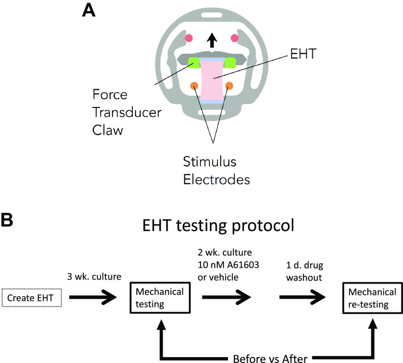
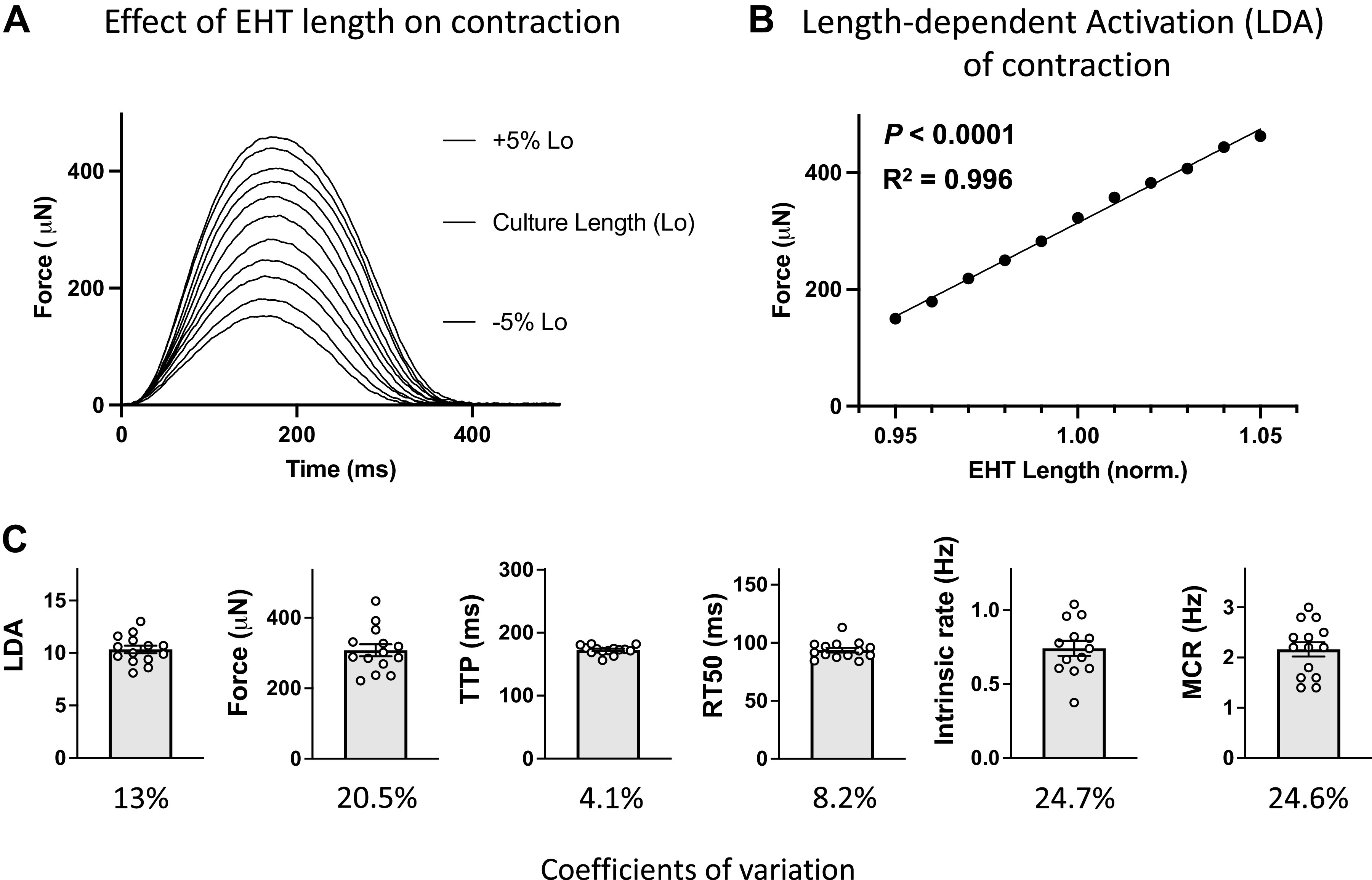
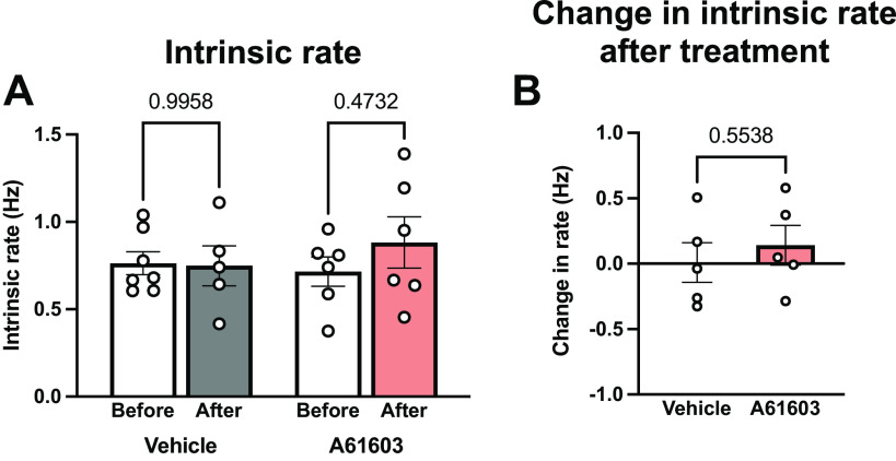
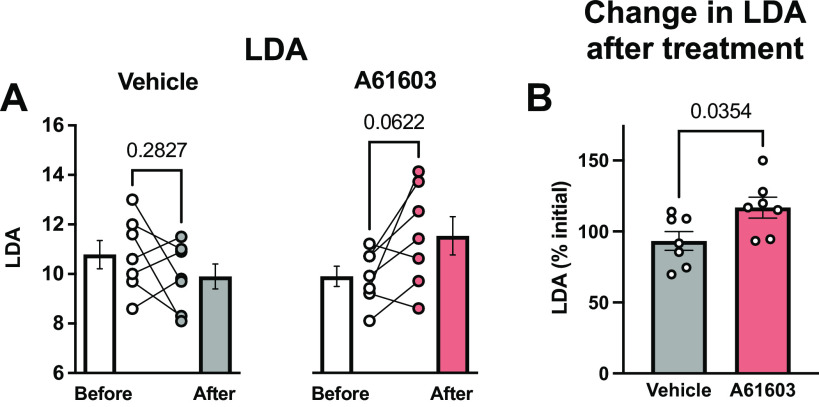
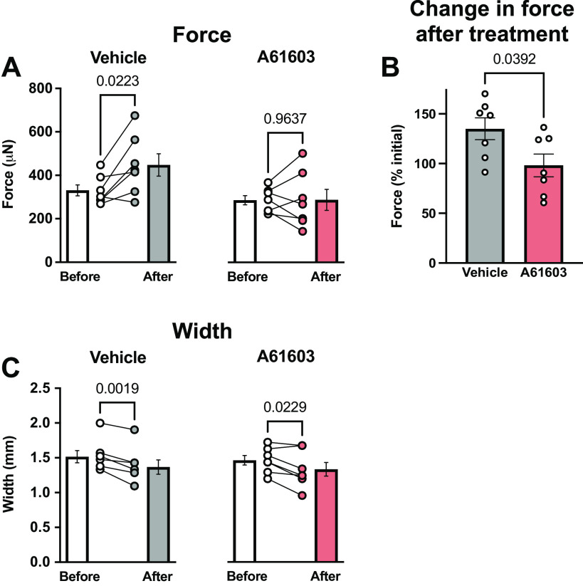
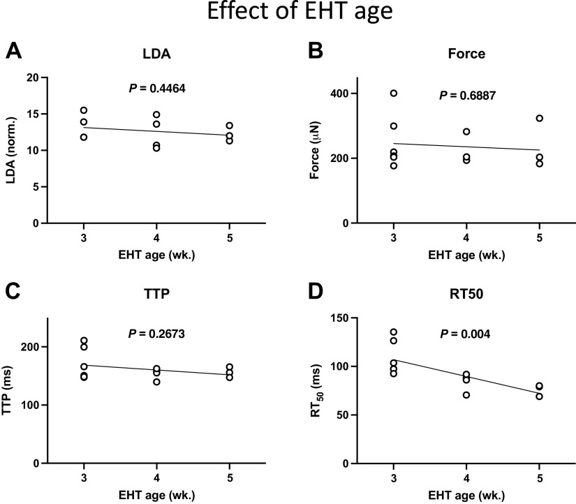
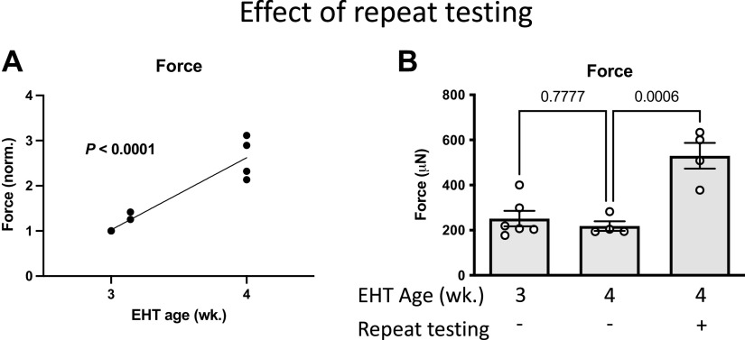
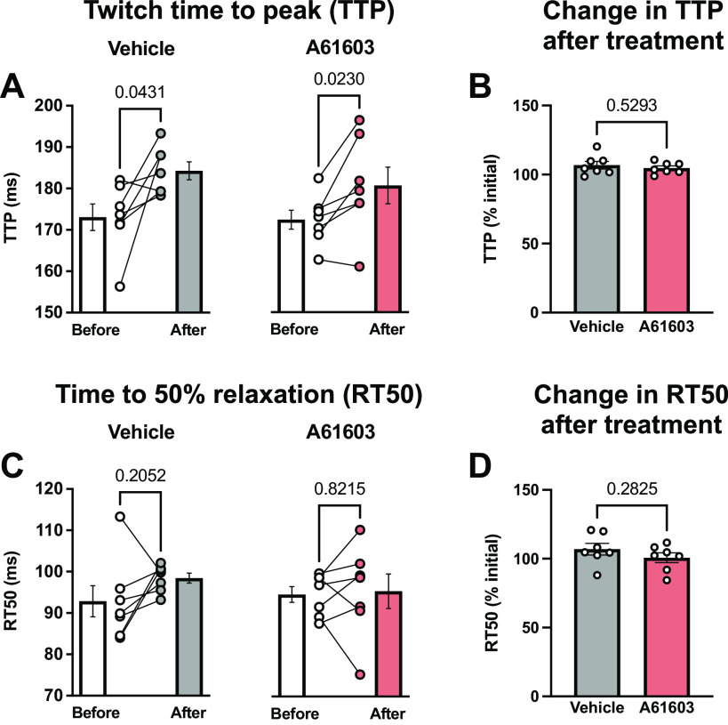
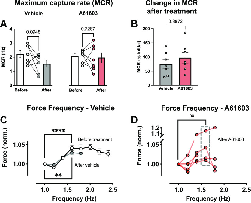
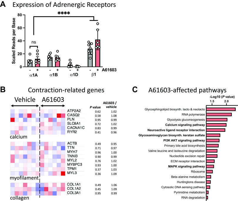
Chronic stimulation of cardiac α1A-adrenergic receptors (α1A-ARs) improves symptoms in multiple preclinical models of heart failure. However, the translational significance remains unclear. Human engineered heart tissues (EHTs) provide a means of quantifying the effects of chronic α1A-AR stimulation on human cardiomyocyte physiology. EHTs were created from thin slices of decellularized pig myocardium seeded with human induced pluripotent stem cell (iPSC)-derived cardiomyocytes and fibroblasts. With a paired experimental design, EHTs were cultured for 3 wk, mechanically tested, cultured again for 2 wk with α1A-AR agonist A61603 (10 nM) or vehicle control, and retested after drug washout for 24 h. Separate control experiments determined the effects of EHT age (3-5 wk) or repeat mechanical testing. We found that chronic A61603 treatment caused a 25% increase of length-dependent activation (LDA) of contraction compared with vehicle treatment (n = 7/group, P = 0.035). EHT force was not increased after chronic A61603 treatment. However, after vehicle treatment, EHT force was increased by 35% relative to baseline testing (n = 7/group, P = 0.022), suggesting EHT maturation. Control experiments suggested that increased EHT force resulted from repeat mechanical testing, not from EHT aging. RNA-seq analysis confirmed that the α1A-AR is expressed in human EHTs and found chronic A61603 treatment affected gene expression in biological pathways known to be activated by α1A-ARs, including the MAP kinase signaling pathway. In conclusion, increased LDA in human EHT after chronic A61603 treatment raises the possibility that chronic stimulation of the α1A-AR might be beneficial for increasing LDA in human myocardium and might be beneficial for treating human heart failure by restoring LDA.NEW & NOTEWORTHY Chronic stimulation of α1A-adrenergic receptors (α1A-ARs) is known to mediate therapeutic effects in animal heart failure models. To investigate the effects of chronic α1A-AR stimulation in human cardiomyocytes, we tested engineered heart tissue (EHT) created with iPSC-derived cardiomyocytes. RNA-seq analysis confirmed human EHT expressed α1A-ARs. Chronic (2 wk) α1A-AR stimulation with A61603 (10 nM) increased length-dependent activation (LDA) of contraction. Chronic α1A-AR stimulation might be beneficial for treating human heart failure by restoring LDA.

Keywords: engineered heart tissue; heart failure; human; induced pluripotent stem cell; α1A-adrenergic receptor.


Conflict of interest statement

Propria, LLC, has a financial interest in MyoPod products/services presented herein. S.G.C. is the founder of, holds equity in, and has received consulting fees from Propria, LLC. The experiments reported in the submitted manuscript were performed at Propria LLC. None of the other authors has any conflicts of interest, financial or otherwise, to disclose.

Figures

Figure 1.
Figure 1.
A: schematic of engineered heart tissue (EHT) mounted in MyoPod cassette. EHT length was varied using an adjustable force transducer claw. B: schematic of the experimental protocol.
Figure 2.
Figure 2.
Representative records and pooled data for contractions of engineered heart tissue (EHT) after 3 wk culture, before drug/vehicle treatment. A: superimposed twitch contractions for EHT at various lengths above and below culture length (Lo). B: relationship between force vs. EHT length: same experiment as in A (means ± SE, n = 3 contractions, errors are smaller than symbols). C: baseline parameters before drug/vehicle treatment for all EHTs in cohort (individual and mean values, n = 14, and coefficients of variation).
Figure 3.
Figure 3.
Chronic A61603 treatment did not change engineered heart tissue (EHT) intrinsic contraction rate. A: intrinsic rate of spontaneous EHT contractions measured before and after chronic (2 wk) treatment with vehicle or A61603 (means ± SE, n = 5–7/group, P values for mixed effects analysis). B: no change in intrinsic rate after treatment relative to before treatment (P value for unpaired t test).
Figure 4.
Figure 4.
Chronic A61603 treatment increased length-dependent activation (LDA). A: LDA measured before and after chronic (2 wk) treatment with vehicle or A61603 (means ± SE, n = 7/group, P values for paired t tests). B: LDA (%initial) was higher after A61603 treatment vs. after vehicle treatment (P value for unpaired t test).
Figure 5.
Figure 5.
Chronic A61603 treatment did not increase engineered heart tissue (EHT) force. A: twitch contraction force measured before and after chronic (2 wk) treatment with vehicle or A61603 (means ± SE, n = 7/group, P values for paired t tests). B: EHT force (%initial) was increased after vehicle treatment but not after A61603 treatment (P value for unpaired t test). C: EHT width before and after treatment with vehicle or A61603 (means ± SE, n = 7/group, P values for paired t tests).
Figure 6.
Figure 6.
Effect of engineered heart tissue (EHT) age on contraction parameters measured on a single occasion (n = 3–6 per age group; P values for linear regressions). A: length-dependent activation (LDA). B: force. C: time to peak force (TTP). D: time to 50% relaxation (RT50).
Figure 7.
Figure 7.
Repeat mechanical testing after 3 wk culture increased engineered heart tissue (EHT) force. A: relationship of EHT force (normalized) to EHT age. First, EHT mechanical testing was at 3 wk (n = 6, same EHT as Fig. 6B), with retesting after 1 day or 1 wk (P value for linear regression). B: effect of both EHT age and repeat testing on EHT force (data from Fig. 6B and Fig. 7A) (P values for unpaired t tests).
Figure 8.
Figure 8.
Effect of chronic A61603 treatment on the timing of contraction and relaxation. A: twitch time to peak (TTP) measured before and after chronic (2 wk) treatment with vehicle or A61603 (means ± SE, n = 7/group, P values for paired t tests). B: TTP after treatment relative to before treatment (P value for unpaired t test). C: time to 50% relaxation (RT50) measured before and after chronic (2 wk) treatment with vehicle or A61603 (means ± SE, n = 7/group, P values for paired t tests). D: RT50 after treatment relative to before treatment (P value for unpaired t test).
Figure 9.
Figure 9.
Effect of chronic A61603 treatment on the response to rapid pacing. A: maximum capture rate (MCR) measured before and after chronic (2 wk) treatment with vehicle or A61603 (means ± SE, n = 7/group, P values for paired t tests). B: MCR after treatment relative to before treatment (P value for unpaired t test). C: relationship between engineered heart tissue (EHT) force (normalized) and stimulation frequency before treatment with vehicle or A61603 (n = 14) and after vehicle treatment (n = 5) (means ± SE, ****P < 0.0001, **P < 0.01, paired t tests). D: force-frequency relation after A61603 treatment (n = 6) (means ± SE, ns, not significant, paired t test).
Figure 10.
Figure 10.
Effects on chronic A61603 treatment on adrenergic receptors and contraction-related gene expression. A: expression of adrenergic receptors in engineered heart tissue (EHT) treated with A61603 (n = 6) or vehicle (n = 7). ****P < 0.0001. B: heat maps for selected contraction-related genes; values scaled from 0.4× (deep blue) to 1.6× (deep red) the average for the vehicle group. C: significantly changed biological pathways associated with prioritized genes in EHT treated with A61603 (n = 6) vs. vehicle (n = 7). Boldface indicates pathways with known links to α1-AR signaling. Origin value on the abscissa corresponds to P = 0.05.

References

    1. Cowley PM, Wang G, Swigart PM, Raghunathan A, Reddy N, Dulam P, Lovett DH, Simpson PC, Baker AJ. Reversal of right ventricular failure by chronic alpha1A-subtype adrenergic agonist therapy. Am J Physiol Heart Circ Physiol 316: H224–H232, 2019. doi:10.1152/ajpheart.00507.2018. - DOI - PMC - PubMed
    1. Cowley PM, Wang G, Joshi S, Swigart PM, Lovett DH, Simpson PC, Baker AJ. alpha1A-Subtype adrenergic agonist therapy for the failing right ventricle. Am J Physiol Heart Circ Physiol 313: H1109–H1118, 2017. doi:10.1152/ajpheart.00153.2017. - DOI - PMC - PubMed
    1. Montgomery MD, Chan T, Swigart PM, Myagmar BE, Dash R, Simpson PC. An alpha-1A adrenergic receptor agonist prevents acute doxorubicin cardiomyopathy in male mice. PLoS One 12: e0168409, 2017. doi:10.1371/journal.pone.0168409. - DOI - PMC - PubMed
    1. Beak J, Huang W, Parker JS, Hicks ST, Patterson C, Simpson PC, Ma A, Jin J, Jensen BC. An oral selective alpha-1A adrenergic receptor agonist prevents doxorubicin cardiotoxicity. JACC Basic Transl Sci 2: 39–53, 2017. doi:10.1016/j.jacbts.2016.10.006. - DOI - PMC - PubMed
    1. Zhang J, Simpson PC, Jensen BC. Cardiac alpha1A-adrenergic receptors: emerging protective roles in cardiovascular diseases. Am J Physiol Heart Circ Physiol 320: H725–H733, 2021. doi:10.1152/ajpheart.00621.2020. - DOI - PMC - PubMed

Publication types

MeSH terms

Substances