. 2023 Jan 17;120(3):e2218899120.
doi: 10.1073/pnas.2218899120. Epub 2023 Jan 13.

prM-reactive antibodies reveal a role for partially mature virions in dengue virus pathogenesis

Affiliations

prM-reactive antibodies reveal a role for partially mature virions in dengue virus pathogenesis

Kimberly A Dowd et al. Proc Natl Acad Sci U S A. .

Abstract

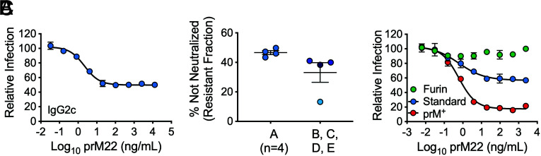
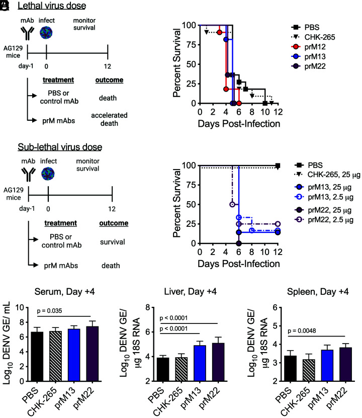
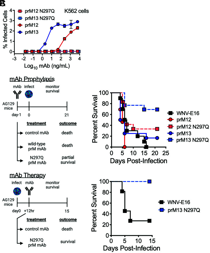
Cleavage of the flavivirus premembrane (prM) structural protein during maturation can be inefficient. The contribution of partially mature flavivirus virions that retain uncleaved prM to pathogenesis during primary infection is unknown. To investigate this question, we characterized the functional properties of newly-generated dengue virus (DENV) prM-reactive monoclonal antibodies (mAbs) in vitro and using a mouse model of DENV disease. Anti-prM mAbs neutralized DENV infection in a virion maturation state-dependent manner. Alanine scanning mutagenesis and cryoelectron microscopy of anti-prM mAbs in complex with immature DENV defined two modes of attachment to a single antigenic site. In vivo, passive transfer of intact anti-prM mAbs resulted in an antibody-dependent enhancement of disease. However, protection against DENV-induced lethality was observed when the transferred mAbs were genetically modified to inhibit their ability to interact with Fcγ receptors. These data establish that in addition to mature forms of the virus, partially mature infectious prM+ virions can also contribute to pathogenesis during primary DENV infections.

Keywords: antibody-dependent enhancement; dengue virus; prM antibody; virion maturation.


Conflict of interest statement

The authors have organizational affiliations to disclose, M.S.D. is a consultant for Inbios, Vir Biotechnology, Moderna, Novavax, Senda Biosciences and Immunome. Yes, the authors have research support to disclose. The Diamond laboratory has received unrelated funding support in sponsored research agreements from Moderna, Vir Biotechnology, and Emergent BioSolutions.

Figures

Fig. 1.
Fig. 1.
Neutralization of DENV2 by a prM-specific mAb. GFP-expressing DENV2 RVPs were incubated with serial dilutions of prM22 for 1 h at 37 °C, followed by infection of Raji-DCSIGNR cells. Infectivity was assessed 48 h later by flow cytometry. (A) Representative dose–response curve of standard DENV2 RVPs (stock A) neutralized by mAb prM22. Error bars indicate the range of duplicate infections. A resistant fraction of nonneutralized virions is apparent at saturating concentrations of antibody. (B) The resistant fraction (calculated as the percentage of virions not neutralized at saturation) is consistent when four independent prM22 neutralization assays are performed with aliquots from the same RVP preparation (stock A) but varies when the assay is repeated with four distinct preparations of RVPs collected from independent transfections (stocks B, C, D, and E). Horizontal line and error bars indicate the mean and SEM, respectively. (C) Representative prM22 neutralization curves of DENV2 RVPs with varying levels of uncleaved prM (furin < standard < prM+). Error bars indicate the range of duplicate infections. Data were confirmed in two additional experiments performed with DENV2 furin and prM+ RVPs only.
Fig. 2.
Fig. 2.
DENV prM alanine scanning to identify amino acids contacted by prM mAbs. (A) The structure of the DENV2 prM protein (cyan) in association with domain II (DII) of the E protein (gray) is shown in a ribbon form from the top and side views relative to the arrangement on the immature virion (adapted from PDB: 3C6E). The conserved hydrophobic fusion loop within E DII is highlighted in green. prM amino acids predicted to be surface exposed are designated in space-filling representation, and residues K26 and E28 are highlighted in green and orange, respectively. (BE) A panel of variant RVPs individually incorporating an alanine at each of the 26 surface-exposed residues was generated. Wild-type (WT) and variant RVPs were incubated with serial dilutions of the indicated mAbs for 1 h at 37 °C, followed by infection of Raji-DCSIGNR cells. Infectivity was assessed 48 h later by flow cytometry. (B) Representative dose–response curves of WT and selected prM variant RVPs (K26A, E28A, and V31A) neutralized by prM12 and prM13 mAbs. Error bars indicate the range of duplicate infections. (C and D) EC50 values for mAb prM12 (C) and mAb prM13 (D) against the panel of 26 variant RVPs incorporating an alanine at the indicated prM residue. Data are expressed as the fold change from the average WT EC50 value (2 to 4 independent experiments per mutant; WT EC50 value calculated from 20 independent experiments for each mAb). The horizontal line and error bars indicate the mean and standard error, respectively. (E) Confirmatory neutralization assays were performed with the indicated mAbs against WT and prM K26A and E28A variant RVPs. EC50 values from 3 to 5 independent experiments are shown. E60 is a maturation state–sensitive E DII-specific antibody used here as a control to demonstrate that the overall antigenicity and prM content of the variants were similar to WT. The horizontal line and error bars represent the mean and SEM, respectively. Statistical differences in log10-transformed mean EC50 values were determined for each mAb using ANOVA with multiple comparison correction; p values are displayed when significant. (F) Lysates from 293T cells transfected to produce WT, prM K26A, or V31A variant DENV2 RVPs were subjected to SDS–PAGE and western blotting analysis with the indicated prM mAbs. HSP90 bands indicate loading of equivalent amounts of cell lysate. 4G2 is a pan-flavivirus–specific control mAb. Data are representative of n = 2 independent experiments for each mAb.
Fig. 3.
Fig. 3.
Cryo-EM single-particle reconstruction of immature DENV2 virions bound to prM12 or prM13 Fabs. (A) Representative cryo-EM micrographs for immature DENV2 virions complexed with saturating concentrations of prM12 (Left) or prM13 (Right) Fabs. (B) The resolution for the three-dimensional (3D) cryo-EM maps was determined using the Fourier shell correlation (FSC) vs. resolution plot. FSC measures the cross-correlation between two 3D maps independently generated from half datasets (gold standard method). Resolution of the maps, assigned when the FSC crosses the 0.143 threshold, was estimated to be 10.2Å and 9.8Å, respectively, for prM12- and prM13-complexed viruses. (C) Surface-shaded radial density maps for immature virion–Fab complexes (prM12 complex, Left, and prM13 complex, Right) viewed down the icosahedral fivefold axis. Low-pass filter was applied to prM12 (10.2 Å) and prM13 (9.8 Å) maps to reduce noise. The color key depicts variation of color as a function of radial distance from the particle center: blue, up to 170Å; cyan, up to 200 Å; green, up to 230 Å; yellow, up to 260 Å; and red, 290 Å and above. The Fab density (red color) is visible on all the three prM molecules constituting an immature asymmetric trimeric spike in both prM12 and prM13 maps. (D) Superimposed density maps of prM12 (gray) and prM13 (green) complexes viewed down the icosahedral fivefold axis reveal differences in the mode of binding of the two Fabs.
Fig. 4.
Fig. 4.
Angle of approach and predicted footprint of prM12 and prM13 Fabs on immature DENV2. (A) Atomic structure of DENV2 prM–E ectodomain (PDB: 3C6E) and modeled structure of prM12 or prM13 Fab were fitted into the cryo-EM density (prM12 complex, Left, and prM13 complex, Right). Only one molecule each of prM–E ectodomain and Fab, corresponding to the P2 position of the asymmetric trimer, is shown. The coloring scheme of E protein domains is per convention (domain 1 [E-dI], red; domain 2 [E-dII], yellow with fusion loop (FL) depicted in orange; and domain 3 [E-dIII], blue). pr is shown in purple, and the heavy and light chains of Fabs are shown in pink and cyan, respectively. Cryo-EM density, within 5 Å radius of the fitted coordinates, is represented as a gray mesh. (B) The different modes of engagement by prM12 and prM13 Fabs are apparent in the superimposed and molecular surface–rendered view of the fitted Fab-bound trimeric spike (Top) and angle of approach (Bottom). prM12 and prM13 Fabs are shown in gray and green, respectively. The pr protein and E protein domains are colored as in A. For clarity, the molecular surfaces of P1 and P3 are omitted in the Bottom panel. Planes drawn using the center of mass of the three E, pr, and Fabs of the trimeric spike are shown as ovals and colored orange, magenta, and gray (prM12)/green (prM13), respectively. (C) The Fab–pr interaction interface for prM12 (Left) and prM13 (Right) is displayed in a ribbon representation. The epitope residues (within ~5 Å of Fabs) for prM12 and prM13 are displayed in gray and green, respectively. Cα of the residue K26 is displayed as a sphere. pr and Fab are colored as in A. CDRH1, CDRH2, CDRH3, CDRL1, CDRL2, and CDRL3 loops are shown in blue, green, red, orange, magenta, and black, respectively. (D) The epitopes of prM12 (Left) and prM13 (Right) are displayed on a stereographic road map of the immature DENV2, which is generated by projecting the prM–E surface residues onto a plane using the program RIVEM. To enable depth perception, the prM–E residues are radially colored relative to distance from the center as shown in the color key: proximal (200 Å), blue, and distal (280 Å), red. The residues that form the epitope are colored in white. The road map is zoomed on the asymmetric unit of the virus (represented as a triangle). The 5-, 3-, and 2-fold axes of symmetry are denoted by pentagon, triangle, and oval shapes, respectively. (E) Sequence alignment of DENV2 strain 16681 prM (zoomed on the prM12 and prM13 epitopes) with other mosquito-transmitted flaviviruses. Residues shaded in red and yellow reflect conserved amino acids: identical (red) and similar (yellow). Amino acid residues predicted to comprise the prM12 epitope are indicated by a gray rectangle above them, while those predicted to form the prM13 epitope are highlighted in green. Secondary structure elements of DENV2 prM (PDB:3C6E) are shown in purple with β, TT, and η referring to β-strand, β-turn, and 310-helix, respectively. The figure was generated using ESPript 3.0.
Fig. 5.
Fig. 5.
Effect of prM-reactive mAbs on DENV2 infection in AG129 mice. (A) Groups of AG129 mice (n = 11, combined from three experiments) were administered 250 µg of the indicated prM mAb or CHIKV-specific isotype-matched control mAb (CHK-265) intraperitoneally 1 d prior to retro-orbital infection with a lethal dose (5 × 105 FFU) of DENV2 D2S20. Animals were monitored for survival. Treatment with prM mAbs, but not CHK-265, resulted in more rapid death compared with PBS control (P < 0.05 for prM12, prM13, or prM22 vs. PBS control, log-rank test). (B) Groups of AG129 mice (n = 6 to 8, combined from two experiments) were administered 25 or 2.5 µg of the indicated mAb intraperitoneally 1 d prior to retro-orbital infection with a sublethal dose (5 × 103 FFU) of DENV2 D2S20. Animals were monitored for survival. Mice receiving prM mAbs, but not CHK-265, displayed evidence of enhanced disease compared with PBS control (P < 0.005 for prM13 or prM22 vs. PBS control, log-rank test). (CE) Groups of AG129 mice (n = 8) were administered 25 µg prM13, prM22, or CHK-265 mAb intraperitoneally 1 d prior to retro-orbital infection with a sublethal dose (5 × 103 FFU) of DENV2 D2S20. Virologic analysis was performed on the serum, spleen, and liver harvested 4 d postinfection by qRT-PCR. The resulting data were analyzed by one-way ANOVA; multiplicity-adjusted P values are denoted when significant (P < 0.05) for comparisons against PBS control animals. GE = genome equivalents. Images in panels A and B were created with BioRender.com.
Fig. 6.
Fig. 6.
Effect of aglycosyl prM-reactive mAbs on DENV2 infection in AG129 mice. (A) WT or aglycosyl (N297Q) chimeric human Fc prM12 and prM13 mAbs were used in an in vitro ADE assay with the DENV2 D2S20 virus stock used to infect mice. After 24 h, infected cells were detected by intracellular staining with an E-specific antibody conjugated to Alexa Fluor 488. Error bars indicate the range of duplicate infections. (B) Groups of AG129 mice (n = 6 to 13, combined from two experiments) were administered 250 µg of the indicated WT or aglycosyl (N297Q) chimeric mAb intraperitoneally 1 d prior to retro-orbital inoculation with a lethal dose (1 × 10⁴ FFU) of DENV2 D2S20. Survival was monitored. WNV-E16 is an isotype-matched control chimeric mAb. N297Q-modified mAbs demonstrated partial protection in comparison with their unmodified counterparts (P < 0.005 for prM12 vs. prM12 N297Q, and prM13 vs. prM13 N297Q comparisons, log-rank test). (C) Groups of AG129 mice (n = 11, combined from two experiments) were infected with a lethal dose (2 × 10⁵ FFU) of DENV2 D2S20 via retro-orbital infection and 12 h later were administered 500 µg of aglycosyl human chimeric prM13 or an isotype control mAb. Survival was monitored. Therapeutic administration of prM13 N297Q was protective (P = 0.0005 for prM13 N297Q vs. isotype control, log-rank test). Images in panels B and C were created with BioRender.com.

References

    1. Gould E. A., Solomon T., Pathogenic flaviviruses. Lancet 371, 500–509 (2008). - PubMed
    1. Pierson T. C., Diamond M. S., The continued threat of emerging flaviviruses. Nat. Microbiol. 5, 796–812 (2020). - PMC - PubMed
    1. Zeng Z., Zhan J., Chen L., Chen H., Cheng S., Global, regional, and national dengue burden from 1990 to 2017: A systematic analysis based on the global burden of disease study 2017. EClin. Med. 32, 100712 (2021). - PMC - PubMed
    1. World Health Organization, Dengue: Guidelines for Diagnosis, Treatment, Prevention and Control: New Edition, World Health Organization, Ed., (World Health Organization, Geneva, 2009). - PubMed
    1. Guzman M. G., Alvarez M., Halstead S. B., Secondary infection as a risk factor for dengue hemorrhagic fever/dengue shock syndrome: An historical perspective and role of antibody-dependent enhancement of infection. Arch. Virol. 158, 1445–1459 (2013). - PubMed

Publication types