. 2023 Feb 14;18(2):449-462.
doi: 10.1016/j.stemcr.2022.12.006. Epub 2023 Jan 12.

Remodeling of H3K9me3 during the pluripotent to totipotent-like state transition

Affiliations

Remodeling of H3K9me3 during the pluripotent to totipotent-like state transition

Hu Li et al. Stem Cell Reports. .

Abstract

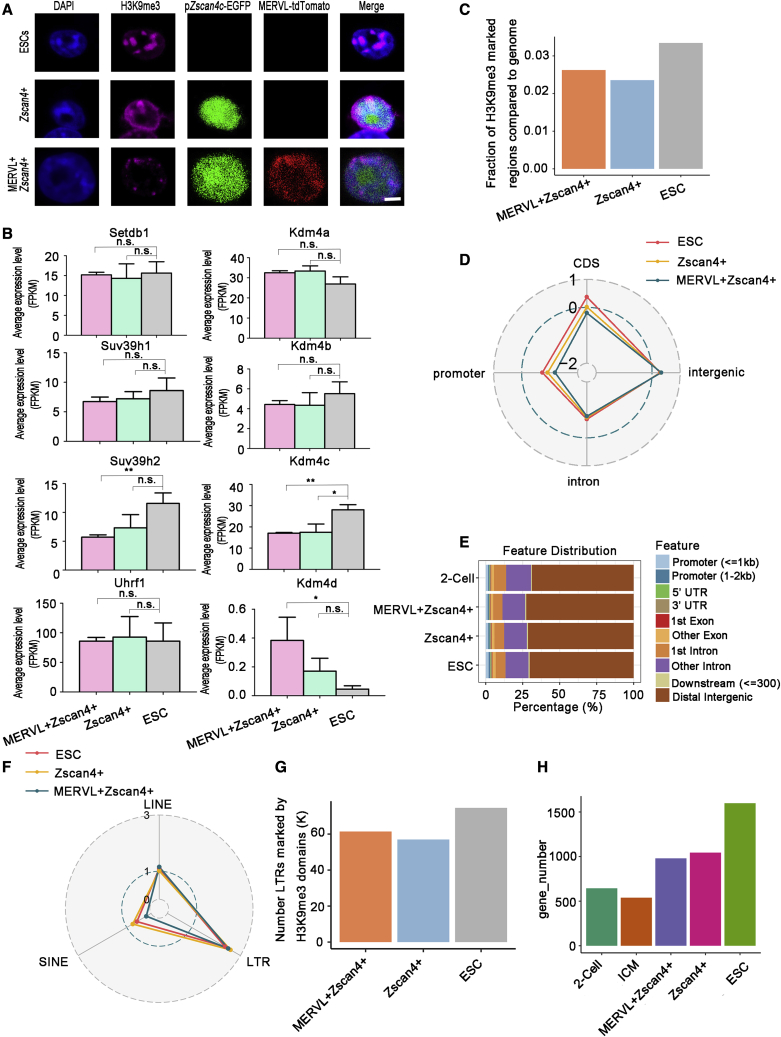
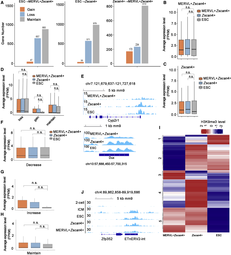
Multiple chromatin modifiers associated with H3K9me3 play important roles in the transition from embryonic stem cells to 2-cell (2C)-like cells. However, it remains elusive how H3K9me3 is remodeled and its association with totipotency. Here, we integrated transcriptome and H3K9me3 profiles to conduct a detailed comparison of 2C embryos and 2C-like cells. Globally, H3K9me3 is highly preserved and H3K9me3 dynamics within the gene locus is not associated with gene expression change during 2C-like transition. Promoter-deposited H3K9me3 plays non-repressive roles in the activation of genes during 2C-like transition. In contrast, transposable elements, residing in the nearby regions of up-regulated genes, undergo extensive elimination of H3K9me3 and are tended to be induced in 2C-like transitions. Furthermore, a large fraction of trophoblast stem cell-specific enhancers undergo loss of H3K9me3 exclusively in MERVL+/Zscan4+ cells. Our study therefore reveals the unique H3K9me3 profiles of 2C-like cells, facilitating the further exploration of totipotency.

Keywords: H3K9me3; pluripotency; totipotency; transposable elements (TEs).


Conflict of interest statement

Conflict of interests The authors declare no competing interests.

Figures

None
Graphical abstract
Figure 1
Figure 1
Genome-wide profiling of H3K9me3 in MERVL+/Zscan4+ cells and Zscan4+ cells (A) Immunostaining analysis with a H3K9me3 antibody. Zscan4 expression was visualized with EGFP (green), and MERVL expression was visualized with tdTomato (red). Cell nuclei were visualized with DAPI. Scale bar, 20 μm. Two independent experiments were performed. (B) Mean RNA expression level (FPKM, fragments per kilobase of transcript per million) of H3K9me3 writers and erasers determined by RNA-seq data (Eckersley-Maslin et al., 2016). n = 3; mean ± SD; n.s., not significant, p < 0.05, ∗∗p < 0.01, ∗∗∗∗p < 0.0001; unpaired Student’s t test. (C) Bar plot showing the fraction of genomic regions covered by H3K9me3 peaks in the indicated cells. The value of the whole fraction (the whole genome) is equal to “1.” (D) Radar chart showing the enrichment score (log ratio of observed/random) of H3K9me3 peaks in promoters, the coding sequence (CDS), intergenic and intron regions. The enrichment score >0 indicates that H3K9me3 domain is enriched in the indicated regions. (E) The genome-wide distribution of H3K9me3 peaks in 2-cell embryos, and the indicated cells. (F) Radar chart showing the enrichment score (log ratio of observed/random) of H3K9me3 peaks in TEs. The enrichment score >0 indicates that H3K9me3 domain is enriched in the indicated regions. (G) Bar plot showing the number of H3K9me3-marked LTR regions in the indicated cells. (H) Bar plot showing the number of H3K9me3-marked promoter regions in 2-cell embryos, ICM, and the indicated cells. See also Figure S1.
Figure 2
Figure 2
H3K9me3 dynamics at gene loci during 2C-like transitions (A) Bar plot showing the number of genes with gain, loss, or maintenance of H3K9me3 marks in promoters during 2C-like transition. (B and C) Boxplot showing the mean RNA expression level (FPKM) of genes loss ICM-specific-H3K9me3 mark during the transition from ESCs to MERVL+/Zscan4+ cells (B) or from ESCs to Zscan4+ cells (C) determined by RNA-seq data (Eckersley-Maslin et al., 2016). n = 3 biological replicates. (D) Boxplot for the mean RNA expression level (FPKM) of genes showing loss, gain or maintenance of promoter H3K9me3 during the transition from ESCs to MERVL+/Zscan4+ cells. n = 3 biological replicates. (E) Integrative genomics viewer of H3K9me3 signals at the Cyp2r1 (top) and Dux (bottom) range in the indicated cells. (F–H) Mean RNA expression level of genes showing decreased (F), increased (G), or maintained (H) intronic H3K9me3 signals in MERVL+/Zscan4+ cells compared with ESCs determined by RNA-seq (Eckersley-Maslin et al., 2016). n = 3 biological replicates. (I) Heatmap showing the dynamics of H3K9me3 domains in the nearby regions (± 20 kb around TSS) of up-regulated genes during 2C-like transition. Each row represents the Z score of H3K9me3 signal subtract input. (J) Integrative genomics viewer of H3K9me3 signals at the Zfp352 region in the indicated cells. The center of the boxplots in Figure 2 represents the median value and the lower and upper lines represent the 25% and 75% quantiles, respectively. Statistical analysis of Figure 2, p value, Wilcoxon rank-sum test, n.s. represents not significant, represents p < 0.05, ∗∗ represents p < 0.01, and ∗∗∗ represents p < 0.001. See also Figure S2.
Figure 3
Figure 3
Expression change and H3K9me3 dynamics in TEs during 2C-like transitions (A) Volcano plots showing differentially expressed TEs between MERVL+/Zscan4+ cells and ESCs (left panel) or Zscan4+ cells and ESCs (right panel). Red dots represent upregulated TEs and blue dots represent downregulated TEs (TE families with RPKM change greater than 2-fold or less than 0.5-fold). See methods in supplemental experimental procedures for details. n = 3 biological replicates. (B) Boxplot showing the average expression levels of TEs (RPKM) located in the RRR regions as defined in (Matoba et al., 2014) determined by RNA-seq (Eckersley-Maslin et al., 2016). n = 3 biological replicates. (C) Bar plots showing the average expression levels (RPKM) of indicated ERVs in the indicated cells determined by RNA-seq (Eckersley-Maslin et al., 2016). n = 3 biological replicates. (D) Bar plots showing the average expression levels of indicated ERVs in preimplantation development determined by RNA-seq (Deng et al., 2014). n = 4 biological replicates. (E) Heatmaps showing H3K9me3 signals (left) and expression levels (right) of TEs in the indicated cells. Left panel, each row represents the Z score of H3K9me3 signal subtract input. Right panel, each row represents the Z score of expression level (RPKM). The heatmap in the right panel was generated using the same order of H3K9me3 clusters. (F) Integrative genomic viewer of H3K9me3 signals at the indicated loci in the indicated cells. (G) Boxplot showing the average expression levels (RPKM) of TEs included in Figure S3L during early embryo development determined by RNA-seq (Deng et al., 2014). n = 4 biological replicates. (H) Bar plot showing the average expression levels of elements belonging to the ERVK subfamilies in the nearby regions of up-regulated genes (included in Figure S3L) or across the whole genome in the indicated cells. n = 3 biological replicates. The center of the boxplots in Figure 3 represents the median value and the lower and upper lines represent the 25% and 75% quantiles, respectively. Statistical analysis of Figure 3, p value, Wilcoxon rank-sum test, n.s. represents not significant, represents p < 0.05, ∗∗ represents p < 0.01, and ∗∗∗ represents p < 0.001. See also Figure S3.
Figure 4
Figure 4
Lineage-specific H3K9me3 is removed in MERVL+/Zscan4+ cells (A) Boxplot showing the H3K9me3 signals (RPKM) at promoters marked with ESC-specific H3K9me3 in the indicated cells. n = 2 biological replicates. (B) Heatmap generated from the cluster analysis of H3K9me3 dynamics in ESC-specific H3K9me3-marked promoters in the indicated cells. Each row represents the Z score of H3K9me3 signal subtract input. (C) Integrative Genomics viewer of H3K9me3 signals around Gpa33 and Gm6578 in the indicated cells. (D) Boxplot showing the H3K9me3 signals of TSC-specific enhancers in the indicated cells. n = 2 biological replicates. (E) Integrative Genomics viewer of H3K9me3 signals in the regions of TSC-specific enhancer in the indicated cells. (F) Boxplot showing the H3K9me3 signals in the region of TSC-specific super-enhancers in the indicated cells. N = 2 biological replicates. (G) Integrative Genomics viewer of H3K9me3 signals in the region of TSC-specific super enhancer in the indicated cells. (H) Radar chart showing the enrichment score (log ratio of observed/random) of H3K9me3 peaks in TEs. (I) Integrative Genomics viewer of H3K9me3 signals in the region of TSC-specific LTR enhancer in the indicated cells. The center of the boxplots in Figure 4 represents the median value and the lower and upper lines represent the 25% and 75% quantiles, respectively. Statistical analysis of Figure 4, p value, Wilcoxon rank-sum test, n.s. represents not significant, represents p < 0.05, ∗∗ represents p < 0.01, and ∗∗∗ represents p < 0.001. See also Figure S4.

References

    1. Akiyama T., Xin L., Oda M., Sharov A.A., Amano M., Piao Y., Cadet J.S., Dudekula D.B., Qian Y., Wang W.D., et al. Transient bursts of Zscan4 expression are accompanied by the rapid derepression of heterochromatin in mouse embryonic stem cells. DNA Res. 2015;22:307–318. - PMC - PubMed
    1. Amano T., Hirata T., Falco G., Monti M., Sharova L.V., Amano M., Sheer S., Hoang H.G., Piao Y., Stagg C.A., et al. Zscan4 restores the developmental potency of embryonic stem cells. Nat. Commun. 2013;4:1966. - PMC - PubMed
    1. Becker J.S., Nicetto D., Zaret K.S. H3K9me3-Dependent heterochromatin: barrier to cell fate changes. Trends Genet. 2016;32:29–41. - PMC - PubMed
    1. Bulut-Karslioglu A., De La Rosa-Velázquez I.A., Ramirez F., Barenboim M., Onishi-Seebacher M., Arand J., Galán C., Winter G.E., Engist B., Gerle B., et al. Suv39h-Dependent H3K9me3 marks intact retrotransposons and silences LINE elements in mouse embryonic stem cells. Mol. Cell. 2014;55:277–290. - PubMed
    1. Burton A., Brochard V., Galan C., Ruiz-Morales E.R., Rovira Q., Rodriguez-Terrones D., Kruse K., Le Gras S., Udayakumar V.S., Chin H.G., et al. Heterochromatin establishment during early mammalian development is regulated by pericentromeric RNA and characterized by non-repressive H3K9me3. Nat. Cell Biol. 2020;22:767–778. - PMC - PubMed

Publication types