. 2023 Jan 25;145(3):1668-1677.
doi: 10.1021/jacs.2c09937. Epub 2023 Jan 14.

Surface-Templated Glycopolymer Nanopatterns Transferred to Hydrogels for Designed Multivalent Carbohydrate-Lectin Interactions across Length Scales

Affiliations

Surface-Templated Glycopolymer Nanopatterns Transferred to Hydrogels for Designed Multivalent Carbohydrate-Lectin Interactions across Length Scales

Anamika Singh et al. J Am Chem Soc. .

Abstract

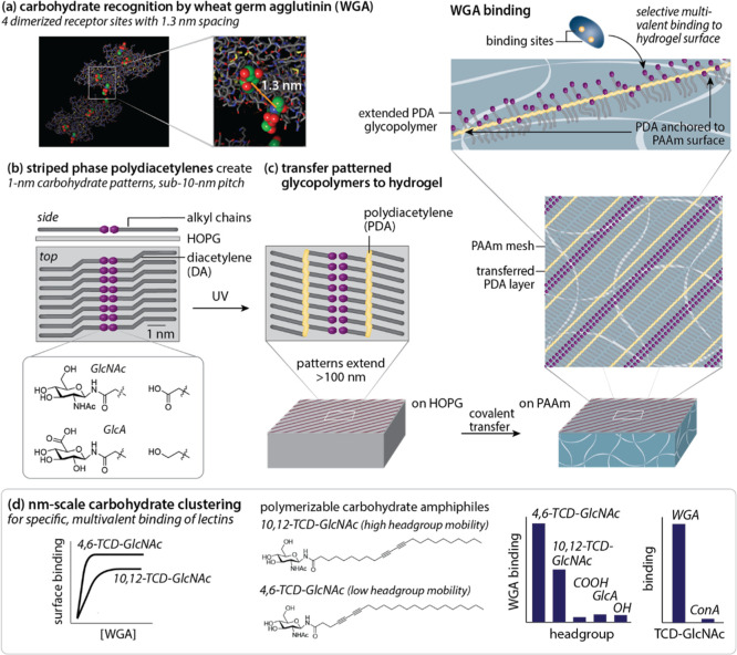
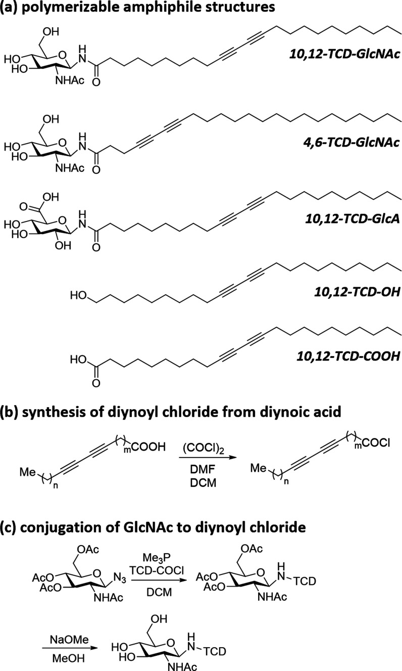
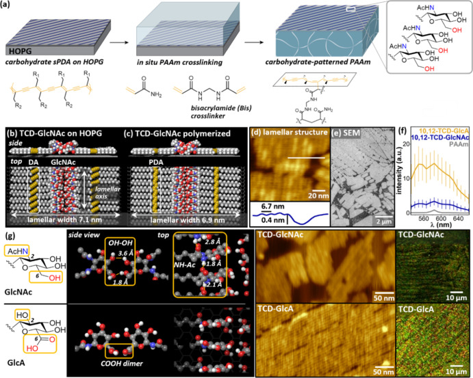
Multivalent interactions between carbohydrates and proteins enable a broad range of selective chemical processes of critical biological importance. Such interactions can extend from the macromolecular scale (1-10 nm) up to much larger scales across a cell or tissue, placing substantial demands on chemically patterned materials aiming to leverage similar interactions in vitro. Here, we show that diyne amphiphiles with carbohydrate headgroups can be assembled on highly oriented pyrolytic graphite (HOPG) to generate nanometer-resolution carbohydrate patterns, with individual linear carbohydrate assemblies up to nearly 1 μm, and microscale geometric patterns. These are then photopolymerized and covalently transferred to the surfaces of hydrogels. This strategy suspends carbohydrate patterns on a relatively rigid polydiacetylene (persistence length ∼ 16 nm), exposed at the top surface of the hydrogel above the bulk pore structure. Transferred patterns of appropriate carbohydrates (e.g., N-acetyl-d-glucosamine, GlcNAc) enable selective, multivalent interactions (KD ∼ 40 nM) with wheat germ agglutinin (WGA), a model lectin that exhibits multivalent binding with appropriately spaced GlcNAc moieties. WGA binding affinity can be further improved (KD ∼ 10 nM) using diacetylenes that shift the polymer backbone closer to the displayed carbohydrate, suggesting that this strategy can be used to modulate carbohydrate presentation at interfaces. Conversely, GlcNAc-patterned surfaces do not induce specific binding of concanavalin A, and surfaces patterned with glucuronic acid, or with simple carboxylic acid or hydroxyl groups, do not induce WGA binding. More broadly, this approach may have utility in designing synthetic glycan-mimetic interfaces with features from molecular to mesoscopic scales, including soft scaffolds for cells.


Conflict of interest statement

The authors declare no competing financial interest.

Figures

Figure 1
Figure 1
(a) Schematic illustrating multivalent GlcNAc recognition by WGA (PDB: 2UVO). (b) Schematic of striped phase assembly and structure on HOPG. (c) Schematic of PDA transfer to PAAm, in relation to PAAm polymerization architecture, and extended glycopolymer display for selective multivalent binding. (d) Illustration of binding selectivity for WGA at striped interfaces based on the choice of carbohydrate amphiphile used for striped phase assembly. Parts of panel (b) adapted with permission from ref (56). Copyright 2022 American Chemical Society.
Scheme 1
Scheme 1. Structures and Syntheses of Diyne Amphiphiles
Figure 2
Figure 2
(a) Schematic of the carbohydrate sPDA pattern transfer to PAAm. (b,c) Molecular models of (b) unpolymerized and (c) polymerized TCD-GlcNAc monolayers on HOPG. (d) AFM images illustrating the lamellar structure of the polymerized TCD-GlcNAc monolayer. (e) SEM image illustrating the presence of microscale domain structure. (f) Fluorescence spectra of TCD-GlcNAc and TCD-GlcA on PAAm. (g) Chemical structures of GlcNAc and GlcA (left panel) and molecular models illustrating headgroup interactions in striped phases of TCD-GlcNAc and TCD-GlcA on HOPG (left center panel). AFM micrographs of TCD-GlcNAc and TCD-GlcA on HOPG (right center panel). Fluorescence micrographs of TCD-GlcNAc and TCD-GlcA on PAAm (right panel). Parts of panel (a) adapted from ref (56) with permission. Copyright 2022 American Chemical Society.
Figure 3
Figure 3
(a–d) Fluorescence images of monolayers after exposure to WGA (5 μg/mL). (e–h) Fluorescence images of monolayer on PAAm. (i,j) Fluorescence spectra of monolayers (i) before and (j) after exposure to WGA (5 μg/mL). (k) WGA binding to monolayers normalized for surface coverage. (l) Fluorescence spectra illustrating minimal increase in adsorption of rhodamine-labeled ConA (5 μg/mL) to TCD-GlcNAc/PAAm (red trace) vs unfunctionalized PAAm (light red). WGA exposed to unfunctionalized PAAm is shown for comparison (light blue). Higher gain settings were used in (l) to adequately resolve spectral differences at low emission intensity, leading to the higher observed fluorescence for the TCD-GlcNAc + WGA trace in comparison with the intensity in (j). (m–o) μCP patterned TCD-GlcNAc (m) on HOPG, (n) after transfer to PAAm, and (o) after transfer to PAAm and exposure to WGA. Inset in (n) uses enhanced contrast to emphasize square pattern.
Figure 4
Figure 4
(a,b) Molecular models and dynamics of headgroup regions in polymerized monolayers of (a) 4,6-TCD-GlcNAc/HOPG and (b) 10,12-TCD-GlcNAc/HOPG. (c) Models following 4 ns dynamics with HOPG removed and PDA frozen, to simulate conditions on sPDAs transferred to PAAm. (d) Distribution of neighboring GlcNAc–GlcNAc distances along each sPDA. Gray tie line at 13.0 Å represents the distance between dimerized WGA binding sites. (e,f) Fluorescence intensities of 4,6-TCD-GlcNAc/PAAm (gold) and 10,12-TCD-GlcNAc (blue) surfaces, without (e) and with (f) exposure to 5 μg/mL WGA. (g) Fluorescence intensities of sPDA/PAAm substrates exposed to specified concentrations of WGA; images in inset were acquired using a widefield epifluorescence microscope, thus intensities are not directly comparable to values in (e,f).

References

    1. Goldstein I. J.; Hayes C. E. The Lectins: Carbohydrate-Binding Proteins of Plants and Animals. Adv. Carbohydr. Chem. Biochem. 1978, 35, 127–340. 10.1016/s0065-2318(08)60220-6. - DOI - PubMed
    1. Drickamer K.; Taylor M. E. Biology of Animal Lectins. Annu. Rev. Cell Biol. 1993, 9, 237–264. 10.1146/annurev.cb.09.110193.001321. - DOI - PubMed
    1. Koehler M.; Delguste M.; Sieben C.; Gillet L.; Alsteens D. Initial Step of Virus Entry: Virion Binding to Cell-Surface Glycans. Annu. Rev. Virol. 2020, 7, 143–165. 10.1146/annurev-virology-122019-070025. - DOI - PubMed
    1. Syed P.; Gidwani K.; Kekki H.; Leivo J.; Pettersson K.; Lamminmäki U. Role of Lectin Microarrays in Cancer Diagnosis. Proteomics 2016, 16, 1257–1265. 10.1002/pmic.201500404. - DOI - PubMed
    1. Purcell S. C.; Godula K. Synthetic Glycoscapes: Addressing the Structural and Functional Complexity of the Glycocalyx. Biol. Pharm. Bull. 2019, 9, 20180080.10.1098/rsfs.2018.0080. - DOI - PMC - PubMed

Publication types