. 2023 Jan 31;42(1):111987.
doi: 10.1016/j.celrep.2022.111987. Epub 2023 Jan 12.

GOT1 regulates CD8+ effector and memory T cell generation

Affiliations

GOT1 regulates CD8+ effector and memory T cell generation

Wei Xu et al. Cell Rep. .

Abstract

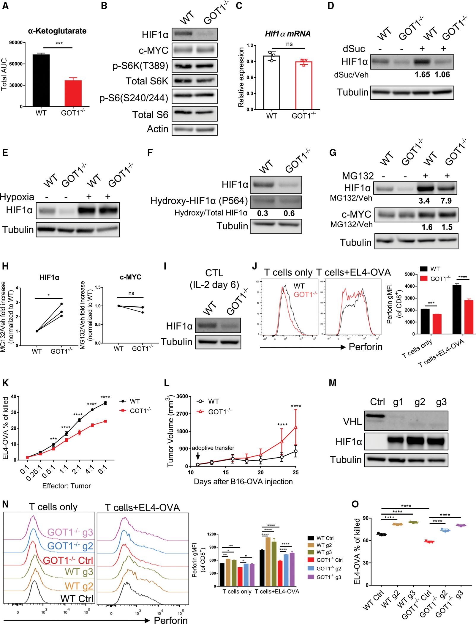
T cell activation, proliferation, function, and differentiation are tightly linked to proper metabolic reprogramming and regulation. By using [U-13C]glucose tracing, we reveal a critical role for GOT1 in promoting CD8+ T cell effector differentiation and function. Mechanistically, GOT1 enhances proliferation by maintaining intracellular redox balance and serine-mediated purine nucleotide biosynthesis. Further, GOT1 promotes the glycolytic programming and cytotoxic function of cytotoxic T lymphocytes via posttranslational regulation of HIF protein, potentially by regulating the levels of α-ketoglutarate. Conversely, genetic deletion of GOT1 promotes the generation of memory CD8+ T cells.

Keywords: CP: Metabolism; GOT1; HIF; NADH/NAD; effector and memory CD8(+) T cell; glucose; glutamate; serine.


Conflict of interest statement

Declaration of interests J.D.P. is a cofounder and equity holder of Dracen Pharmaceuticals. C.H.P. and J.D.P. are current employees of Calico LLC.

Figures

Figure 1.
Figure 1.. Increased metabolic flux from α-ketoglutarate to glutamate upon CD8+ T cell activation is mediated by increased expression of GOT1
(A) Naive or 24-h plate-bound anti-CD3 and soluble anti-CD28 activated OT-I CD8+ T cells were pulsed with [U-13C]glucose for 4–6 h before collecting for mass spectrometry analysis. Percent contribution of carbon flux from [U-13C]glucose to glutamate is shown. (B) Graph describing the intersection of glutamine and glucose metabolism. (C) OT-I CD8+ T cells were activated with plate-bound anti-CD3 and soluble anti-CD28 in the presence of vehicle (Veh), AOA (250 μM), or EGCG (500 μM) for 24 h. Cells were then pulsed with [U-13C]glucose for 4–6 h before collecting for mass spectrometry analysis. Percent contribution of carbon flux from [U-13C]glucose to glutamate is shown. *comparison between Veh and AOA, +comparison between Veh and EGCG. (D and E) 24-h plate-bound anti-CD3 and soluble anti-CD28 activated WT and GOT1−/− OT-I CD8+ T cells were pulsed with [U-13C]glucose (D) or [U-13C]glutamine (E) for 4–6 h before collecting for mass spectrometry analysis. Abundance of (left) and percent contribution (right) of carbon flux from [U-13C]-glucose (D) or [U-13C]glutamine (E) to glutamate are shown. (F) Isolated OT-I or P14 CD8+ T cells were activated using plate-bound anti-CD3 and soluble anti-CD28. Immunoblot measurements of GOT1 and GLUD1 at indicated time points are shown. Actin served as the loading control. (G) In vitro effector (IL-2) and memory (IL-7/15) OT-I or P14 CD8+ T cells were generated as previously described. Immunoblot measurements of GOT1 and GLUD1 (left) and statistical analysis (right) are shown. Actin served as the loading control. (H) Naive (Tn) Thy1.1+ P14 CD8+ T cells were adoptively transferred into WT recipients and infected with LCMV Armstrong. Thy1.1+ CD8+ T cells were FACS sorted on day 7 as effector T cells (Teff) and on day 60 as memory T cells (Tmem). Immunoblot measurements of GOT1 and GLUD1 (left) and statistical analysis (right) are shown. Actin served as the loading control. *p < 0.05, **p < 0.01, ***p < 0.001, ****p < 0.0001; ns, not significant. Two-way ANOVA with Sidak’s multiple comparisons test (A, C, D, and E) and paired t test (G and H). Data are representative of at least three independent experiments. Data are represented as mean ± SD. Also see Figure S1.
Figure 2.
Figure 2.. GOT1 supports serine biosynthesis and promotes CD8+ T cell proliferation under serine-restricted conditions
(A) Abundance of non-essential amino acids (NEAAs) from targeted metabolomics analysis of 24-h activated WT and GOT1−/− OT-I CD8+ T cells. (B) 24-h plate-bound anti-CD3 and soluble anti-CD28 activated WT or GOT1−/− OT-I CD8+ T cells were pulsed with [U-13C]glucose for 4–6 h before collecting for mass spectrometry analysis. Percent contribution of carbon flux from [U-13C]glucose to serine is shown. (C) Flow cytometry analysis of proliferation of WT or GOT1−/− P14 CD8+ T cells cultured under 0, 0.1, and 0.3 mM serine conditions at indicated time points. (D) Flow cytometry analysis of proliferation of separately cultured or co-cultured WT and GOT1−/− P14 CD8+ T cells in serine-free media at indicated time points. (E) Targeted metabolomics analysis of blank media and media after 10 h of WT OT-I CD8+ T cell culture. Volcano plot (left) and abundance of pyruvate (right) are shown. (F) WT and GOT1−/− P14 CD8+ T cells were activated with plate-bound anti-CD3 and soluble anti-CD28 without or with 1 mM pyruvate (Pyr) in serine-free media. Flow cytometry analysis of proliferation at indicated time points is shown. (G) Abundance of NADH (left) and NAD+ (middle) from targeted metabolomics analysis of 24-h activated WT and GOT1−/− OT-I CD8+ T cells without or with 0.3 mM serine. NAD+/NADH ratios are shown (right). (H) WT and GOT1−/− P14 CD8+ T cells were activated with plate-bound anti-CD3 and soluble anti-CD28 without or with 1 mM α-ketobutyrate (αKB) in serine-free media. Flow cytometry analysis of proliferation at indicated time points is shown. (I) WT and GOT1−/− P14 CD8+ T cells were activated with plate-bound anti-CD3 and soluble anti-CD28 without or with 1 mM sodium formate, 200 μM hypoxanthine, or 50 μM adenine in serine-free media. Flow cytometry analysis of proliferation is shown. *p < 0.05, **p < 0.01, ***p < 0.001, ****p < 0.0001; ns, not significant. Two-way ANOVA with Sidak’s multiple comparisons test (A, B, and G). Data are representative of three independent experiments. Data are represented as mean ± SD. Also see Figure S2.
Figure 3.
Figure 3.. GOT1 posttranslationally regulates HIF1α expression and the cytotoxic function of CTLs
(A) Abundance of α-ketoglutarate from targeted metabolomics analysis of 24-h activated WT and GOT1−/− OT-I CD8+ T cells. (B) WT and GOT1−/− OT-I CD8+ T cells were activated using plate-bound anti-CD3 and soluble anti-CD28 for 24 h. Immunoblot measurements of HIF1α, c-MYC, and mTORC1 activity indicated by p-S6K and p-S6 are shown. Total S6K, total S6, and actin served as the loading control. (C) Real-time PCR analysis of Hif1α mRNA levels in 24-h plate-bound anti-CD3 and soluble anti-CD28 activated WT and GOT1−/− OT-I CD8+ T cells. (D) WT and GOT1−/− OT-I CD8+ T cells were activated with plate-bound anti-CD3 and soluble anti-CD28 without (Veh) or with 1 mM dimethyl-succinate (dSuc) for 24 h. Immunoblot measurements of HIF1α are shown. Tubulin served as the loading control. (E) WT and GOT1−/− OT-I CD8+ T cells were activated with plate-bound anti-CD3 and soluble anti-CD28 under normoxia for 21 h. Cells were then incubated under normoxia or hypoxia for an additional 3 h. Immunoblot measurements of HIF1α are shown. Tubulin served as the loading control. (F) Immunoblot measurements of HIF1α and hydroxy-HIF1α at proline 564 site in 24-h activated WT and GOT1−/− OT-I CD8+ T cells. Tubulin served as the loading control. (G and H) WT and GOT1−/− OT-I CD8+ T cells were activated with plate-bound anti-CD3 and soluble anti-CD28 for 24 h followed by 2 h of vehicle (Veh) or MG132 (10 μM) treatment. (G) Immunoblot measurements of HIF1α and c-MYC are shown. Tubulin served as the loading control. (H) Compared with vehicle control, fold increase after MG132 treatment was analyzed. (I) OT-I or P14 CTLs were generated as previously described. Immunoblot measurements of HIF1α in WT and GOT1−/− CTLs are shown. Tubulin served as the loading control. (J) Flow cytometry comparison of perforin production between WT and GOT1−/− OT-I CTLs alone or co-cultured with EL4-OVA for 6 h. Flow plots (left panel) and statistical analysis of geometric mean fluorescence intensity (gMFI, right) are shown. (K) Percentages of killed EL4-OVA by WT or GOT1−/− CTLs at indicated effector:tumor ratios. (L) WT and GOT1−/− OT-I CTLs were adoptively transferred into B16-OVA tumor bearing mice on day 11 after inoculation. Measurements of tumor volume are shown. (M) Naive WT CD8+ T cells were isolated and electroporated with control sgRNA (Ctrl) or sgRNAs targeting Vhl (g1, g2, g3) complexed with Cas9 endonuclease. Cells were recovered and activated with plate-bound anti-CD3 and soluble anti-CD28 and expanded in IL-2 until day 6. Immunoblot measurements of VHL and HIF1α are shown. Tubulin served as the loading control. (N and O) Naive WT and GOT1−/− OT-I CD8+ T cells were isolated and electroporated with control sgRNA (Ctrl) or sgRNAs targeting Vhl (g2, g3) complexed with Cas9 endonuclease. Cells were activated and expanded in IL-2 until day 6, and co-incubated without or with EL4-OVA at 1:1 ratio to assess perforin production (N), or at effector:tumor 4:1 ratio to assess the percentages of tumor cells killed (O). *p < 0.05, **p < 0.01, ***p < 0.001, ****p < 0.0001; ns, not significant. Unpaired t test (A and C), paired t test (H), two-way ANOVA with Sidak’s multiple comparisons test (J, K, and L), two-way ANOVA with Tukey’s multiple comparisons test (N), one-way ANOVA with Tukey’s multiple comparisons test (O). Data are representative of at least three independent experiments. Data are represented as mean ± SD. Also see Figure S3.
Figure 4.
Figure 4.. Deletion of GOT1 promotes memory CD8+ T cell generation
(A) Flow cytometry comparison of CD62L expression (left) and real-time PCR analysis of Sell mRNA (right) between WT and GOT1−/− CTLs. (B) WT and GOT1−/− CTLs with different congenic markers were mixed at 1:1 ratio and co-adoptively transferred into WT recipients. Flow cytometry analysis of percentages of WT and GOT1−/− CTLs before adoptive transfer (left) or from the spleen and lymph nodes 48-h after adoptive transfer (right). (C–G) GOT1−/− CD8+ T cells show enhanced memory generation in an adoptive transfer model. (C) Experimental scheme. Naive WT and GOT1−/− OT-I CD8+ T cells were adoptively transferred into WT recipients separately and infected with LMOVA. (D) Percentages of antigen specific Thy1.1+ cells of CD8+ T cells from spleen on day 7. (E) Percentages of KLRG1+CD127 and KLRG1CD127+ cells of Thy1.1+ T cells from spleen on day 7. (F) Percentages of IL-2+ cells of Thy1.1+ T cells post OVAI peptide stimulation ex vivo from spleen on day 7. (G) Percentages of antigen specific Thy1.1+ cells of CD8+ T cells from blood on day 70 (left) and from spleen 5 days post Vaccinia-OVA (VacOVA) rechallenge (right). (H–J) GOT1−/− CD8+ T cells show enhanced memory generation in a co-adoptive transfer model. (H) Experimental scheme. WT and GOT1−/− OT-I CD8+ T cells with different congenic markers were mixed at 1:1 ratio, co-transferred into WT recipients, and infected with LMOVA. (I) Flow cytometry analysis of percentages of WT and GOT1−/− OT-I CD8+ T cells before transfer (left) and from blood at indicated time points after adoptive transfer (right). (J) Percentages of WT and GOT1−/− OT-I CD8+ T cells from spleen and lymph nodes on day 60. *p < 0.05, **p < 0.01, ***p < 0.001, ****p < 0.0001; ns, not significant. Unpaired t test (A), paired t test (B, J), Mann-Whitney t test (D, E, F, and G), two-way ANOVA with Sidak’s multiple comparisons test (I). Data are representative of three independent experiments (A–B, D–G, and I–J). Data are represented as mean ± SD.

References

    1. Wang R, and Green DR (2012). Metabolic reprogramming and metabolic dependency in T cells. Immunol. Rev. 249, 14–26. 10.1111/j.1600-065X.2012.01155.x. - DOI - PMC - PubMed
    1. Geltink RIK, Kyle RL, and Pearce EL (2018). Unraveling the complex interplay between T cell metabolism and function. Annu. Rev. Immunol. 36, 461–488. 10.1146/annurev-immunol-042617-053019. - DOI - PMC - PubMed
    1. van der Windt GJW, and Pearce EL (2012). Metabolic switching and fuel choice during T-cell differentiation and memory development. Immunol. Rev. 249, 27–42. 10.1111/j.1600-065X.2012.01150.x. - DOI - PMC - PubMed
    1. Wang R, Dillon CP, Shi LZ, Milasta S, Carter R, Finkelstein D, McCormick LL, Fitzgerald P, Chi H, Munger J, and Green DR (2011). The transcription factor Myc controls metabolic reprogramming upon T lymphocyte activation. Immunity 35, 871–882. 10.1016/j.immuni.2011.09.021. - DOI - PMC - PubMed
    1. Ma EH, Bantug G, Griss T, Condotta S, Johnson RM, Samborska B, Mainolfi N, Suri V, Guak H, Balmer ML, et al. (2017). Serine is an essential metabolite for effector T cell expansion. Cell Metab. 25, 482. 10.1016/j.cmet.2017.01.014. - DOI - PubMed

Publication types

MeSH terms

LinkOut - more resources