. 2023 Jan 31;42(1):111955.
doi: 10.1016/j.celrep.2022.111955. Epub 2023 Jan 4.

Cell-specific cargo delivery using synthetic bacterial spores

Affiliations

Cell-specific cargo delivery using synthetic bacterial spores

Minsuk Kong et al. Cell Rep. .

Abstract

Delivery of cancer therapeutics to non-specific sites decreases treatment efficacy while increasing toxicity. In ovarian cancer, overexpression of the cell surface marker HER2, which several therapeutics target, relates to poor prognosis. We recently reported the assembly of biocompatible bacterial spore-like particles, termed "SSHELs." Here, we modify SSHELs with an affibody directed against HER2 and load them with the chemotherapeutic agent doxorubicin. Drug-loaded SSHELs reduce tumor growth and increase survival with lower toxicity in a mouse tumor xenograft model compared with free drug and with liposomal doxorubicin by preferentially accumulating in the tumor mass. Target cells actively internalize and then traffic bound SSHELs to acidic compartments, whereupon the cargo is released to the cytosol in a pH-dependent manner. We propose that SSHELs represent a versatile strategy for targeted drug delivery, especially in cancer settings.

Keywords: Bacillus subtilis; CP: Cancer; Doxil; SpoIVA; SpoVM; drug delivery; microparticle; nanoparticle; spore; sporulation; synthetic biology.


Conflict of interest statement

Declaration of interests K.S.R. and I.W. are inventors on a patent describing SSHEL technology that has been assigned to the US government.

Figures

Figure 1.
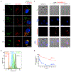
Figure 1.. Dox delivery via SSHELs targeting HER2+ cells reduces tumor burden
For a Figure360 author presentation of this figure, see https://doi.org/10.1016/j.celrep.2022.111955. (A)SSHEL assembly schematic. 1-μm mesoporous silica beads (gray) were loaded with dox (red) and covered with a lipid bilayer (yellow), then SpoVM peptide (blue), and polymerized SpoIVA protein (green). Inset: SpoIVA with an engineered Cys conjugated to TCO covalently attached to tetrazine-conjugated anti-HER2 affibody (blue star). (B) Micrograph of SSHELs displaying labeled affibodies, visualized using (left) DIC or (right) fluorescence from affibody. (C) Fluorescence from SSHELs displaying labeled affibody (green) measured using flow cytometry or beads displaying known quantity of molecules of equivalent soluble fluorochrome (MESF; gray). (D) Dox-SSHELs made with labeled SpoIVA. Top left: DIC; top right: SpoIVA; bottom left: fluorescence from dox; bottom right: overlay, dox and SpoIVA. Scale bars: 1 μm. (E and F) Tumor size of female athymic nude mice implanted subcutaneously (s.c.) with SKOV3 ovarian cancer cells and treated intravenously (i.v.) with PBS (black), (E) 3 mg/kg or (F) 6 mg/kg dox (red), Dox-SSHELαHER2 (green), or SSHELαHER2 (blue) at days post xenograft indicated with arrows. Data points: mean values; errors: SD; n = 7 mice. p values: *<0.05; ****<0.001. See also Figures S1A–S1D.
Figure 2.
Figure 2.. Dox delivery via SSHELs reduces toxicity
(A)Tumor size of athymic nude mice implanted s.c. with SKOV3 cells and treated i.v. with PBS (black), 3 mg/kg liposomal dox (blue), Dox-SSHEL αHER2 (green), or unencapsulated dox with empty SSHELs (red) at indicated days post xenograft. Data points: mean values; errors: SD. (B) Survival curves of mice in (A). i and ii indicate two separate trials (“Trial 3” and “Trial 4”). (C) Number of mice (n = 12) displaying ulcerated tumor or skin descaling when treated with liposomal dox (blue) or Dox-SSHELαHER2 (green) on indicated day. (D and E) Biodistribution of labeled SSHELαHER2 in three mice harboring SKOV3 tumors 24 h post-injection. (D) Fluorescence intensity histograms of cells harvested from indicated organs from each mouse (green, blue, and orange) or untreated mouse (red). (E) Mean fluorescence intensities of cells harvested from indicated organs in (D). Errors: SD. See also Figures S1E, S1F, S2, and S3.
Figure 3.
Figure 3.. SSHEL αHER2 specifically binds to and kills HER2+ cells
(A–F) Labeled SSHELαHER2 incubated at different multiplicities of incubation (MOI) with HER2+ (A, SKBR3 breast cancer, B, SKOV3) or HER2 (C, MDA-MB-231, D, MCF7) cells; binding assessed by flow cytometry. (E) SKBR3 or (F) SKOV3 cells preincubated with excess anti-HER2 affibody, then incubated with SSHELαHER2 at MOI = 100. (G–I) Micrographs of (G) SKBR3, (H) SKOV3, or (I) MDA-MB-231 incubated with labeled SSHELαHER2. Overlay: DIC (gray) and fluorescence from SSHELαHER2 (green). Scale bar: 20 μm. (J–M”) Micrographs of zebrafish with labeled blood vessels (blue), injected in mid-brain parenchyma with SKBR3 with labeled lysosomes (green), and later injected with labeled SSHELαHER2 (pink). (J) Overlay of blood vessels and SSHELαHER2; (K) bright-field image; (L) overlay of SSHELαHER2 and bright field; (M) overlay of SKBR3 lysosomes and SSHELαHER2. (J’–M’) Magnification of area indicated in (J–M). (J”–M”) Magnification of area indicated in (J’–M’). Arrows: SSHELαHER2 bound to SKBR3 surface; arrowhead: colocalization of SSHELαHER2 and SKBR3 lysosome. (N–S) Cell viability of HER2+ (SKBR3, red; SKOV3, orange) and HER2 (MCF7, blue; MDA-MB-231, light blue) cells incubated with (N) Dox- SSHELαHER2, (O) free dox, (P) SSHEL, (Q) affibody, (R) Dox-SSHELBSA, or (S) SSHELαHER2. Data points: mean values; errors: SD (n = 3). (T and U) Caspase 3/7 activities of (T) SKBR3 and (U) SKOV3 after treatment with free dox (120 nM), unloaded SSHELαHER2 (MOI = 200), or dox-loaded-SSHELαHER2 (MOI = 200, 120 nM doxorubicin). Data are mean fold-change compared with PBS-treated cells; errors: SD (n = 3). See also Figure S4.
Figure 4.
Figure 4.. Internalization of Dox-SSHELαHER2 and trafficking to acidic compartment releases the encased dox
(A) Micrograph of SKOV3 labeled with LysoTracker and DAPI (to visualize acidic organelles and DNA, respectively), incubated with labeled SSHELαHER2 at indicated time points. Left: SSHELαHER2 (green); center: LysoTracker (red); right: overlay of SSHELαHER2, LysoTracker, and DAPI (blue); yellow: colocalization of SSHELαHER2 and LysoTracker. Scale bar: 20 μm. (B) SKOV3 cells without (red) or with labeled SSHELαHER2 (MOI = 100) (dark green), or with actin inhibitor latrunculin A (blue), dynamin inhibitor MiTMAB (light green), or both (orange), after which internalization of SSHELαHER2 was assessed by flow cytometry. (C) Micrographs of DAPI-labeled SKBR3 (blue) incubated with Dox-SSHELαHER2 and visualized using intrinsic fluorescence (red) of dox. Arrows: SSHEL-encased dox; arrowheads: cytosolic dox. Scale bar: 50 μm. (D) pH-dependent release of dox in vitro particles reported as a percentage of initial amount of encapsulated dox at pH = 7.4 (red) or pH = 5.0 (blue). Data points: mean; errors: SD (n = 3).

References

    1. Raj S, Khurana S, Choudhari R, Kesari KK, Kamal MA, Garg N, Ruokolainen J, Das BC, and Kumar D (2021). Specific targeting cancer cells with nanoparticles and drug delivery in cancer therapy. Semin. Cancer Biol 69, 166–177. 10.1016/j.semcancer.2019.11.002. - DOI - PubMed
    1. Seymour LW (1992). Passive tumor targeting of soluble macromolecules and drug conjugates. Crit. Rev. Ther. Drug Carrier Syst 9, 135–187. - PubMed
    1. Lammers T, Kiessling F, Hennink WE, and Storm G (2012). Drug targeting to tumors: principles, pitfalls and (pre-) clinical progress. J. Control. Release 161, 175–187. 10.1016/j.jconrel.2011.09.063. - DOI - PubMed
    1. Morales-Cruz M, Delgado Y, Castillo B, Figueroa CM, Molina AM, Torres A, Milián M, and Griebenow K (2019). Smart targeting to improve cancer therapeutics. Drug Des. Devel. Ther 13, 3753–3772. 10.2147/DDDT.S219489. - DOI - PMC - PubMed
    1. Oieni J, Levy L, Letko Khait N, Yosef L, Schoen B, Fliman M, Shalom-Luxenburg H, Malkah Dayan N, D’Atri D, Cohen Anavy N, and Machluf M (2020). Nano-Ghosts: biomimetic membranal vesicles, technology and characterization. Methods 177, 126–134. 10.1016/j.ymeth.2019.11.013. - DOI - PubMed

Publication types

Substances