. 2023 Jan 31;42(1):111930.
doi: 10.1016/j.celrep.2022.111930. Epub 2023 Jan 5.

Chemokine CXCL4 interactions with extracellular matrix proteoglycans mediate widespread immune cell recruitment independent of chemokine receptors

Affiliations

Chemokine CXCL4 interactions with extracellular matrix proteoglycans mediate widespread immune cell recruitment independent of chemokine receptors

Anna L Gray et al. Cell Rep. .

Abstract

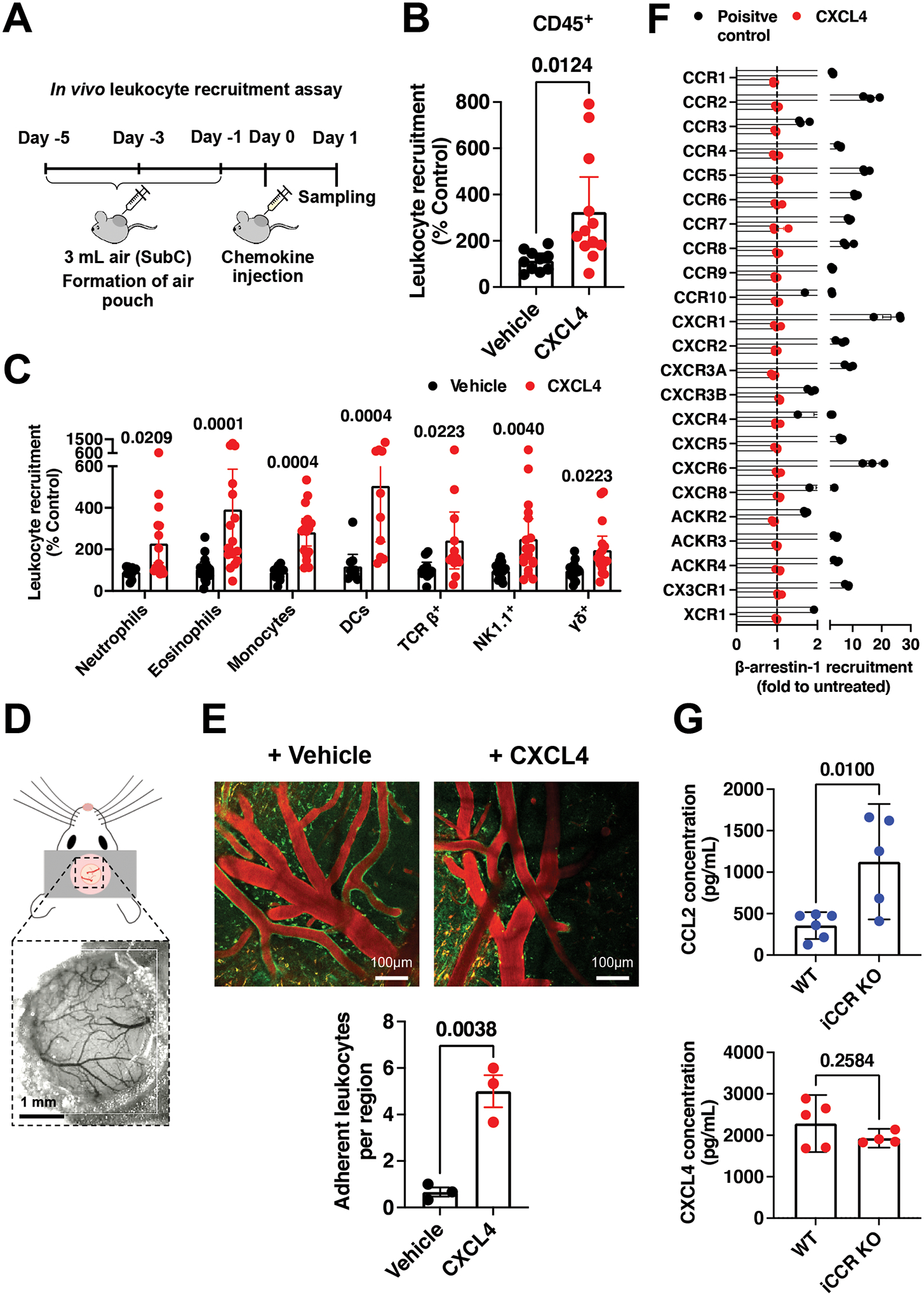
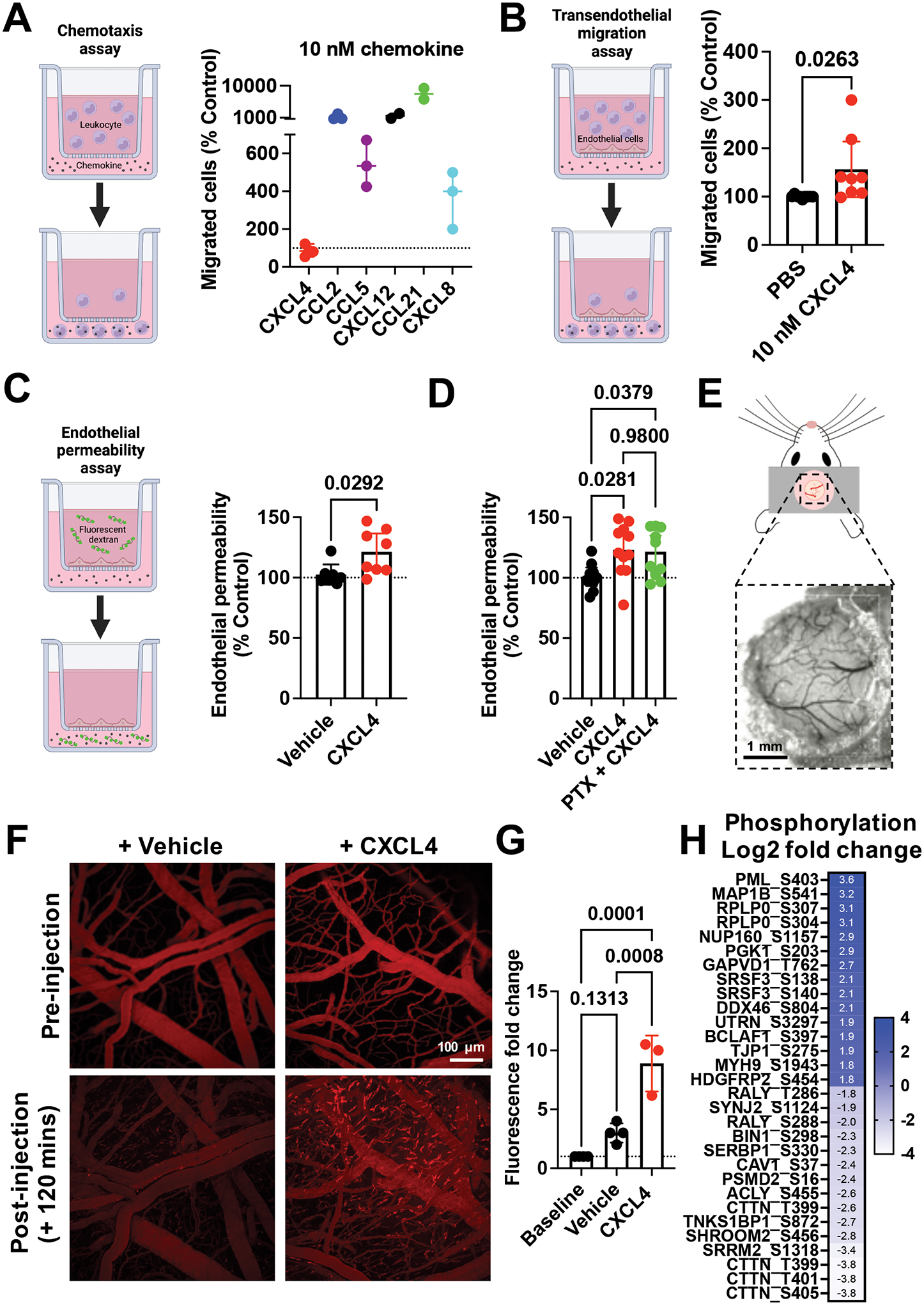
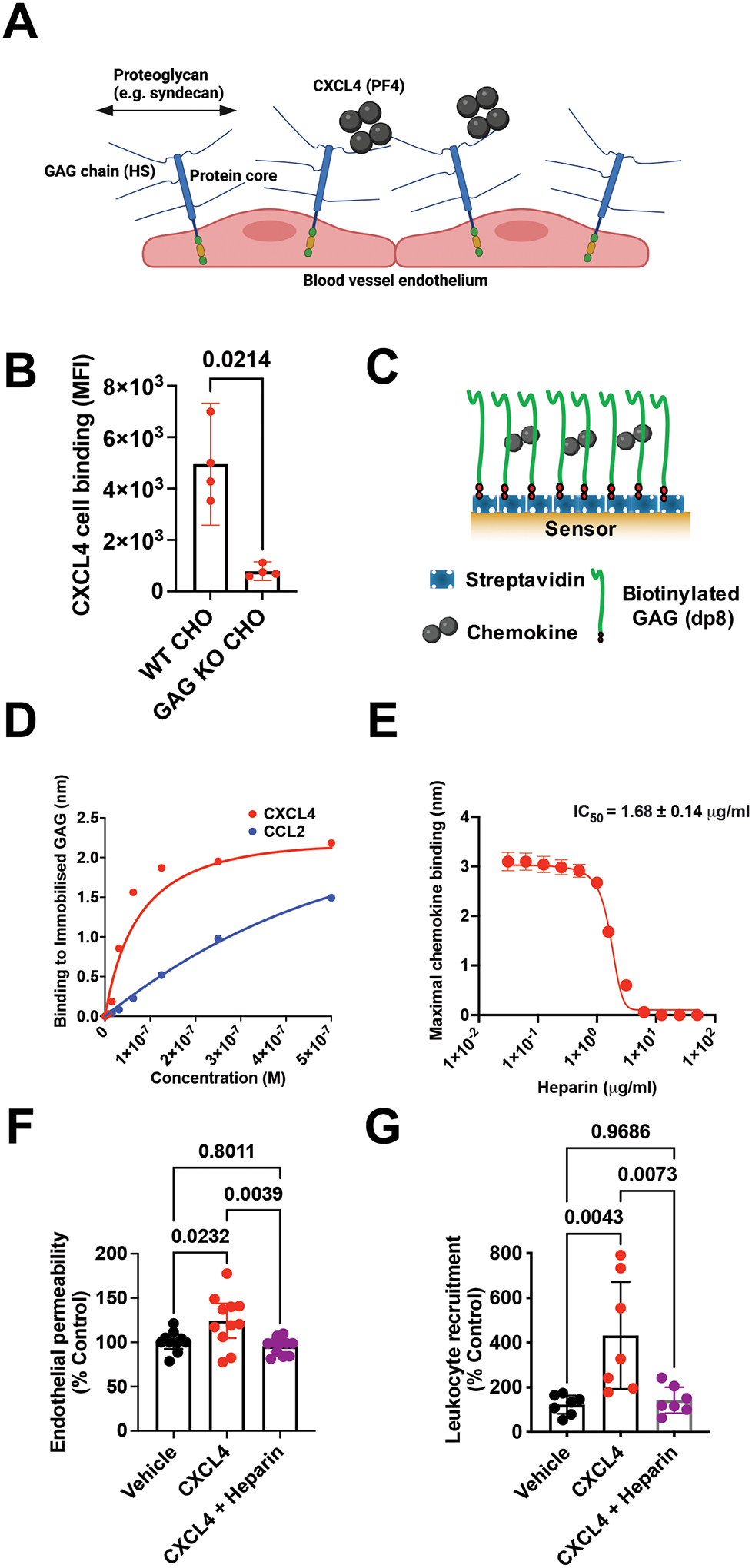
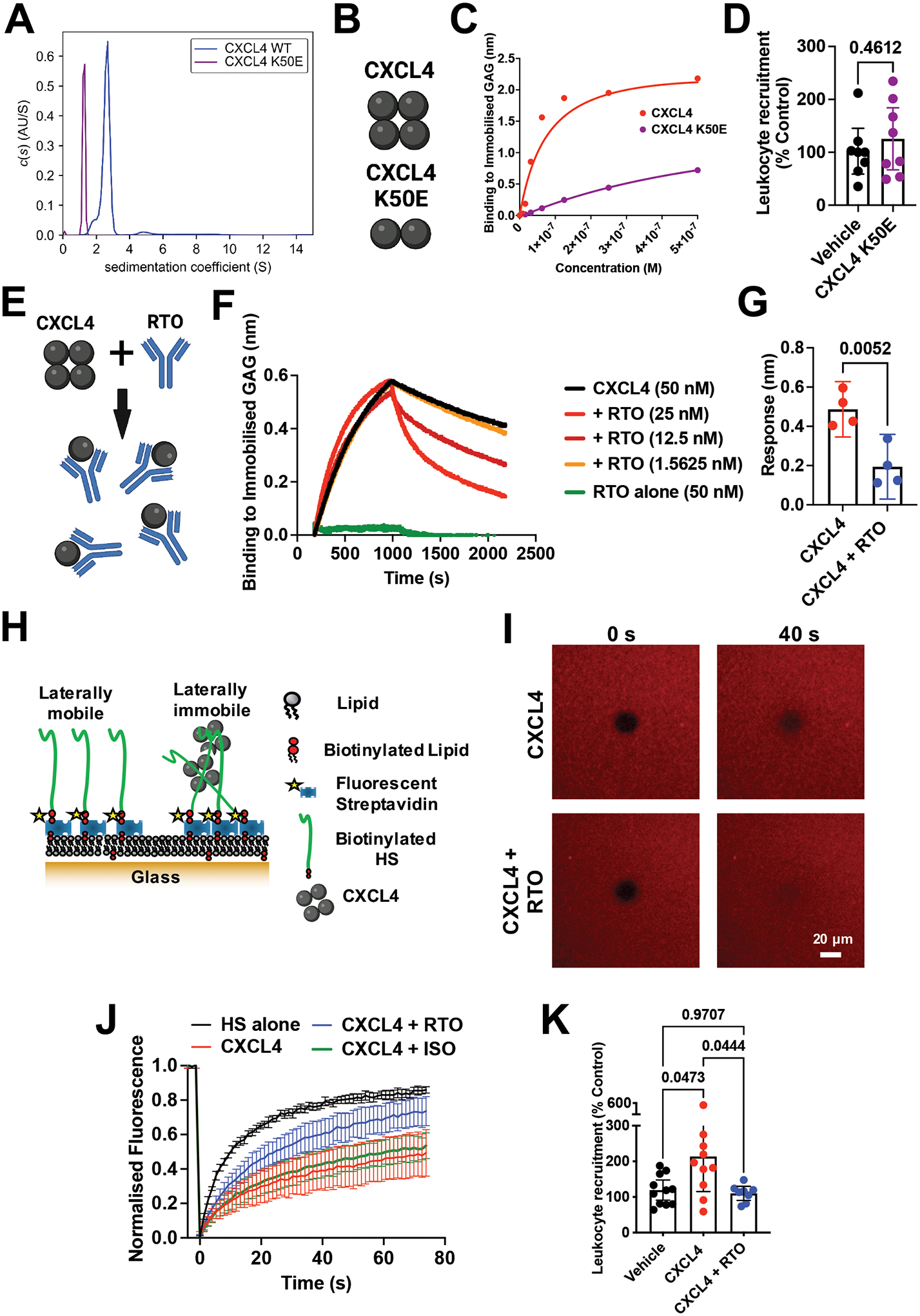
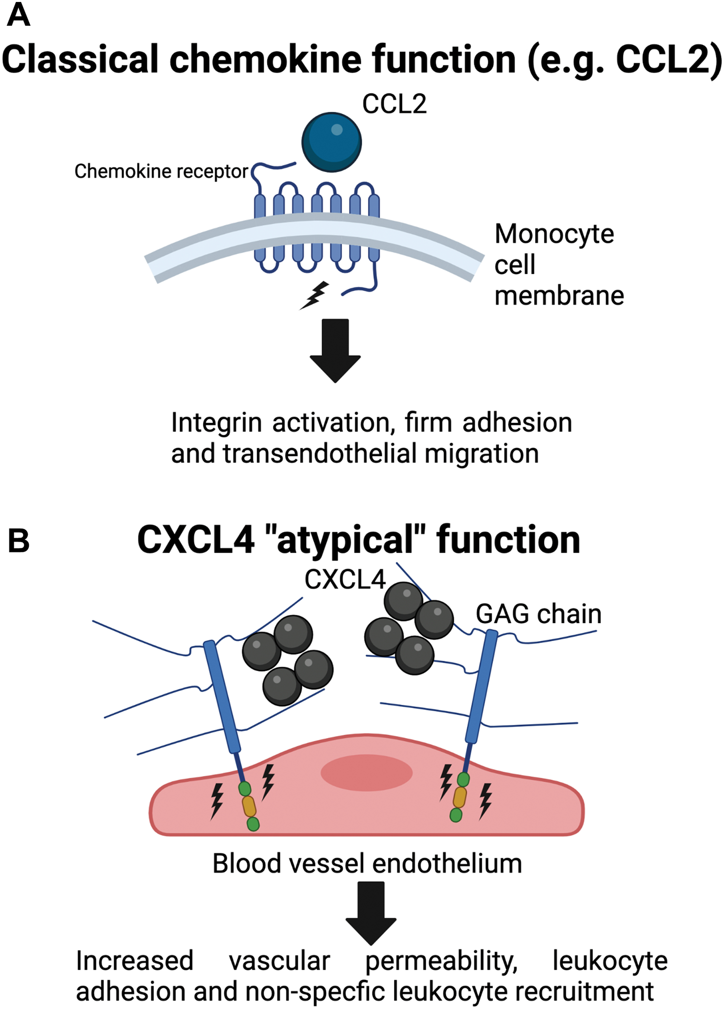
Leukocyte recruitment from the vasculature into tissues is a crucial component of the immune system but is also key to inflammatory disease. Chemokines are central to this process but have yet to be therapeutically targeted during inflammation due to a lack of mechanistic understanding. Specifically, CXCL4 (Platelet Factor 4, PF4) has no established receptor that explains its function. Here, we use biophysical, in vitro, and in vivo techniques to determine the mechanism underlying CXCL4-mediated leukocyte recruitment. We demonstrate that CXCL4 binds to glycosaminoglycan (GAG) sugars on proteoglycans within the endothelial extracellular matrix, resulting in increased adhesion of leukocytes to the vasculature, increased vascular permeability, and non-specific recruitment of a range of leukocytes. Furthermore, GAG sulfation confers selectivity onto chemokine localization. These findings present mechanistic insights into chemokine biology and provide future therapeutic targets.

Keywords: CP: Immunology; CXCL4; PF4; chemokine; extracellular matrix; glycosaminoglycan; leukocyte; proteoglycan; recruitment; trafficking.


Conflict of interest statement

Declaration of interests The authors declare no competing interests.

Figures

Figure 1.
Figure 1.. CXCL4 recruits a wide range of different leukocytes in vivo.
(A) Schematic of the in vivo leukocyte recruitment assay. (B) CD45+ cell counts 24 hr after CXCL4 injection. (C) Quantification of different leukocytes recruited by CXCL4. (D) Schematic of cranial window implantation for in vivo leukocyte adhesion analysis. (E) In vivo analysis of leukocyte (labelled green) adhesion to the walls of the vasculature (labelled red) following injection of CXCL4 or vehicle control. (F) Agonist activity of CXCL4 (100 nM) towards 19 classical and 3 atypical chemokine receptors evaluated in a β-arrestin-1 recruitment assay. For all receptors, one known agonist chemokine (100 nM) listed in the IUPHAR repository of chemokine receptor ligands was added as positive control. (G) CCL2 and CXCL4 quantification in the serum of wild type or iCCR (CCR1, 2, 3 and 5) KO inflamed mice. All plots are mean with 95% confidence intervals and represent at least two separate experiments, data have been pooled and each dot in B, C, E and G represents an individual mouse and each dot in F represents the mean of an individual experiment. B and C are normalised to vehicle controls. Results in F are expressed as fold change relative to untreated controls and are presented as mean of three independent experiments. Individual p values are shown, B, E and G analysed using an unpaired t-test and C analysed using a one-way ANOVA with a post-hoc Sidak analysis of log-transformed data.
Figure 2.
Figure 2.. CXCL4 increases endothelial permeability in a receptor independent manner and mediates endothelial intracellular signalling.
(A) Chemokine mediated chemotaxis of relevant receptor expressing cells; CXCL4, CCL2 or CCL5 (monocytes), CXCL12 (CXCR4+ Jurkat cells), CCL21 (CCR7+ L1.2 cells) and CXCL8 (CXCR2+ neutrophils). (B) Transendothelial migration of human monocytes towards CXCL4. (C) Transwell endothelial permeability in the absence and presence of CXCL4, (D) CXCL4 alone or in combination with pertussis toxin. (E) Schematic of cranial window implantation for in vivo vascular permeability analysis. (F) In vivo analysis of leakage of intravenously injected fluorescent dextran from the vasculature into the meninges following intravenous injection of CXCL4 or vehicle control. (G) Quantification of (F). (H) Heat map of the Log2 fold change of indicated protein phosphorylation sites from endothelial cells stimulated with CXCL4, relative to vehicle controls. All plots are mean with 95% confidence intervals, represent at least two separate experiments where data have been pooled. Each dot in A-D represents an individual transwell and each dot in G represents an individual mouse. Data in A-D and G are normalised to vehicle controls. Individual p values are shown, and C analysed using an unpaired t-test, D and G using a one-way ANOVA with a post-hoc Tukey analysis.
Figure 3.
Figure 3.. CXCL4 function is mediated by its interaction with glycosaminoglycans (GAGs).
(A) Schematic of proteoglycans forming the luminal endothelial glycocalyx. (B) CXCL4 binding to CHO cells with and without surface GAGs. (C) Schematic of the biophysical BLI GAG-binding assay. (D) Maximum signal of chemokine:dp8 binding at different chemokine concentrations in the BLI assay. (E) Maximum CXCL4 (100 nM) binding to dp8 signal following pre-incubation with increasing concentrations of heparin. (F) Endothelial permeability (Transwell) assay with and without CXCL4 and exogenous heparin. (G) In vivo leukocyte (CD45+) recruitment (air pouch) to CXCL4. B, F and G are mean with 95% confidence intervals and represent at least two separate experiments where data have been pooled. D and E are representative of at least two separate experiments. Each dot in B and F represents a technical replicate and each dot in G represents an individual mouse. F and G are normalised to vehicle controls. Individual p values are shown, B analysed using an unpaired t-test, F and G analysed using a one-way ANOVA with a post-hoc Tukey analysis.
Figure 4.
Figure 4.. CXCL4 oligomerisation drives leukocyte recruitment.
(A) AUC analysis of CXCL4 and the mutant K50E to show sedimentation coefficients (indicative of size). (B) Schematic of oligomerisation states of CXCL4 or the CXCL4 K50E mutant. (C) BLI analysis of CXCL4 K50E binding to immobilised heparin dp8. (D) In vivo leukocyte (CD45+) recruitment (air pouch) to CXCL4 K50E mutant. (E) Schematic of RTO antibody binding to CXCL4 monomer to inhibit oligomerisation. (F) CXCL4 (50 nM) binding to dp8 was monitored in the absence and presence (at a range of concentrations) of RTO antibody. (G) Final CXCL4:dp8 signal (after washing) from (F) is plotted with and without pre-incubation with RTO antibody (25 nM). (H) Schematic of FRAP assay to analyse HS cross-linking. (I) FRAP analysis of GAG in-plane mobility with CXCL4 alone or in combination with the RTO antibody, images post bleaching at indicated times. (J) Fluorescence recovery over time. (K) CXCL4 mediated leukocyte recruitment (CD45+) with and without RTO antibody. D, G, J and K are mean with 95% confidence intervals and represent at least two separate experiments where data have been pooled. Each dot in D and K represents an individual mouse and each dot in G represents a technical replicate. A, C, F, I and J are representative of at least two separate experiments. Data in D and K are normalised to vehicle controls. Individual p values are shown, G analysed using an unpaired t-test and K analysed using a one-way ANOVA with a post-hoc Tukey analysis.
Figure 5.
Figure 5.. GAG sulphation mediates chemokine interaction selectivity and cellular localisation.
(A) Overview of enzymes involved in the biosynthesis of HS and CS GAGs. Elongation proceeds from right to left, a tetrasaccharide linker is elongated with either HS or CS disaccharide repeats into long linear polysaccharides. HS is further modified by addition of sulphate groups at the N-, 2-O, 6-O or 3-O (rarely) positions, this process is catalysed by the indicated sulphotransferases. (B) Chemokine binding to WT CHO cells or CHO cells with no GAGs (KO B4galt7), no CS (KO Csgalnact1/2/Chsy1) or no HS (KO Extl3). (C) Heat map analysis of chemokine binding to CHO cells with KO/KI of sulfotransferases acting on HS, where data is normalised to WT cells. (D) Heatmap analysis of maximum BLI signal of chemokine (500 nM) binding to differentially de-sulphated heparin fragments, data normalised to binding on fully-sulphated heparin. (E) Raw signal of chemokine binding to 2-O de-sulphated heparin in the BLI assay. Chemokine mediated in vivo leukocyte recruitment with and without 2-O de-sulphated heparin (F) CD45+ cell counts (CXCL4) or (G) monocyte (Ly6C+) cell counts (CCL2). B, E, F and G are mean with 95% confidence intervals and represent at least two separate experiments where data have been pooled; each dot in B and E represents a technical replicate and each dot in F and G represents an individual mouse. Data in F and G are normalised to vehicle controls. Individual p values are shown, E analysed using an unpaired t test. F and G analysed using a one-way ANOVA with a post-hoc Tukey analysis.
Figure 6.
Figure 6.. CXCL4 binds to endothelial GAG sugars, resulting in an increase of vascular permeability and non-specific leukocyte recruitment.
(A) Classical chemokines, e.g. CCL2, facilitate leukocyte recruitment by binding to seven transmembrane receptors on circulating leukocytes leading to signalling, integrin activation and firm adhesion of the leukocyte to the endothelium. (B) We propose that CXCL4 binds and re-models (as a tetramer) endothelial GAG sugars within the glycocalyx. This produces increased vascular permeability and leukocyte adhesion to endothelial cells possibly via signalling through the proteoglycan, facilitating “non-specific” recruitment of a range of different leukocytes from the vasculature and into inflamed tissues.

References

    1. Nourshargh S, and Alon R (2014). Leukocyte migration into inflamed tissues. Immunity 41, 694–707. 10.1016/j.immuni.2014.10.008. - DOI - PubMed
    1. Bachelerie F, Ben-Baruch A, Burkhardt AM, Combadière C, Farber JM, Graham GJ, Horuk R, Sparre-Ulrich AH, Locati M, Luster AD, et al. (2014). International Union of Basic and Clinical Pharmacology. [corrected]. LXXXIX. Update on the extended family of chemokine receptors and introducing a new nomenclature for atypical chemokine receptors. Pharmacological Reviews 66, 1–79. 10.1124/pr.113.007724. - DOI - PMC - PubMed
    1. Schall TJ, and Proudfoot AEI (2011). Overcoming hurdles in developing successful drugs targeting chemokine receptors. Nat Rev Immunol 11, 355–363. 10.1038/nri2972. - DOI - PubMed
    1. Proudfoot AEI, Bonvin P, and Power CA (2015). Targeting chemokines: Pathogens can, why can’t we? Cytokine 74, 259–267. 10.1016/j.cyto.2015.02.011. - DOI - PubMed
    1. von Hundelshausen P, and Schmitt MMN (2014). Platelets and their chemokines in atherosclerosis-clinical applications. Front Physiol 5, 294. 10.3389/fphys.2014.00294. - DOI - PMC - PubMed

Publication types