. 2023:678:145-192.
doi: 10.1016/bs.mie.2022.09.018. Epub 2022 Nov 3.

Reconstruction of 3D density from solution scattering

Affiliations

Reconstruction of 3D density from solution scattering

Thomas D Grant. Methods Enzymol. 2023.

Abstract

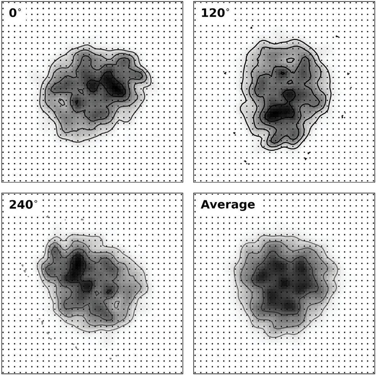
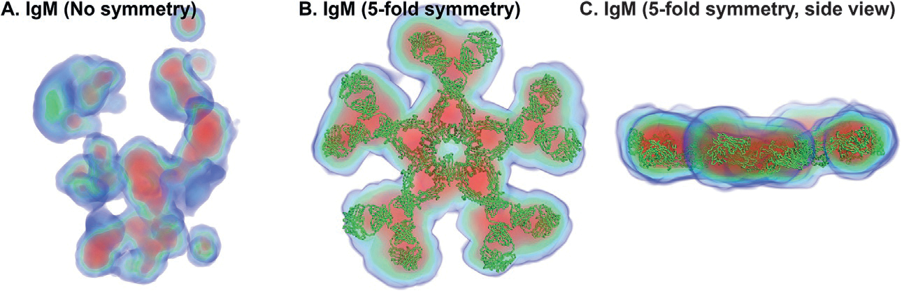
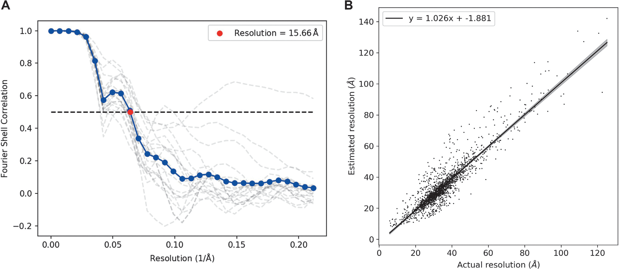
Ab initio modeling methods have proven to be powerful means of interpreting solution scattering data. In the absence of atomic models, or complementary to them, ab initio modeling approaches can be used for generating low-resolution particle envelopes using only solution scattering profiles. Recently, a new ab initio reconstruction algorithm has been introduced to the scientific community, called DENSS. DENSS is unique among ab initio modeling algorithms in that it solves the inverse scattering problem, i.e., the 1D scattering intensities are directly used to determine the 3D particle density. The reconstruction of particle density has several advantages over conventional uniform density modeling approaches, including the ability to reconstruct a much wider range of particle types and the ability to visualize low-resolution density fluctuations inside the particle envelope. In this chapter we will discuss the theory behind this new approach, how to use DENSS, and how to interpret the results. Several examples with experimental and simulated data will be provided.

Keywords: Ab initio; Density; Imaging; Modeling; Phase retrieval; Small angle scattering.


Figures

Fig. 1
Fig. 1
Iterative structure factor retrieval scheme. (A) The starting 3D density for the iterative retrieval cycle is shown as a flattened image and contour map on a 2D grid for illustration purposes. Each step begins with the solvent flattened density output by the last step, except for the first step which begins with random density (not shown). (B) Using a forward FFT the structure factors, F, are calculated from the starting density (for illustration purposes only the amplitudes are shown). (C) The 3D intensities are then calculated as the square of the amplitudes. The dashed circles outline the concentric q shells used to bin grid points for subsequent spherical averaging. (D) The 1D spherically averaged intensities (denoted by I3DΩ) are calculated and compared with the experimental 1D scattering profile, and used to calculate scale factors for each q shell. (E) Each spherical shell of structure factors is scaled using the scale factor, k, calculated in the previous step, resulting in a new set of structure factors. (F) The inverse FFT is performed to obtain a new density map whose corresponding scattering profile now matches the experimental 1D scattering profile. Real space restraints such as solvent flattening are then applied to the new density and the cycle is repeated.
Fig. 2
Fig. 2
Shrinkwrap procedure. (A) The noisy 3D density is shown as a flattened 2D image and a contour map on a 2D grid of voxels, represented by black dots. For illustration purposes the noise is artificially increased. (B) A low-pass Gaussian filter is applied to a copy of the original density. (C) The object support, calculated as the set of filtered voxels above a given threshold, is shown outlined by a solid black line. (D) Solvent flattening is performed by setting the density of voxels outside of the support to zero.
Fig. 3
Fig. 3
Fitting experimental data with denss.fit_data.py GUI. A screenshot of the interactive GUI of denss.fit_data.py is shown. The upper left panel shows the experimental scattering profile in black dots, the fit to the data as a red line, and the intensities at the Shannon points as blue circles. The Residuals between the experimental data and the fit are shown below the intensities. The upper right panel shows the P(r) curve corresponding to the fit. Sliders for adjusting Dmax and α are shown on the lower left. Various parameters describing the size of the particle calculated from the fit are displayed on the bottom right.
Fig. 4
Fig. 4
Single reconstructions of density using DENSS. (A) A single reconstruction of BSA from experimental data using the default SLOW mode of DENSS. (B) A single reconstruction of BSA in FAST mode. (C) A single reconstruction of apoferritin from experimental data using SLOW mode. (D) A cross-section of the density displayed in (C) showing the interior cavity in apoferritin. (E) A single reconstruction of PCNA in SLOW mode showing the distinctive central hole. (F) A single reconstruction of PCNA in FAST mode. All density maps are displayed using the Volume representation in PyMOL colored according to density value (blue: 1σ, green: 5σ, red: 10σ). Atomic models taken from the SASBDB entry are displayed in gray cartoon representation.
Fig. 5
Fig. 5
DENSS reconstruction of a micelle with negative contrast. The density map reconstructed by DENSS using experimental data of a DDM micelle using either MEMBRANE mode (A) or SLOW mode (B). Density is displayed as an isocontour surface at +1σ (gray transparent surface) and 1σ (pink surface). Note that the negative contrast is only seen in MEMBRANE mode.
Fig. 6
Fig. 6
Evaluating the success of DENSS reconstructions. (A) A plot generated by denss.py shows the three metrics used to assess convergence including χ2, Rg, and support volume. Each value is plotted on a separate y-axis as a function of the number of steps completed of the iterative reconstruction procedure. (B) A plot generated by denss.py showing the quality of the fit of the calculated scattering profile from the final density map compared to the experimental data. As described above, the experimental data (black dots) are first fitted using a smooth curve (black dashed line, e.g., using denss.fit_data.py). The fit is then interpolated to the q values corresponding to the concentric q shells in reciprocal space (blue circles). The final calculated scattering profile from the DENSS reconstruction is shown as red circles, and the residuals between the interpolated experimental intensities and the calculated intensities is shown below.
Fig. 7
Fig. 7
Symmetry averaging procedure for threefold symmetry. During the symmetry averaging step, the initial density (0°) is rotated by 120° and 240°. The three symmetry related copies are then averaged to produce a final density with threefold symmetry.
Fig. 8
Fig. 8
Symmetry averaging significantly improves DENSS reconstructions of lgM. (A) DENSS reconstruction of IgM using default SLOW mode. (B) DENSS reconstruction adding fivefold symmetry. (C) Side view of density shown in (B). Density maps are displayed using the Volume representation in PyMOL colored according to density value (blue: 1σ, green: 5σ, red: 10σ). The atomic model was taken from the PDB entry 2RCJ and is displayed in green ribbon representation.
Fig. 9
Fig. 9
Averaging of multiple individual DENSS reconstructions of BSA. Ten individual density reconstructions generated by denss.all.py are numbered and shown on the left. The final reconstruction from denss.all.py after alignment and averaging of all 20 individual reconstructions is shown on the right. All density maps are displayed using the Volume representation in PyMOL colored according to density value (blue: 1σ, green: 5σ, red: 10σ). The atomic model is taken from the SASBDB entry and displayed in gray cartoon representation.
Fig. 10
Fig. 10
Resolution estimation from DENSS. (A) An example of the Fourier Shell Correlation vs resolution plot, output by denss.all.py, is shown. FSC curves for each of the 20 reconstructions compared to the reference are shown as gray dashed lines, and the averaged FSC is shown as a blue line. The estimated resolution, where the averaged FSC curve crosses 0.5, is shown by the red circle. (B) ~1900 Simulated scattering curves from PDB entries were used by DENSS to generate averaged density maps using default settings. The resolution estimated by denss.all.py is shown on the y-axis. For each reconstruction, the averaged density map was aligned to the known structure using denss. align.py and the actual resolution was calculated using the denss.pdb2mrc.py and denss.calcfsc.py scripts, plotted on the x-axis. A linear trend was fit to the data and is shown by the solid black line (95% confidence intervals shown by the shaded gray region). The equation of the line is given in the legend, showing a slope of 1.026 and intercept of −1.881, demonstrating that the estimated resolution by DENSS closely approximates the true resolution.
Fig. 11
Fig. 11
Graphical viewers showing density maps of BSA from DENSS. (A) Density in UCSF Chimera shown as a gray transparent isosurface at 1σ. The aligned atomic model is shown in cartoon representation. (B) Density displayed using the Volume representation in PyMOL colored according to density value (blue: 1σ, green: 5σ, red: 10σ). The atomic model in gray cartoon representation. (C) Density displayed as isomesh in COOT contoured at 1σ. The atomic model is shown using the lines representation.
Fig. 12
Fig. 12
DENSS reconstruction of GLIC protein from SANS data. GLIC was reconstructed using the denss.all.py script with C5 symmetry applied using default parameters. The density map is displayed using the Volume representation in PyMOL colored according to density value (blue: 1σ, green: 5σ, red: 10σ). The atomic model is shown in gray cartoon representation. The density maps on the right have each been rotated by 90°.

Similar articles

Cited by

References

    1. Afonine PV, Poon BK, Read RJ, Sobolev OV, Terwilliger TC, Urzhumtsev A, et al. (2018). Real-space refinement in PHENIX for cryo-EM and crystallography. Acta Crystallographica. Section D: Structural Biology, 74(Pt. 6), 531–544. 10.1107/S2059798318006551. - DOI - PMC - PubMed
    1. Berman HM, Westbrook J, Feng Z, Gilliland G, Bhat TN, Weissig H, et al. (2000). The protein data bank. Nucleic Acids Research, 28(1), 235–242. 10.1093/nar/28.1.235. - DOI - PMC - PubMed
    1. Bernadó P, Mylonas E, Petoukhov MV, Blackledge M, & Svergun DI (2007). Structural characterization of flexible proteins using small-angle X-ray scattering. Journal of the American Chemical Society, 129(17), 5656–5664. - PubMed
    1. Beyett TS, Fraley AE, Labudde E, Patra D, Coleman RC, Eguchi A, et al. (2019). Perturbation of the interactions of calmodulin with GRK5 using a natural product chemical probe. Proceedings of the National Academy of Sciences of the United States of America, 116(32), 15895–15900. 10.1073/pnas.1818547116. - DOI - PMC - PubMed
    1. Brown PJ, Fox AG, Maslen EN, O’Keefe MA, & Willis BTM (2006). Intensity of diffracted intensities. International Tables for Crystallography, 554–595.

Publication types

LinkOut - more resources