. 2023 Jan 14;13(1):757.
doi: 10.1038/s41598-022-27328-y.

SARS-CoV-2 infection augments species- and age-specific predispositions in cotton rats

Affiliations

SARS-CoV-2 infection augments species- and age-specific predispositions in cotton rats

Marina S Boukhvalova et al. Sci Rep. .

Abstract

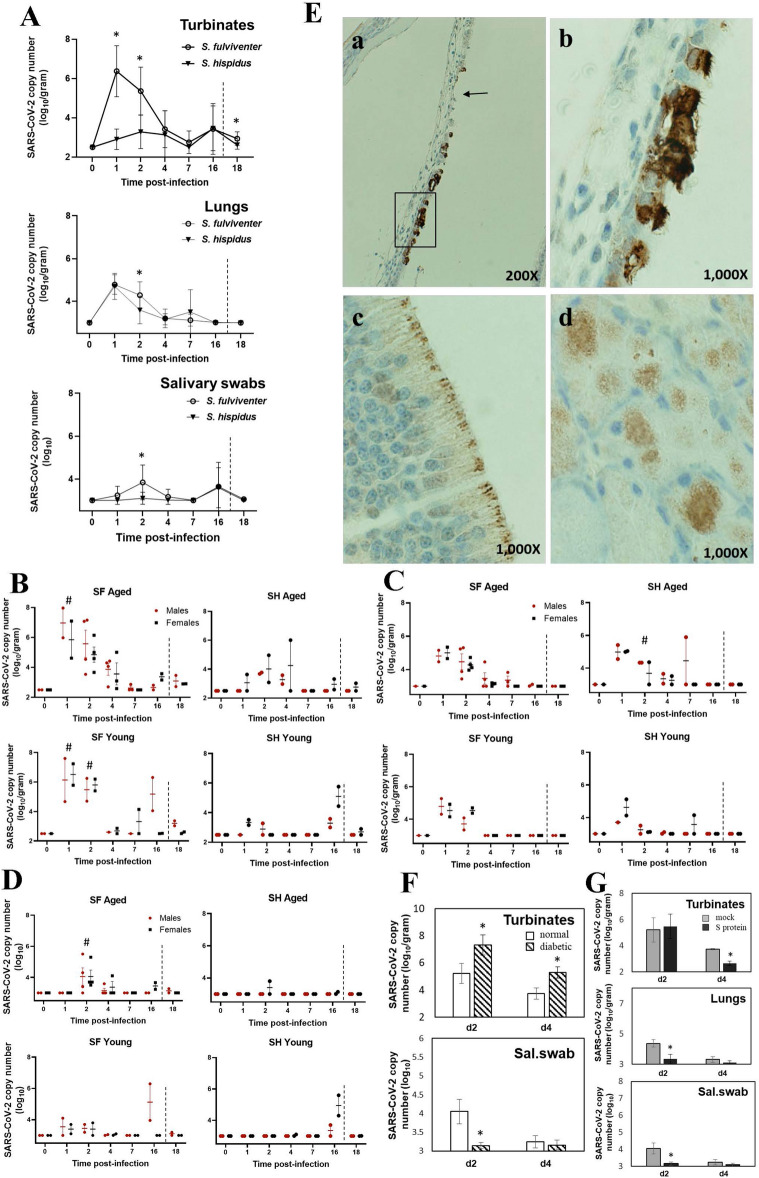
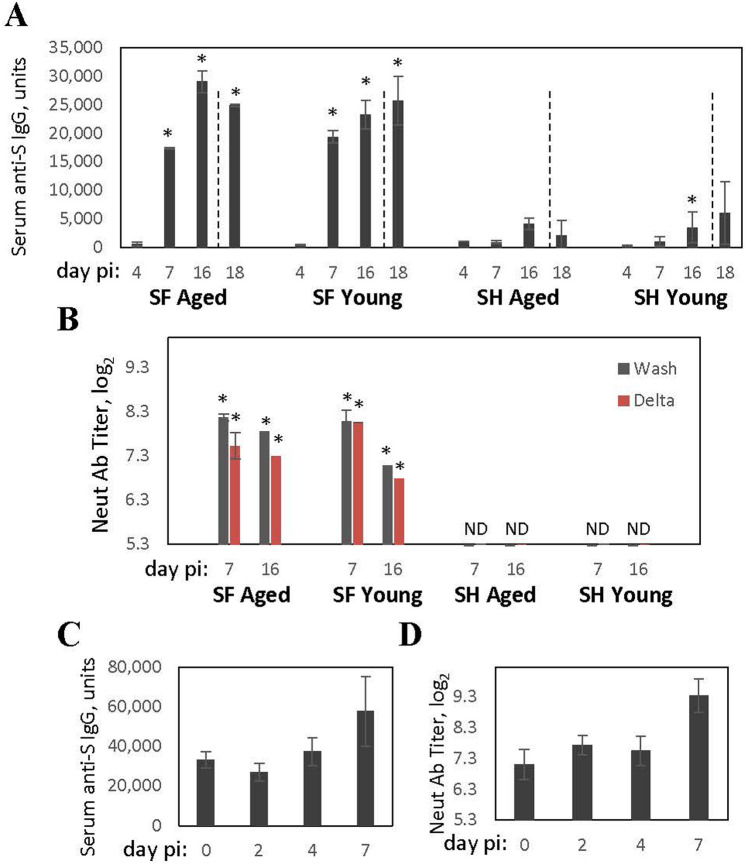
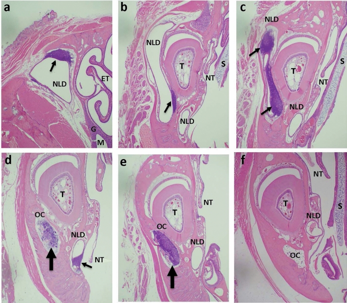
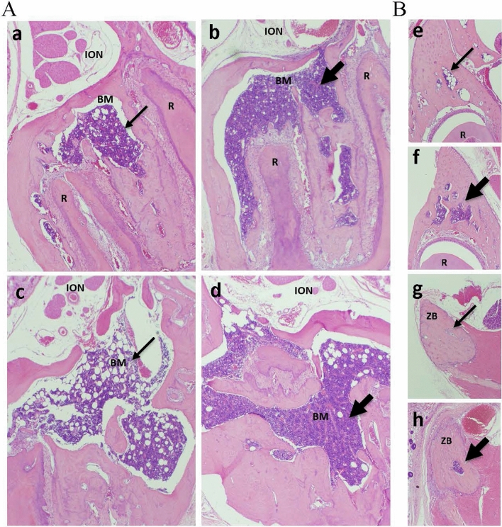
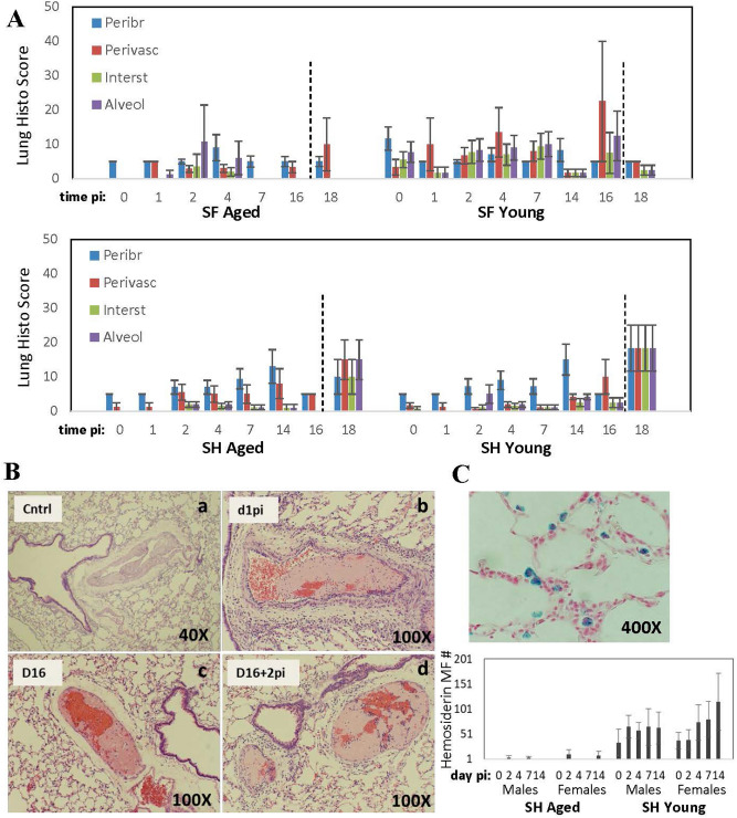
Heterogeneity of COVID-19 manifestations in human population is vast, for reasons unknown. Cotton rats are a clinically relevant small animal model of human respiratory viral infections. Here, we demonstrate for the first time that SARS-CoV-2 infection in cotton rats affects multiple organs and systems, targeting species- and age-specific biological processes. Infection of S. fulviventer, which developed a neutralizing antibody response and were more susceptible to SARS-CoV-2 replication in the upper respiratory tract, was accompanied by hyperplasia of lacrimal drainage-associated lymphoid tissue (LDALT), a first known report of mucosa-associated lymphoid tissue activation at the portal of SARS-CoV-2 entry. Although less permissive to viral replication, S. hispidus showed hyperplasia of bone marrow in the facial bones and increased pulmonary thrombosis in aged males. Augmentation of these features by SARS-CoV-2 infection suggests a virus-induced breach in regulatory mechanisms which could be devastating for people of all ages with underlying conditions and in particular for elderly with a multitude of ongoing disorders.


Conflict of interest statement

The authors declare that the research was conducted in the absence of any commercial or financial relationships that could be construed as a potential conflict of interest. Although the authors view each of these as non-competing financial interests, we report that KMB is an active member of the Human Cell Atlas and has been a Scientific Advisor at Arcato Laboratories, Inc. We also report that MSB and JCGB are part owners of Sigmovir Biosystems, Inc.

Figures

Figure 1
Figure 1
Viral replication in SARS-CoV-2-infected cotton rats. (A) S. fulviventer (SF) and S. hispidus (SH) were inoculated with SARS-CoV-2 and sacrificed on days 1–16 post-infection for analysis of viral load in turbinates, lungs, and salivary swabs by qPCR. A group of animals was re-challenged with SARS-CoV-2 on day 16 post-infection and sacrificed 2 days later (day 18, dashed line indicates re-infection). The data for males and females of different ages are presented altogether. Seven to twelve animals per time point. *p < 0.05 compared to SH. (BD) Viral loads shown separately for male or female, aged or young animals for turbinates (B), lungs (C) or salivary swabs (D). The mean ± SE is shown for each time point. #p < 0.05 compared to SH of the same age category (males and females together). (E) Nasal sections of SF infected with SARS-CoV-2 and sacrificed on day 1 post-infection were stained for SARS-CoV-2 S protein. Virus was detectable in the respiratory epithelium of maxilloturbinate (a,b). Panel a shows damaged area of respiratory epithelium (arrow) that already does not contain viral antigens and another area of epithelium (boxed) intensely stained that is further magnified in panel b. Olfactory epithelium (c) and submucosal glands surrounding the maxillary sinus (d) were also positive for SARS-CoV-2. (F) SF with diabetes and normal SF were challenged with SARS-CoV-2 and sacrificed on days 2 and 4 post-challenge for analysis of viral load in the turbinates and salivary (Sal.) swabs by qPCR. The mean ± SE is shown for each group of cotton rats (4–8 animals per group, males and females). *p < 0.05 compared to normal animals. (G) SF were vaccinated with the purified S protein of SARS-CoV-2 adjuvanted with alum, boosted, and infected with SARS-CoV-2. Control animals were mock-immunized with alum, infected with SARS-CoV-2, and sacrificed in parallel with the S-protein-immunized infected animals on days 2 and 4 post-infection for analysis of viral load in turbinates, lungs, and salivary swabs by qPCR. Results represent the mean ± SE for each group of animals (4–8 animals per group, males and females). *p < 0.05 compared to mock-immunized, SARS-CoV-2-infected animals, sacrificed on the same day.
Figure 2
Figure 2
Antibody response of cotton rats to SARS-CoV-2-infection. (A) Aged and young cotton rats SF and SH were infected with SARS-CoV-2, and sera was collected for analysis of binding IgG against SARS-CoV-2 S protein on days 4, 7, and 16 after infection. A group of animals was re-challenged with SARS-CoV-2 on day 16 post-infection and sera was collected 2 days later (day 18, dashed line indicates re-infection). (B) Neutralizing antibodies in the serum of aged and young infected SF or SH on days 7 and 16 post-infection. Neutralization was measured against homologous (Washington, Wash, dark grey bars) and heterologous (Delta, red bars) strains of SARS-CoV-2. No neutralizing antibody activity was detected against either virus in sera collected from SARS-CoV-2 infected SH (ND). Results represent the mean ± SE shown for each group (males and females). *p < 0.05 compared to uninfected animals. (C) Binding IgG and (D) neutralizing antibodies against Washington strain in sera of cotton rats immunized with S protein and sampled immediately prior to SARS-CoV-2 infection (day 0) or on days 2, 4, and 7 after SARS-CoV-2 infection.
Figure 3
Figure 3
Lacrimal drainage-associated lymphoid tissue (LDALT) hyperplasia after SARS-CoV-2 infection in cotton rats SF. Nasal/paranasal sections from SARS-CoV-2-infected (ae) or control uninfected (f) SF at the level of the second palatal ridge (a) or at the level of incisive papilla (bf). (a) The caudal portion of nasolacrimal duct (NLD) of infected SF on day 7 post-infection showing LDALT (arrow) at the top corner of NLD. Ethmoid turbinates (ET), maxillary sinus (M), and submucosal glands (G) are visible. (b) The same animal as in panel a, but with the head cut at the level of incisive papilla. NLD is visible in two locations: lateral to the incisor tooth (T) and below it. LDALT is seen only in the portion of the duct lateral to the tooth (arrow). The septum (S) and nasoturbinates (NT) are visible in this sample. (c) An example of a more intense activation of LDALT (arrows) in a different SF on day 14 post-infection in a cut position similar to that shown in panel b. (d, e) Lymphoid hyperplasia leading to the “occlusion” (thick arrow) of the osseous canal (OC) housing NLD in infected SF on day 4 (d) or day 7 (e) post-infection. A ventral section of NLD shows signs of LDALT activation as well (d, thin arrow). (f) The section from a control, uninfected SF showing lack of LDALT hyperplasia and lack of occlusion of OC. H&E stain, 20X magnification.
Figure 4
Figure 4
Bone marrow (BM) hyperplasia in SARS-CoV-2-infected cotton rats SH. (A) BM hyperplasia in maxilla at the molar tooth level cut. Young (a,b) and aged (c,d) SH males were mock-challenged with PBS (a,c) or infected with SARS-CoV-2 and sacrificed on day 7 (b) or day 4 (d) post-infection. BM, bone marrow; R, tooth root; ION, infraorbital nerve. (B) BM in additional facial bone areas: in maxilla at the incisor papilla level cut (e,f) and in the zygomatic bone (ZB) (g,h). Young SH male mock-challenged with PBS (e,g) or infected with SARS-CoV-2 and sacrificed 7 days later (f,h). Thick arrows point to hyperplastic BM, thin arrows point to normal BM. H&E, 40X.
Figure 5
Figure 5
Pulmonary histopathology in cotton rats infected with SARS-CoV-2. (A) Peribronchiolitis (Peribr), perivasculitis (Perivasc), interstitial inflammation (Interst) and alveolitis (Alveol) were evaluated in H&E-stained lung samples from aged and young SF and SH infected with SARS-CoV-2 (or re-infected) as described in the legend to Fig. 1. Dashed line indicates re-infection. Results are cumulative of two independent studies (4–10 samples per time point, males and females). (B) Pulmonary thrombosis in aged male SH. (a) A hypertrophic blood vessel wall in uninfected (Cntrl) animal, (b–d) acute thrombi in SARS-CoV-2-infected animals on days 1, 16, and 16 + 2 (after re-infection). (C) Hemosiderin-positive macrophages in the lungs of young SH on day 14 post-infection (Prussian Blue stain) and graphic summary of positive cells in aged and young SH on various days after SARS-CoV-2 infection (no hemosiderin-positive macrophages were detected in SF).

Similar articles

Cited by

References

    1. WHO Coronavirus (COVID-19) Dashboard, https://covid19.who.int/
    1. de Wit E, et al. SARS and MERS: Recent insights into emerging coronaviruses. Nat Rev Microbiol. 2016;14:523–534. doi: 10.1038/nrmicro.2016.81. - DOI - PMC - PubMed
    1. Song Z, et al. From SARS to MERS, thrusting coronaviruses into the spotlight. Viruses. 2019;11:59. doi: 10.3390/v11010059. - DOI - PMC - PubMed
    1. Johns Hopkins Unuversity. Coronavirus Resource Center. Mortality analyses. https://coronavirus.jhu.edu/data/mortality Accessed July 214, 2022.
    1. Grasselli G, et al. Baseline characteristics and outcomes of 1591 patients infected with SARS-CoV-2 admitted to ICUs of the Lombardy region. Italy. JAMA. 2020;323:1574–1581. doi: 10.1001/jama.2020.5394. - DOI - PMC - PubMed

Publication types