. 2023 Apr 5;31(4):970-985.
doi: 10.1016/j.ymthe.2023.01.014. Epub 2023 Jan 14.

Use of 2,6-diaminopurine as a potent suppressor of UGA premature stop codons in cystic fibrosis

Affiliations

Use of 2,6-diaminopurine as a potent suppressor of UGA premature stop codons in cystic fibrosis

Catherine Leroy et al. Mol Ther. .

Abstract

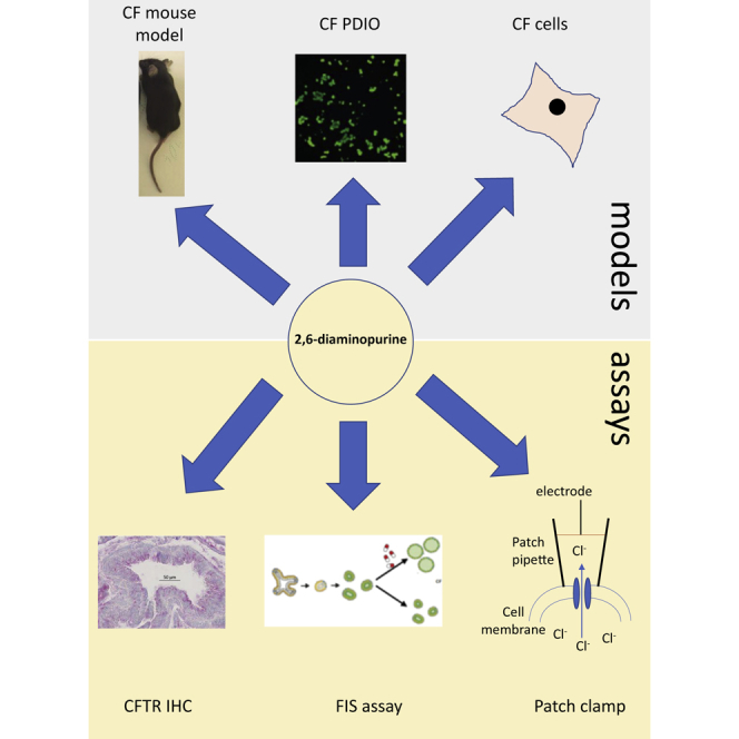
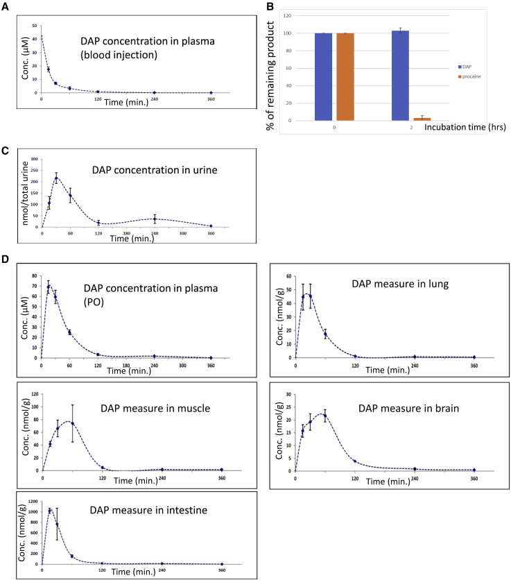
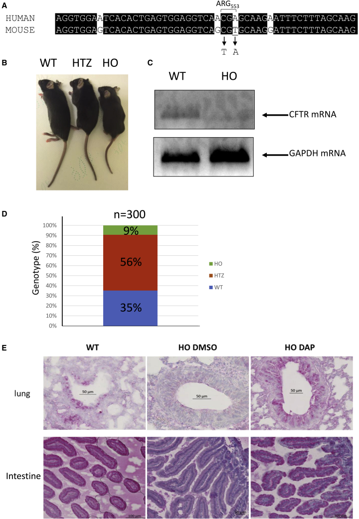
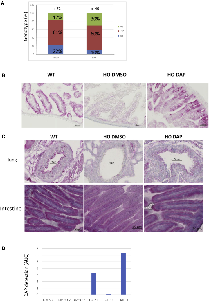
Nonsense mutations are responsible for around 10% of cases of genetic diseases, including cystic fibrosis. 2,6-diaminopurine (DAP) has recently been shown to promote efficient readthrough of UGA premature stop codons. In this study, we show that DAP can correct a nonsense mutation in the Cftr gene in vivo in a new CF mouse model, in utero, and through breastfeeding, thanks, notably, to adequate pharmacokinetic properties. DAP turns out to be very stable in plasma and is distributed throughout the body. The ability of DAP to correct various endogenous UGA nonsense mutations in the CFTR gene and to restore its function in mice, in organoids derived from murine or patient cells, and in cells from patients with cystic fibrosis reveals the potential of such readthrough-stimulating molecules in developing a therapeutic approach. The fact that correction by DAP of certain nonsense mutations reaches a clinically relevant level, as judged from previous studies, makes the use of this compound all the more attractive.

Keywords: 2,6-diaminopurine; cystic fibrosis; mouse model; nonsense mutation; readthrough molecule.


Conflict of interest statement

Declaration of interests J.B. is inventor on a patent related to the FIS assay and received financial royalties from 2017 onward. S.A., C. Bailly, S.R., and F.L. are inventors on a patent demonstrating that DAP is a readthrough molecule useful for the treatment of genetic diseases related to nonsense mutations.

Figures

None
Graphical abstract
Figure 1
Figure 1
Pharmacokinetic parameters of DAP in CD-1 mouse (A) DAP plasma levels after blood injection of DAP at 8.1 mg/kg. Plasma concentrations of DAP were measured 30, 60, 120, 240, and 360 min post-injection. (B) Measure of DAP stability in plasma. DAP or procaine (positive control) was incubated for 2 h in plasma to assess its stability. (C) Measure by mass spectrometry of the concentration of DAP in urine after DAP injection. (D) DAP plasma levels after PO exposure of 29 mg/kg DAP (upper left), and biodistribution of DAP after PO exposure. DAP was measured by mass spectrometry in lung, muscle, brain tissues, and small intestine. Three mice were used for each time point. Error bars show the SD.
Figure 2
Figure 2
The CFTR-NS mouse model (A) Alignment of the human and mouse sequences in the region containing arginine 553. The two nucleotides changed in the murine sequence to generate the UGA stop codon are underlined. (B) Picture showing a homozygous wild-type mouse (WT), a mouse heterozygous for the nonsense mutation (HTZ), and a mouse homozygous for the nonsense mutation (HO). (C) Measurement of CFTR mRNA level in the lungs of wild-type (WT) and CFTR-NS (HO) mice by RT-PCR. Measurement of the GAPDH mRNA level serves as a loading control. (D) Proportion of the three genotypes at weaning. (E) Detection of CFTR (in red) by IHC in lung (around the bronchi) and intestinal tissues (duodenum) from WT and HO mice exposed PO to DMSO or DAP. All the IHCs are representative of at least five mice.
Figure 3
Figure 3
CFTR rescue by DAP in the CFTR-NS mouse model (A) Proportions of the three genotypes among live newborns. Mothers were exposed to DMSO or DAP by daily PO exposure during gestation. The counts are from four mothers treated with DMSO and four mothers treated with DAP for one to five litters and the total number of newborn mice analyzed is indicated on the top of each bar. A statistical analysis resulted in a p value lower than 0.01, indicating a strong trust in these results (Monte-Carlo method). (B) CFTR (in red) in the intestine (duodenum), detected by IHC applied to tissue from WT and HO newborns, from breastfed pups whose mothers were exposed to DMSO or DAP by daily PO exposure during and after gestation, and from adults (WT and HO) exposed to 3 days of DMSO or DAP by daily PO exposure. (C) CFTR (in red) detected by IHC in lung (around the bronchi) and intestinal (duodenum) tissues from 2-week-breastfed WT and HO pups whose mothers were exposed PO daily to DMSO or DAP. All the IHCs are representative of at least five mice. (D) Measurement of the presence of DAP in the contents of the stomach of newborns. Each sample is a mix of the contents of the entire litter.
Figure 4
Figure 4
DAP rescues CFTR function in mouse organoids (A) Schematic representation of the forskolin-induced swelling (FIS) assay, in which CFTR function can be assessed in mouse- or patient-derived intestinal organoids. (B) Organoids derived from CFTR-NS mouse intestinal cells were exposed to DMSO, DAP, or G418, and CFTR function was assessed by FIS. All the results are representative of three experiments. The p values were calculated by one-way ANOVA: ∗∗∗p < 0.005.
Figure 5
Figure 5
Rescue of CFTR function in CF patient cells and in 16HBE14o− cells carrying a nonsense mutation in both Cftr alleles (A) CFTR function was assessed in CF patient cells by means of an SPQ assay after DMSO (red), G418 (green), or DAP (blue) treatment. Arrows indicate addition of the forskolin-cAMP cocktail to the cell medium. The tested genotypes are W1282X/W1282X (patient 1), G542X/G542X (patients 2 and 3), and WT (lower right). (B) Sample single-channel activity of the CFTR Cl currents in the 16HBE14o− expressing wild-type (WT) CFTR without (left) or following pretreatment with 40 μM forskolin and 400 μM IBMX for 30 min, prior to patching the cells (right). Note the absence of activity in patches made to non-stimulated cells and the presence of a characteristic CFTR activity following forskolin/IBMX treatment. (C) Single-channel activity of CFTR Cl currents in 16HBE14o− cells expressing mutant CFTR. To stimulate CFTR currents, cells were pretreated with 40 μM forskolin and 400 μM IBMX for 30 min, prior to patching the cells. Sample traces were acquired in excised outside patches of forskolin/IBMX-pretreated cells expressing, from the top to the bottom, W1282X, G542X, and R553X CFTR. Note the absence of activity in the non-treated (left side) cells and presence of the characteristic CFTR activity in the cells treated with DAP. C and O denote closed and open states of the ion channel. (D–F) Whole-cell currents in isolated cells from WT (D), HO DAP (E), and HO DMSO (F) mice. Whole-cell currents were acquired in NMDG-Cl-based solutions as described. After whole-cell mode was established, the cells were left undisturbed for 5 min to allow replacement of intracellular K+ and Na+ with NMDG, to register basal current levels with Cl as the major ion current carrier. This was followed by application of 40 μM forskolin and 400 μM IBMX to stimulate CFTR activity. Panels compare representative IV (current-voltage) relationships (left) as well as the time course of the CFTR activity stimulation with forskolin/IBMX (right). Note an increase in current following forskolin/IBMX application in WT and HO DAP mice and the absence of the effect in the HO DMSO case.
Figure 6
Figure 6
DAP rescues CFTR function in patient-cell-derived organoids (A) FIS assay on organoids derived from rectal cells of CF patients homozygous for the W1282X mutation. Organoids were exposed to DMSO, DAP, G418, or ELX-02 for 24 or 48 h. (B) Cell viability measured by alamarBlue incorporation into organoids derived from rectal cells of CF patients homozygous for the W1282X mutation. The organoids were exposed to DMSO or DAP. (C) FIS assay on four such organoids exposed to DMSO or DAP. (D) Measurement by qRT-PCR of the level of CFTR mRNA in organoids 1 and 2 to evaluate the efficacy of NMD in the presence of DMSO, an inhibitor of NMD (SMG1i), or DAP at 25, 50, or 100 μM. (E) Western blot demonstrating the absence of physiological stop codon readthrough by DAP at 25, 50, or 100 μM. Calu-6 cells were exposed to DMSO, DAP, or G418 previously to extract protein and perform a western blot analysis. The three leftmost lanes are serial dilutions of U2OS cell extract. Asterisks show degradation products or non-specific bands. The place to which the isoforms having undergone a readthrough of the physiological stop codon should migrate is indicated on the right side of the gel by a bracket. (F) FIS assay on CF patient-cell-derived organoids carrying UGA nonsense mutation on both CFTR alleles. (G) FIS assay on CF patient-cell-derived organoids carrying a UGA nonsense mutation on one CFTR allele and a different mutation from a nonsense mutation (F508del or 1717-1G>A) on the other CFTR allele. (H) FIS assay on CF patient-cell-derived organoids carrying a mutation different from UGA nonsense mutation on both CFTR alleles (F508del or 1717-1G>A). The dotted line corresponds to the amplitude of swelling obtained with organoids homozygous for the F508del mutation upon treatment with lumacaftor-ivacaftor. Error bar shows the SD, p values were calculated with Student’s t test: ∗p < 0.05, ∗∗p < 0.01, ∗∗∗p < 0.001, n.s., not significant. Averages of three experiments with three technical replicates each are shown.

Comment in

References

    1. Mort M., Ivanov D., Cooper D.N., Chuzhanova N.A. A meta-analysis of nonsense mutations causing human genetic disease. Hum. Mutat. 2008;29:1037–1047. - PubMed
    1. Gupta P., Li Y.R. Upf proteins: highly conserved factors involved in nonsense mRNA mediated decay. Mol. Biol. Rep. 2018;45:39–55. - PubMed
    1. Lejeune F. Nonsense-mediated mRNA decay at the crossroads of many cellular pathways. BMB Rep. 2017;50:175–185. - PMC - PubMed
    1. Kurosaki T., Maquat L.E. Nonsense-mediated mRNA decay in humans at a glance. J. Cell Sci. Feb. 2016;129:461–467. - PMC - PubMed
    1. Karousis E.D., Nasif S., Muhlemann O. Nonsense-mediated mRNA decay: novel mechanistic insights and biological impact. Wiley Interdiscip. Rev. RNA. 2016;7:661–682. - PMC - PubMed

Publication types

MeSH terms

Substances

LinkOut - more resources