Review
. 2023 Mar;1865(3):184121.
doi: 10.1016/j.bbamem.2023.184121. Epub 2023 Jan 13.

Membrane reshaping by protein condensates

Affiliations
Review

Membrane reshaping by protein condensates

Samsuzzoha Mondal et al. Biochim Biophys Acta Biomembr. 2023 Mar.

Abstract

Proteins can organize into dynamic, functionally important assemblies on fluid membrane surfaces. Phase separation has emerged as an important mechanism for forming such protein assemblies on the membrane during cell signaling, endocytosis, and cytoskeleton regulation. Protein-protein phase separation thus adds novel fluid mosaics to the classical Singer and Nicolson model. Protein condensates formed in this process can modulate membrane morphologies. This is evident from recent reports of protein condensate-driven membrane reshaping in processes such as endocytosis, autophagosome formation, and protein storage vacuole morphogenesis in plants. Lateral phase separation (on the membrane surface) of peripheral curvature coupling proteins can modulate such membrane morphological transitions. Additionally, three-dimensional protein phase separation can result in droplets that through adhesion can affect membrane shape changes. How do these condensate-driven curvature generation mechanisms contrast with the classically recognized scaffolding and amphipathic helix insertion activities of specific membrane remodeling proteins? A salient feature of these condensate-driven membrane activities is that they depend upon both macroscopic features (such as interfacial energies of the condensate, membrane, and cytosol) as well as microscopic, molecular-level interactions (such as protein-lipid binding). This review highlights the current understanding of the mechanisms underlying curvature generation by protein condensates in various biological pathways.

Keywords: Condensates; Endocytosis; Membrane curvature; Phase separation.


Conflict of interest statement

Declaration of competing interest The authors declare that they have no known competing financial interests or personal relationships that could have appeared to influence the work reported in this paper.

Figures
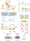
Figure 1.
Figure 1.. Protein condensates can regulate membrane curvature by various mechanisms.
A. Elastic and capillary interactions in droplet / membrane adhesion. During wetting of an elastic surface such as a membrane by a liquid like droplet (yellow) a three phase contact line forms. The effective contact angle α is a function of membrane tension, droplet surface tension, and droplet-membrane interfacial tension (γmem, γdrop, and γdm respectively) whereas the intrinsic contact angle θ is related to the material properties of the droplet and membrane [23]. For a droplet of radius R capillary forces apply a torque of ~2πγdropR2 that pulls the membrane against its bending rigidity κ which acts in the opposite direction relative to the capillary forces [9]. These two opposing effects would deform the membrane on a length scale of the elasto-capillary length, LEC = √(κ/γdrop), shown as the radius of curvature of the red circle in the right panel. B. Scaled membrane tension σ, defined as σ = γmem/γdrop, determines lens-shaped (top) versus engulfed (completely wrapped, bottom) end-states of droplet-membrane interactions. The effective contact angles α and β can be used to describe the droplet/membrane morphologies. For σ > 1, partial wetting of membrane by the droplet gives rise to a lens shaped stage (α < 180°, β > 90°). For σ < 1, the membrane can wrap around the droplet surface, leading to a partially wrapped stage (α < 180°, β < 90°) followed by complete engulfment of the droplets [18]. C. Viscoelastic model of membrane bending by endocytic condensates in yeast cells [7]. Top panel, the endocytic condensate droplet (yellow) formed between membrane (mem, grey) and cytosol (c, light blue) is assumed [7] to maximize its contact area with the cytosol, which increases the cytosolic indentation depth (h՛ > h). If that droplet area increase occurs while keeping the droplet volume constant, an invagination with depth of δ would be created at the membrane-droplet interface, which induces membrane invagination. In this model, energy is proposed to be gained from the work of adhesion at the droplet-cytosol (dark blue) and the droplet-membrane (cyan) interfaces (Ua|d-c and Ua|d-m respectively, green arrows). Bottom panel, the total energy cost is a sum of elastic penalties (red arrows) to cause cytosolic and membrane deformation (Ue|c and Ue|m respectively), surface energy penalties to form extra droplet/cytosol and droplet/membrane interfacial area (Ui|d-c and Ui|d-m respectively) [29], and viscosity penalty to displace the cytosolic material (Uv|c). The invagination depth (δ) depends on the balance of total energy gain (ψδ) and total energy cost (Φδε+1), where ε depends on the deformation geometry, and ψ and Φ summarize material properties determining gain and cost, respectively. D. During autophagy, protein droplets containing p62 cause wetting of autophagosomal double-membrane sheets. Top, the shape of a double-membrane sheet formed between the droplet (yellow) and cytosol (blue) surfaces can be explained by assuming two different spontaneous curvatures, one at the cytosolic surface (mc), and the other at the droplet surface (md), and a curvature asymmetry can be defined as mcd ≡ mcmd. The double-membrane may close either toward the droplet (when mcd ≥ 0) or toward the cytosol (when mcd < 0), to choose a specific cargo. Bottom, an enlarged section from the top panel to show protein-protein interactions, such as p62 present in droplet and LC3 present on membrane surface, can determine the direction of autophagosome enclosure [5]. E. Generation of membrane buds and tubules from GUVs enclosing a phase separated aqueous two-phase system. Efflux of water from GUVs causes phase separation of the enclosed polymers and also generates excess membrane area. The excess membrane area release can lead to formation of membrane buds and tubules by an interplay between wetting behavior of the phase separated system and membrane spontaneous curvature. F. Membrane bound BAR-protein, endophilin, can lead to adhesion between two adjacent membranes when present with its multivalent binding partner lamellipodin [4]. Adhesion of formed membrane tubules on the flat membrane surface (top) can increase membrane tension by adhesion and wrapping transitions (bottom), suppressing tubule and bud formation. G. Anchored IDRs (red and blue chains) on a flat membrane show decreasing segment density (shaded area) at increasing distance from the membrane. In such conditions, a phase separating IDR would form less sticker-sticker interactions (right panel) further away from the membrane. If the membrane bends toward the IDR, more sticker-sticker interactions will take place (left panel). The increase in binding interactions would compensate for the compressive stress generated on the membrane. If the membrane and anchored chains exhibit attractive interactions (green highlighted regions) it will provide additional effects to bend the membrane toward the IDRs [22].

References

    1. Shin Y, Brangwynne CP, Liquid phase condensation in cell physiology and disease, Science 357(6357) (2017). - PubMed
    1. Su XL, Ditlev JA, Hui EF, Xing WM, Banjade S, Okrut J, King DS, Taunton J, Rosen MK, Vale RD, Phase separation of signaling molecules promotes T cell receptor signal transduction, Science 352(6285) (2016) 595–599. - PMC - PubMed
    1. Day KJ, Kago G, Wang L, Richter JB, Hayden CC, Lafer EM, Stachowiak JC, Liquid-like protein interactions catalyse assembly of endocytic vesicles, Nat Cell Biol 23(4) (2021) 366–376. - PMC - PubMed
    1. Mondal S, Narayan K, Botterbusch S, Powers I, Zheng J, James HP, Jin R, Baumgart T, Multivalent interactions between molecular components involved in fast endophilin mediated endocytosis drive protein phase separation, Nature Communications 13(1) (2022) 5017. - PMC - PubMed
    1. Agudo-Canalejo J, Schultz SW, Chino H, Migliano SM, Saito C, Koyama-Honda I, Stenmark H, Brech A, May AI, Mizushima N, Knorr RL, Wetting regulates autophagy of phase-separated compartments and the cytosol, Nature 591(7848) (2021) 142–146. - PubMed

Publication types

MeSH terms

Substances

LinkOut - more resources