Review
. 2023 Jan 16;21(1):14.
doi: 10.1186/s12951-023-01778-6.

Therapeutic potential and mechanisms of mesenchymal stem cell-derived exosomes as bioactive materials in tendon-bone healing

Affiliations
Review

Therapeutic potential and mechanisms of mesenchymal stem cell-derived exosomes as bioactive materials in tendon-bone healing

Jiaxuan Zou et al. J Nanobiotechnology. .

Abstract

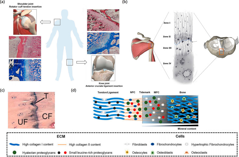
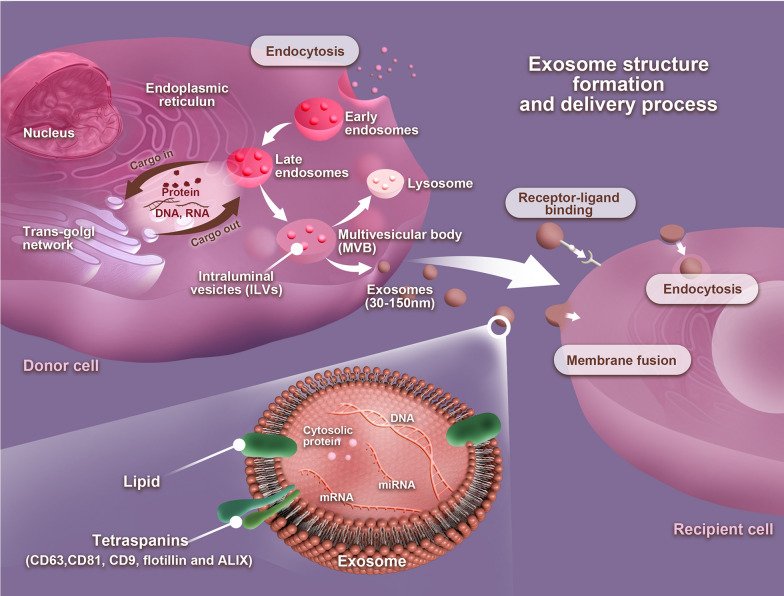
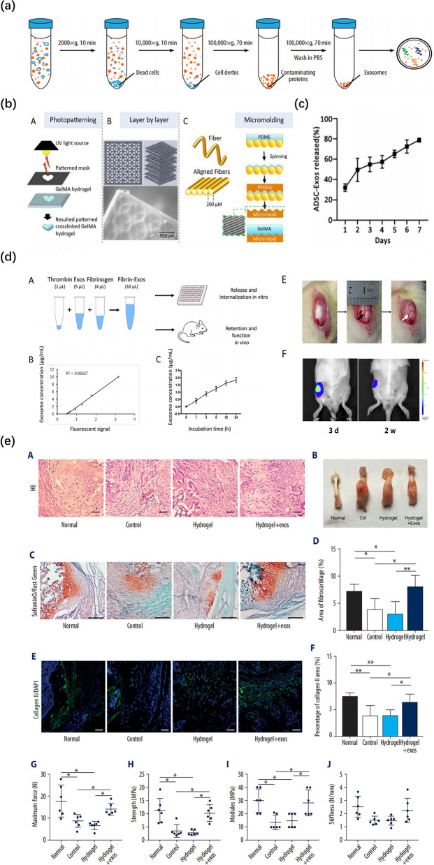
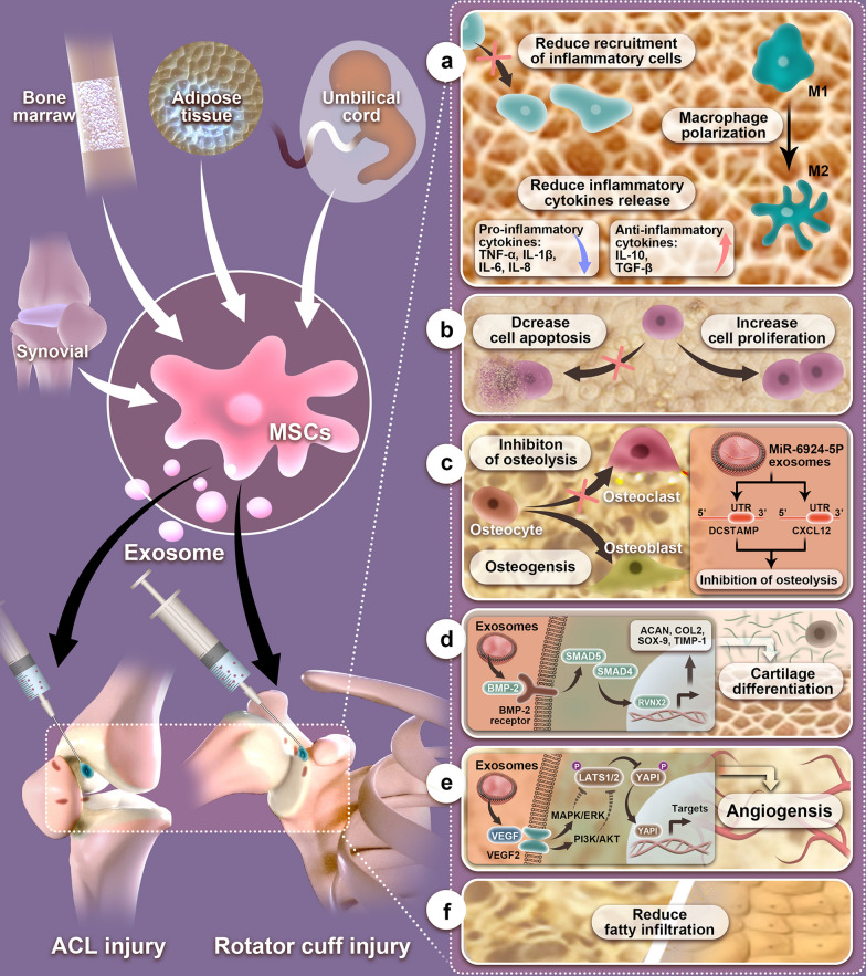
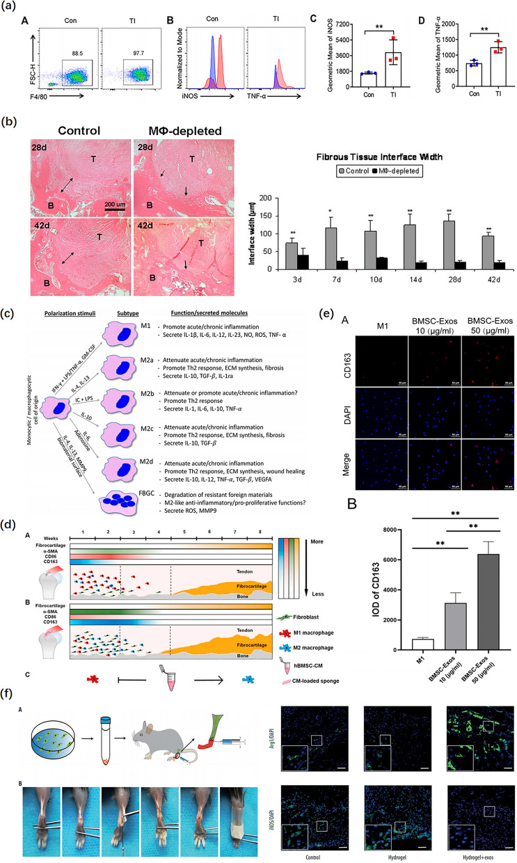
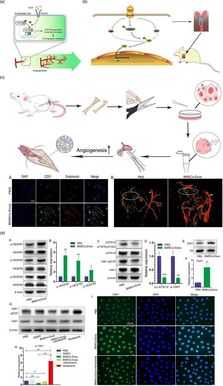
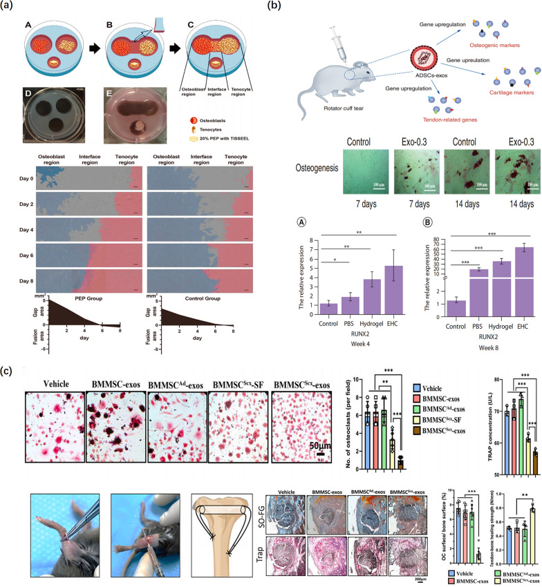
Tendon-bone insertion (TBI) injuries, such as anterior cruciate ligament injury and rotator cuff injury, are the most common soft tissue injuries. In most situations, surgical tendon/ligament reconstruction is necessary for treating such injuries. However, a significant number of cases failed because healing of the enthesis occurs through scar tissue formation rather than the regeneration of transitional tissue. In recent years, the therapeutic potential of mesenchymal stem cells (MSCs) has been well documented in animal and clinical studies, such as chronic paraplegia, non-ischemic heart failure, and osteoarthritis of the knee. MSCs are multipotent stem cells, which have self-renewability and the ability to differentiate into a wide variety of cells such as chondrocytes, osteoblasts, and adipocytes. Numerous studies have suggested that MSCs could promote angiogenesis and cell proliferation, reduce inflammation, and produce a large number of bioactive molecules involved in the repair. These effects are likely mediated by the paracrine mechanisms of MSCs, particularly through the release of exosomes. Exosomes, nano-sized extracellular vesicles (EVs) with a lipid bilayer and a membrane structure, are naturally released by various cell types. They play an essential role in intercellular communication by transferring bioactive lipids, proteins, and nucleic acids, such as mRNAs and miRNAs, between cells to influence the physiological and pathological processes of recipient cells. Exosomes have been shown to facilitate tissue repair and regeneration. Herein, we discuss the prospective applications of MSC-derived exosomes in TBI injuries. We also review the roles of MSC-EVs and the underlying mechanisms of their effects on promoting tendon-bone healing. At last, we discuss the present challenges and future research directions.

Keywords: Biomaterials; Drug delivery; Exosomes; Mesenchymal stem cells; Nanocarriers; Nanomedicine; Tendon–bone healing.


Conflict of interest statement

The authors declare that they have no competing interests.

Figures

Fig. 1
Fig. 1
Physical Structure of tendon-bone insertion. a The histological staining of tendon/ligament-to-bone insertion (H&E, and Masson staining). Reproduced with permission [46]. b Zone I consists of the ligament. Zone II comprises nonmineralized fibrocartilage. Zone III is composed of mineralized cartilage. Zone IV consists of bone. Tidemark between Zone II and Zone III (black arrow) is shown. Reproduced with permission [7]. c The tidemark stained with H&E. Reproduced with permission [46]. d The schematic of tendon/ligament-to-bone insertion. RCT indicates rotator cuff tendon; ACL indicates anterior cruciate ligament; NFC indicates non-mineralized fibrocartilage; MFC indicates mineralized fibrocartilage; UF indicates uncalcified fibrocartilage; CF indicates calcified fibrocartilage; T indicates tidemark; ECM indicates extracellular matrix. Reproduced with permission [46]
Fig. 2
Fig. 2
Exosome structure, formation, and delivery process. The plasma membrane invaginates to form early endosomes and then gradually forms late endosomes. Multivesicular bodies (MVBs) form by inward budding of the late endosomal limiting membrane. The MVB can either fuse with lysosomes or autophagosomes to be degraded or fuse with the plasma membrane to the release of late endosomal contents, i.e., intraluminal vesicles (ILVs) with a variety of information into the extracellular space to form exosomes. In the extracellular space, exosomes can be taken up by recipient cells via three patterns: receptor–ligand binding, endocytosis, or membrane fusion
Fig. 3
Fig. 3
Exosomes combined with hydrogel promoted tendon-bone healing. a Schematic diagram of the isolation of exosomes. Reproduced with permission [105]. b Schematic representation of photopatterning of GelMA using a pre-patterned photomask. Stacked layers of patterned GelMA hydrogels fabricated using a micro-mirror projection stereolithography system. Schematic representation of a fiber-assisted micromolding technique for the production of parallel microgrooved surfaces that serve as a template for micropatterning GelMA. Reproduced with permission [106]. c Profile of ADSC-Exos released from the GelMA. Reproduced with permission [12]. d Release in vitro and retention in vivo of the BMSCs-exos embedded in fibrin-exo. The black arrow indicates the patellar window defect; The white arrow indicates the implanted fibrin-exo/fibrin-vesicle. Reproduced with permission [97]. e BMSC-Exos combined with hydrogel promoted fibrocartilage regeneration and enhanced the biomechanical properties in tendon-bone healing (H&E, safranin O-fast green, DAPI, and collagen type II staining). Reproduced with permission [105]
Fig. 4
Fig. 4
Mechanisms of exosomes on TBI injuries. a Exosomes could attenuate early inflammatory response via reducing inflammatory, polarizing macrophage from a pro-inflammatory M1 phenotype to a pro-regenerative M2 phenotype, and reducing inflammatory cytokines release. b Exosomes could enhance cell proliferation and reduce apoptosis. c Exosomes rich in miR-6924-5p could directly inhibit osteoclast formation by binding to the 3′-untranslated regions (3′ UTRs) of OCSTAMP and CXCL12. d Exosome-delivered BMP-2 promotes cartilage differentiation via Smad/RUNX2 signaling pathway. Activation of BMP-2/Smad signaling induces Runx2 expression through the activation of Smad4 and Smad5, after which Runx2 induces the expression of cartilage differentiation-related proteins, Aggrecan, Collagen II, SOX-9, and TIMP-1. e Exosomes promote angiogenesis by activating the VEGF and Hippo signaling pathways. When VEGF binds to the vascular endothelial growth factor receptor (VEGFR), the VEGF‐VEGFR interaction inhibits LATS1/2 and YAP1 phosphorylation by activating the PI3K and MAPK signaling pathways. The leads to increased expression of YAP1 in the nucleus inducing angiogenesis. f Exosomes could reduce fatty infiltration
Fig. 5
Fig. 5
The effect of exosomes on the regulation of the transition of macrophages from M1 to M2 during tendon-bone healing. a An Increase in the pro-inflammatory phenotype of macrophages after a mouse tendon-bone injury (Flow cytometric analysis). Reproduced with permission [122]. b At all time points, the width of the fibrous tissue interface was significantly reduced following macrophage depletion (H&E staining). Reproduced with permission [112]. c Classification of macrophages by their inducing stimulus. Reproduced with permission [118]. d Brief summary of tendon-bone healing after rotator cuff repair in rats. CM indicates conditioned medium. Reproduced with permission [32]. e The BMSC-Exos polarized macrophages from the M1 phenotype into M2 (DAPI and CD163 staining). Reproduced with permission [91]. f The mouse tendon-bone reconstruction model and surgical procedure. BMSC-Exos induce M2 macrophage polarization during tendon-bone healing (DAPI, Arg1, and iNOS staining). Reproduced with permission [105]
Fig. 6
Fig. 6
The effect of exosomes on enhancing angiogenesis. a YAP/TAZ orchestrate VEGF signaling during developmental angiogenesis. Reproduced with permission [126]. b VEGF‐induced angiogenesis via the VEGF–VEGFR–Hippo signaling pathway axis and promoted rotator tendon‐bone healing in rats. Reproduced with permission [128]. c BMSC-Exos promoted angiogenesis around the tendon-bone interface of the rotator cuff in rats (DAPI, CD31, endomucin staining, and angiography). Reproduced with permission [102]. d Functional effects of BMSC-Exos on angiogenesis-related signaling pathways in HUVECs (Western blot analysis, DAPI, and YAP1 staining). Reproduced with permission [102]
Fig. 7
Fig. 7
The effect of exosomes on the regulation of bone metabolism. a Schematic representation of the co-culture model. Cell growth and migration were significantly increased in all regions following exposure to PEP. PEP indicates purified exosome product. Reproduced with permission [73]. b The tendon-derived stem cell differentiation is enhanced by adipose-derived stem cell exosomes (Alizarin Red S staining). Real-time PCR analysis showed the EHC significantly induced upregulation of osteogenic marker RUNX2. EHC: Adipose-derived stem cell exosome–hydrogel complex. Reproduced with permission [103]. c Schematic diagram of the tendon-bone healing model. BMMSCScx-exos inhibit osteoclastogenesis and improve tendon-bone healing. (TRAP, and safranin O-fast green staining). BMMSCScx indicates Scx-overexpressing PDGFRα (+) BMMSCs; BMMSCAd indicates the control group; SF indicates soluble factors. Reproduced with permission [89]

Similar articles

Cited by

References

    1. Wang Y, He G, Guo Y, Tang H, Shi Y, Bian X, et al. Exosomes from tendon stem cells promote injury tendon healing through balancing synthesis and degradation of the tendon extracellular matrix. J Cell Mol Med. 2019;23:5475–5485. doi: 10.1111/jcmm.14430. - DOI - PMC - PubMed
    1. Benjamin M, Toumi H, Ralphs JR, Bydder G, Best TM, Milz S. Where tendons and ligaments meet bone: attachment sites (‘entheses’) in relation to exercise and/or mechanical load. J Anat. 2006;208:471–490. doi: 10.1111/j.1469-7580.2006.00540.x. - DOI - PMC - PubMed
    1. Saveh-Shemshaki N, Nair LS, Laurencin CT. Nanofiber-based matrices for rotator cuff regenerative engineering. Acta Biomater. 2019;94:64–81. doi: 10.1016/j.actbio.2019.05.041. - DOI - PubMed
    1. Silvers-Granelli HJ, Bizzini M, Arundale A, Mandelbaum BR, Snyder-Mackler L. Does the FIFA 11+ injury prevention program reduce the incidence of ACL injury in male soccer players? Clin Orthop. 2017;475:2447–2455. doi: 10.1007/s11999-017-5342-5. - DOI - PMC - PubMed
    1. Chen H, Li S, Xiao H, Wu B, Zhou L, Hu J, et al. Effect of exercise intensity on the healing of the bone-tendon interface: a mouse rotator cuff injury model study. Am J Sports Med. 2021;49:2064–2073. doi: 10.1177/03635465211011751. - DOI - PubMed