. 2023 Jan;18(1):310-321.
doi: 10.1016/j.jds.2022.04.024. Epub 2022 Jun 4.

Anti-apoptosis and anti-inflammation activity of circ_0097010 downregulation in lipopolysaccharide-stimulated periodontal ligament cells by miR-769-5p/Krüppel like factor 6 axis

Affiliations

Anti-apoptosis and anti-inflammation activity of circ_0097010 downregulation in lipopolysaccharide-stimulated periodontal ligament cells by miR-769-5p/Krüppel like factor 6 axis

Dan-Dan Sun et al. J Dent Sci. 2023 Jan.

Abstract

Background/purpose: Periodontitis is a prevalent infectious inflammatory disease. Growing evidence has revealed important roles for circular RNAs (circRNAs) and circRNA sponge activity in periodontitis. Here, we elucidated the precise part of circ_0097010 in periodontitis pathogenesis.

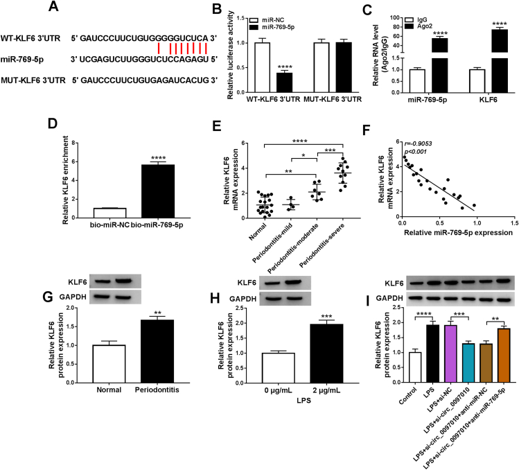
Materials and methods: Human periodontal ligament cells (hPDLCs) were exposed to lipopolysaccharide (LPS). Cell viability, proliferation and apoptosis were evaluated by CCK-8 assay, EdU incorporation assay and flow cytometry, respectively. Circ_0097010, microRNA (miR)-769-5p and Krüppel like factor 6 (KLF6) were quantified by qRT-PCR and Western blot. Interleukin 6 (IL-6) level, tumor necrosis factor-α (TNF-α) secretion, superoxide dismutase (SOD) activity and malondialdehyde (MDA) level were detected by enzyme-linked immunosorbent assay (ELISA). Dual-luciferase reporter, RNA immunoprecipitation (RIP) and RNA pull-down assays were used to confirm the direct relationship between miR-769-5p and circ_0097010 or KLF6.

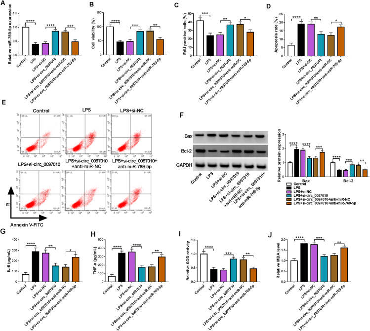
Results: Our data showed that LPS repressed cell proliferation and induced cell apoptosis and inflammation in hPDLCs. Circ_0097010 was upregulated in periodontitis samples and LPS-exposed hPDLCs. Downregulation of circ_0097010 exerted anti-apoptosis and anti-inflammation functions in LPS-exposed hPDLCs. Mechanistically, circ_0097010 acted as a miR-769-5p sponge, and reduced abundance of miR-769-5p reversed the anti-apoptosis and anti-inflammation effects of circ_0097010 suppression. KLF6 was a direct miR-769-5p target, and miR-769-5p-mediated inhibition of KLF6 possessed anti-apoptosis and anti-inflammation functions in LPS-induced hPDLCs. Moreover, circ_0097010 controlled KLF6 expression by miR-769-5p.

Conclusion: These data identify circ_0097010 as a key regulator of LPS-induced inflammation and apoptosis in hPDLCs and highlight a novel mechanism of circ_0097010 regulation through miR-769-5p/KLF6 axis.

Keywords: Circ_0097010; Inflammation; Krüppel like factor 6 (KLF6); MiR-769-5p; Periodontitis.


Conflict of interest statement

The authors have no conflicts of interest relevant to this article.

Figures

Figure 1
Figure 1
LPS impairs cell proliferation and induces apoptosis and inflammation in hPDLCs. (A) CCK-8 cell viability assay with hPDLCs after stimulation with the indicated doses of LPS for 24 h. (B–I) EdU cell proliferation assay (B), flow cytometry for cell apoptosis (C and D), Bax and Bcl-2 expression levels by Western blot (E), the production levels of IL-6 and TNF-α by ELISA using the assay kits (F and G), SOD activity and MDA level using the assay kits (H and I) in hPDLCs exposed to 0 or 2 μg/mL of LPS for 24 h. ∗∗P < 0.01, ∗∗∗P < 0.001, ∗∗∗∗P < 0.0001.
Figure 2
Figure 2
Downregulation of circ_0097010 mitigates LPS-driven anti-proliferation, pro-apoptosis and pro-inflammation effects on hPDLCs. (A) Relative circ_0097010 expression by qRT-PCR in gingival tissue samples of patients with periodontitis (n = 22) and healthy volunteers (n = 19). (B) Effect of RNase R digestion on the levels of circ_0079010 and GAPDH. (C–K) hPDLCs were transiently introduced with or without si-circ_0079010 or si-NC before stimulation with 0 or 2 μg/mL of LPS for 24 h. (C) qRT-PCR of relative circ_0079010 expression in hPDLCs after treatment as indicated. (D) CCK-8 cell viability assay with hPDLCs treated as indicated. (E) Assessment of cell proliferation by EdU assay after the indicated treatment. (F) Detection of cell apoptosis by flow cytometry based on the staining with Annexin V and PI. (G) Western blot of Bax and Bcl-2 levels in hPDLCs after the indicated treatment. (H and I) The production levels of IL-6 and TNF-α in treated hPDLCs by ELISA using the assay kits. (J and K) SOD activity and MDA level in treated hPDLCs using the assay kits. ∗∗P < 0.01, ∗∗∗P < 0.001, ∗∗∗∗P < 0.0001.
Figure 3
Figure 3
Circ_0097010 sponges miR-769-5p. (A) Sequence of miR-769-5p, the potential miR-769-5p complementary sites at circ_0097010, and the mutated target sequence in circ_0097010. (B) qRT-PCR showing the transfection efficiency of miR-769-5p mimic. (C) Luciferase activity of the wild-type (WT) or mutant (MUT) reporter constructs after transfection with miR-769-5p mimic or miR-NC mimic. (D) RIP experiments on extracts of hPDLCs using an antibody against Ago2 or IgG. (E) RNA pull-down assays on extracts of hPDLCs using bio-miR-769-5p or bio-miR-NC. (F) Relative miR-769-5p expression by qRT-PCR in gingival tissue samples of patients with periodontitis (n = 22) and healthy volunteers (n = 19). (G) Expression correlation analysis of miR-769-5p and circ_0079010 in periodontitis samples (n = 22) using Pearson's correlation coefficient. (H) qRT-PCR of miR-769-5p in hPDLCs after exposure to 0 or 2 μg/mL of LPS for 24 h. ∗∗∗P < 0.001, ∗∗∗∗P < 0.0001.
Figure 4
Figure 4
Decrease of miR-769-5p reverses circ_0097010 suppression-mediated anti-apoptosis and anti-inflammation functions in LPS-exposed hPDLCs. (A–J) hPDLCs were exposed to 0 or 2 μg/mL of LPS for 24 h after transfection with or without anti-miR-769-5p + si-circ_0097010, anti-miR-NC + si-circ_0097010, si-circ_0097010 or si-NC. (A) Relative miR-769-5p expression by qRT-PCR in treated hPDLCs. (B) CCK-8 cell viability assay with hPDLCs after the indicated transfection. (C) EdU cell proliferation assay with treated hPDLCs. (D and E) Cell apoptosis measurement by flow cytometry using staining with Annexin V and PI. (F) Western blot of the levels of Bax and Bcl-2 in hPDLCs after the indicated transfection. (G and H) ELISA of IL-6 and TNF-α levels in treated hPDLCs using the assay kits. (I and J) SOD activity and MDA level in treated hPDLCs using the assay kits. ∗P < 0.05, ∗∗P < 0.01, ∗∗∗P < 0.001, ∗∗∗∗P < 0.0001.
Figure 5
Figure 5
KLF6 is a direct miR-769-5p target and circ_0097010 controls KLF6 expression by miR-769-5p. (A) Alignment between miR-769-5p and the 3′UTR of human KLF6 and the mutations in the target sequence. (B) Luciferase activity of the wild-type (WT) or mutant (MUT) KLF6 3′UTR reporter constructs transfected into hPDLCs in the presence of miR-796-5p mimic or mimic control. (C) RIP experiments on extracts of hPDLCs using an antibody against Ago2 or IgG. (D) RNA pull-down assays on extracts of hPDLCs using bio-miR-769-5p or bio-miR-NC. (E) Quantification of KLF6 mRNA expression by qRT-PCR in gingival tissue samples of patients with periodontitis (n = 22) and healthy volunteers (n = 19). (F) Correlation analysis between KLF6 mRNA and miR-769-5p expression in periodontitis samples (n = 22) using Pearson's correlation coefficient. (G) Western blot of KLF6 protein in gingival tissue samples of patients with periodontitis (n = 3) and healthy volunteers (n = 3). (H) KLF6 protein expression by Western blot of lysates from hPDLCs after exposure to 0 or 2 μg/mL of LPS for 24 h. (I) Representative Western blot showing KLF6 protein level in LPS-treated hPDLCs after transfection by anti-miR-769-5p + si-circ_0097010, anti-miR-NC + si-circ_0097010, si-circ_0097010 or si-NC. ∗∗P < 0.01, ∗∗∗P < 0.001, ∗∗∗∗P < 0.0001.
Figure 6
Figure 6
KLF6 is a functional effector of miR-769-5p. (A–J) hPDLCs were transfected with or without miR-769-5p mimic, miR-769-5p mimic + KLF6 expression plasmid, miR-769-5p mimic + pcDNA control or miR-NC mimic before exposure to 0 or 2 μg/mL of LPS for 24 h. (A) Relative KLF6 protein level normalized to GAPDH by Western blot in treated hPDLCs. (B) CCK-8 cell viability assay with hPDLCs after the indicated treatment. (C) EdU cell proliferation assay with hPDLCs after the indicated treatment. (D and E) Assessment of cell apoptosis by flow cytometry based on Annexin V-FITC and PI staining. (F) Western blot of Bax and Bcl-2 expression levels in treated hPDLCs. (G and H) The production levels of IL-6 and TNF-α in treated hPDLCs using ELISA with the assay kits. (I and J) Measurement of SOD activity and MDA level using the assay kits. ∗P < 0.05, ∗∗P < 0.01, ∗∗∗P < 0.001, ∗∗∗∗P < 0.0001.
Supplement Figure 1
Supplement Figure 1
Overexpression of circ_0097010 exacerbates LPS-driven anti-proliferation, pro-apoptosis and pro-inflammation effects on hPDLCs. (A-H) hPDLCs were transiently introduced with or without circ_0097010 expression plasmid or pCD5-ciR control plasmid before stimulation with 0 or 2 μg/mL of LPS for 24 h. (A) qRT-PCR of circ_0079010 in hPDLCs after treatment as indicated. (B) CCK-8 cell viability assay with hPDLCs treated as indicated. (C) Cell proliferation by EdU assay after the indicated treatment. (D) Cell apoptosis by flow cytometry based on the staining with Annexin V and PI. (E and F) The production levels of IL-6 and TNF-α in treated hPDLCs by ELISA using the assay kits. (G and H) SOD activity and MDA level in treated hPDLCs using the assay kits. ∗∗P < 0.01, ∗∗∗P < 0.001, ∗∗∗∗P < 0.0001.
Supplement Figure 2
Supplement Figure 2
Effects of KLF6 knockdown on cell proliferation, apoptosis and inflammation in LPS-induced hPDLCs. (A-J) hPDLCs were transfected with or without si-KLF6 or si-NC before exposure to 0 or 2 μg/mL of LPS for 24 h and checked for KLF6 protein level by western blot (A), cell viability by CCK-8 assay (B), cell proliferation by EdU assay (C), cell apoptosis by flow cytometry (D and E), Bax and Bcl-2 levels by western blot (F), IL-6 and TNF-α production levels by ELISA (G and H), and SOD activity and MDA level using the assay kits (I and J). ∗P < 0.05, ∗∗P < 0.01, ∗∗∗P < 0.001, ∗∗∗∗P < 0.0001.

Similar articles

Cited by

References

    1. Bartold P.M. Lifestyle and periodontitis: the emergence of personalized periodontics. Periodontol 2000. 2018;78:7–11. - PubMed
    1. Kwon T., Lamster I.B., Levin L. Current concepts in the management of periodontitis. Int Dent J. 2021;71:462–476. - PMC - PubMed
    1. Sanz M., Marco Del Castillo A., Jepsen S., et al. Periodontitis and cardiovascular diseases: consensus report. J Clin Periodontol. 2020;47:268–288. - PMC - PubMed
    1. Baeza M., Morales A., Cisterna C., et al. Effect of periodontal treatment in patients with periodontitis and diabetes: systematic review and meta-analysis. J Appl Oral Sci. 2020;28 - PMC - PubMed
    1. Kristensen L.S., Andersen M.S., Stagsted L.V.W., Ebbesen K.K., Hansen T.B., Kjems J. The biogenesis, biology and characterization of circular RNAs. Nat Rev Genet. 2019;20:675–691. - PubMed

LinkOut - more resources