. 2022 Dec 28;8(1):846-856.
doi: 10.1021/acsomega.2c06212. eCollection 2023 Jan 10.

S-Nitrosoglutathione Reduces the Density of Staphylococcus aureus Biofilms Established on Human Airway Epithelial Cells

Affiliations

S-Nitrosoglutathione Reduces the Density of Staphylococcus aureus Biofilms Established on Human Airway Epithelial Cells

Alex Wolf et al. ACS Omega. .

Abstract

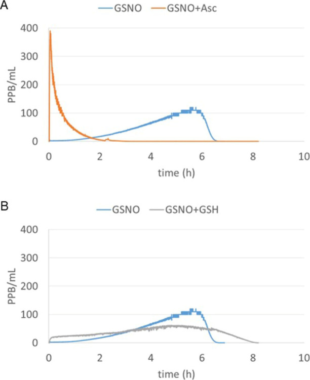
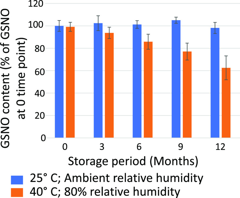
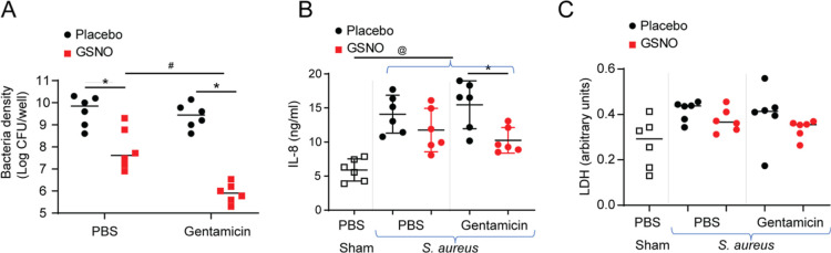
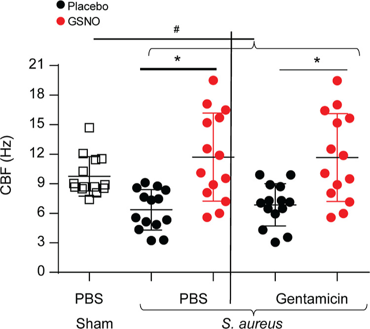
Patients with chronic rhinosinusitis (CRS) often show persistent colonization by bacteria in the form of biofilms which are resistant to antibiotic treatment. One of the most commonly isolated bacteria in CRS is Staphylococcus aureus (S. aureus). Nitric oxide (NO) is a potent antimicrobial agent and disperses biofilms efficiently. We hypothesized that S-nitrosoglutathione (GSNO), an endogenous NO carrier/donor, synergizes with gentamicin to disperse and reduce the bacterial biofilm density. We prepared GSNO formulations which are stable up to 12 months at room temperature and show the maximum amount of NO release within 1 h. We examined the effects of this GSNO formulation on the S. aureus biofilm established on the apical surface of the mucociliary-differentiated airway epithelial cell cultures regenerated from airway basal (stem) cells from cystic fibrosis (CF) and CRS patients. We demonstrate that for CF cells, which are defective in producing NO, treatment with GSNO at 100 μM increased the NO levels on the apical surface and reduced the biofilm bacterial density by 2 log units without stimulating pro-inflammatory effects or inducing epithelial cell death. In combination with gentamicin, GSNO further enhanced the killing of biofilm bacteria. Compared to placebo, GSNO significantly increased the ciliary beat frequency (CBF) in both infected and uninfected CF cell cultures. The combination of GSNO and gentamicin also reduced the bacterial density of biofilms grown on sinonasal epithelial cells from CRS patients and improved the CBF. These findings demonstrate that GSNO in combination with gentamicin may effectively reduce the density of biofilm bacteria in CRS patients. GSNO treatment may also enhance the mucociliary clearance by improving the CBF.


Conflict of interest statement

The authors declare the following competing financial interest(s): AW and GM are employees of the NOTA Labs MEM serves as Chief Technology Officer for NOTA Labs MZ, MBH, and US are scientific officers for NOTA Labs

Figures

Figure 1
Figure 1
(A) NO release over time from a 100 μM GSNO solution in 0.16 M NaCl/0.03 M NaHCO3 in the absence (blue trace) or presence (orange trace) of 100 μM sodium ascorbate. (B) NO release over time from the 100 μM GSNO solution in 0.16 M NaCl/0.03 M NaHCO3 in the absence (blue trace) or presence (gray trace) of 100 μM GSH. NO release was measured by purging the solution continuously with nitrogen and measuring the NO release in the gas stream by chemiluminescence.
Figure 2
Figure 2
Stability study of the GSNO/Asc (1:1) formulation (in 0.16 M NaCl/0.035 M NaHCO3/10 μM Na2EDTA). Samples were packaged under 8–11% RH in a controlled atmosphere glovebox. Samples were stored at 25 °C and ambient humidity (25–55% RH) and at elevated 40 °C and high humidity (80% RH). The GSNO content was determined by measuring the UV–vis absorbance at 334.
Figure 3
Figure 3
S. aureus forms biofilms on the apical surface of mucociliary-differentiated bronchial epithelial cells. The apical surface of the mucociliary-differentiated CF bronchial epithelial cell culture was infected with S. aureus SA1692 and incubated for 24 h. The cell culture was fixed and subjected to SEM. (A) SEM showing the SA biofilm, mucus, and cilia on the apical surface of the cell culture. (B) Magnified view of the SA biofilm. The images are representative of three experiments.
Figure 4
Figure 4
Treatment with GSNO reduces the biofilm bacterial density on the apical surface of CF bronchial epithelial cells. Mucociliary-differentiated CF bronchial epithelial cells were infected apically with S. aureus isolates SA1696, SA1692, SANP, or a mixture of all three isolates and incubated for 24 h. The cells were then treated apically with GSNO (50, 100, or 250 μM) or appropriate placebo formulations 3 times during 24 h. Four hours after the last treatment, the apical surface was gently rinsed with PBS, and then the cells were lysed in 0.1% Triton X-100. The viable bacterial density in the cell lysates was determined by dilution plating. (A–D) The data represent the range with the median calculated from two or three independent experiments with two–three replicates (*p = ≤ 0.05; Mann–Whitney test, different from respective placebo-treated cultures). (E,F) Cell cultures were infected with a mixture of three S. aureus isolates and treated with placebo or 100 μM GSNO as above. The cultures were immunostained with antibodies to S. aureus (green) and ZO-1 (red) and counterstained with DAPI (blue) (E) and observed under a confocal microscope. In some experiments, the cell cultures were fixed in glutaraldehyde and processed for SEM (F). Images are representative of three experiments.
Figure 5
Figure 5
Gentamicin synergizes with GSNO to reduce the bacterial density (A) and IL-8 (B). CF bronchial epithelial cells were infected with a mixture of three S. aureus isolates or sham-infected with PBS and incubated for 24 h. Then, the cultures were apically treated with placebo, 5 μg of gentamicin, 100 μM GSNO, or a combination of GSNO and gentamicin 3 times over a 24 h period. Four hours after the last treatment, the basolateral medium was collected, the apical surface was gently rinsed with PBS, and then the cells were lysed in 0.1% Triton X-100. (A) The viable bacterial density in the cell lysates was determined by dilution plating. IL-8 (B) and LDH (C) were determined in the basolateral medium. The data represent the range with the median calculated from three independent experiments with two replicates. Statistical significance was pinpointed by ANOVA on ranks with the Kruskal–Wallis H test (*p = ≤ 0.05; different from the respective placebo-treated cultures, #p = ≤ 0.05; different from the GSNO-treated cultures, @p = ≤ 0.05; different from the sham-infected cultures).
Figure 6
Figure 6
GSNO enhances the CBF in CF bronchial epithelial cell cultures. Mucociliary-differentiated CF bronchial epithelial cells were infected with a mixture of three S. aureus isolates or sham. After 24 h of incubation, the cultures were apically treated with PBS, gentamicin, GSNO, and a combination of GSNO and gentamicin 3 times over a 24 h period. Four hours after the last treatment, high-speed video microscopy was performed, and the CBF was analyzed. The data represent the range with the median calculated from three independent experiments with 3–6 replicates (*p = ≤ 0.05; ANOVA, different from respective placebo treated cultures, #p = ≤0.05; ANOVA on ranks with the Kruskal–Wallis H test, different from sham-infected cultures).
Figure 7
Figure 7
Effect of gentamicin and GSNO in reducing the biofilm bacterial density, IL-8, and CBF of sinonasal epithelial cells. Mucociliary-differentiated sinonasal epithelial cell cultures from two CRS patients were infected with a mixture of three S. aureus isolates or sham. After 24 h of incubation, the cultures were apically treated with placebo, gentamicin, GSNO, or a combination of GSNO and gentamicin 3 times over a 24 h period. Three hours after the last treatment, the basolateral medium was collected, and IL-8 was analyzed by ELISA. The cells were subjected to high-speed video microscopy, and the CBF was analyzed. The cells were then washed with PBS and lysed in 0.1% Triton X-100, and the cell lysates were dilution-plated to determine the bacterial density. (A,C,E) Cells from patient 1 and (B,D,E) cells from patient 2. (A,B) Bacterial density determined in cell lysates. (C,D) IL-8 levels in the basolateral medium. (E,F) CBF. Data in (A–D) represent the range with the median from three replicate wells and in (E,F) represent the mean with SD from triplicates (*p = ≤ 0.05; ANOVA, different from respective placebo-treated cultures, #p = ≤ 0.05; ANOVA, different from GSNO-treated cultures, @p = ≤ 0.05; ANOVA, different from sham-infected cultures).

Similar articles

Cited by

References

    1. Pleis J. R.; Ward B. W.; Lucas J. W. Summary health statistics for U.S. adults: National Health Interview Survey, 2009. Vital Health Statistics 2010, 249, 1–207. - PubMed
    1. Schiller J. S.; Lucas J. W.; Ward B. W.; Peregoy J. A. Summary health statistics for U.S. adults: National Health Interview Survey, 2010. Vital Health Statistics 2012, 252, 1–207. - PubMed
    1. Caulley L.; Thavorn K.; Rudmik L.; Cameron C.; Kilty S. J. Direct costs of adult chronic rhinosinusitis by using 4 methods of estimation: Results of the US Medical Expenditure Panel Survey. J. Allergy Clin. Immunol. 2015, 136, 1517–1522. 10.1016/j.jaci.2015.08.037. - DOI - PubMed
    1. Rudmik L. Economics of Chronic Rhinosinusitis. Curr. Allergy Asthma Rep. 2017, 17, 20.10.1007/s11882-017-0690-5. - DOI - PubMed
    1. Kern R. C.; Conley D. B.; Walsh W.; Chandra R.; Kato A.; Tripathi-Peters A.; Grammer L. C.; Schleimer R. P. Perspectives on the etiology of chronic rhinosinusitis: an immune barrier hypothesis. Am. J. Rhinol. 2008, 22, 549–559. 10.2500/ajr.2008.22.3228. - DOI - PMC - PubMed