

Submassive Pulmonary Embolism in the Setting of Intracerebral Hemorrhage: A Case of Suction Thrombectomy
- PMID: 36644103
- PMCID: PMC9833621
- DOI: 10.7759/cureus.32432
Submassive Pulmonary Embolism in the Setting of Intracerebral Hemorrhage: A Case of Suction Thrombectomy
Abstract
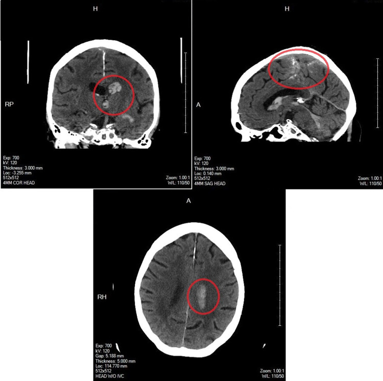
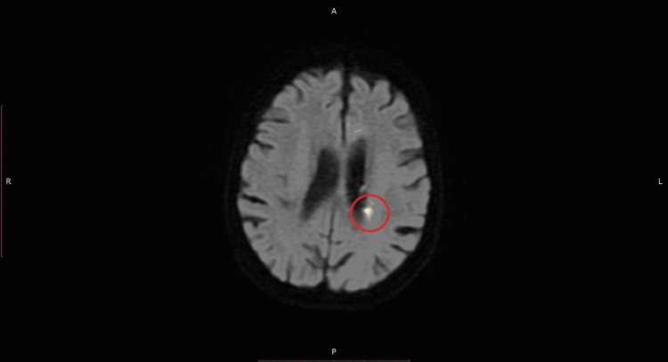
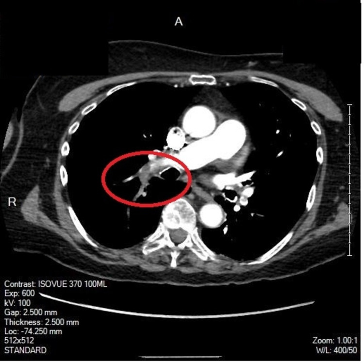
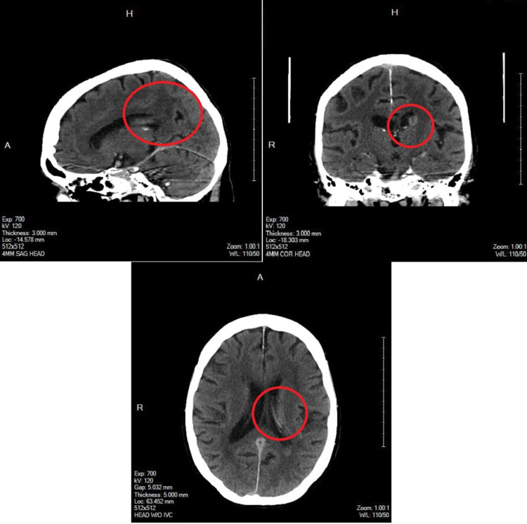
Pulmonary embolism (PE) in the setting of intracerebral hemorrhage (ICH) is an unfortunate, challenging, and highly morbid clinical problem. Interventional strategies have lower associated bleeding risks than the standby for PE treatment: systemic anticoagulation. Despite this benefit, there are few examples in the literature of its utilization in the management of PE in the setting of ICH. This present case provides an example of the successful utilization of suction thrombectomy to manage PE in the setting of ICH. An 80-year-old female presented to an outside hospital with complaints of dizziness, headache, nausea, and vomiting of abrupt onset one hour before arrival. Computed tomography (CT) of the head with CT Angiography (CTA) of the head and neck was performed and demonstrated hemorrhage in all ventricles; most prominently within the left lateral ventricle. Magnetic Resonance Imaging (MRI) of the brain suggested that the cause of her hemorrhage was reperfusion injury after a small acute infarction in the left internal capsule in the setting of anticoagulant use. Ten days after her diagnosis of ICH, a submassive PE was diagnosed with a class IV pulmonary embolism severity index (PESI). An interdisciplinary evaluation was conducted between hospitalist medicine, neurology, neurosurgery, and interventional radiology. A successful suction thrombectomy was performed on hospital day 11. No new neurologic deficits were appreciated post-procedure. The patient's heart rate remained elevated but improved. Blood pressure remained controlled. The patient was weaned off oxygen to room air. Neurosurgery assessed the patient to be of acceptable risk for discharge with the further deferment of anticoagulation until repeat CT head six weeks after discharge. The patient was discharged on hospital day 14. Treating PE in the setting of ICH is without clear guidelines. The appropriate treatment modality is reliant upon the clinical judgment and the individual details of each case. In this case, a high PESI with imaging demonstrating a stable hematoma without evidence of new blood resulted in the decision to use a suction thrombectomy. More research is needed to develop consistent evidence-based guidelines for this clinical challenge.
Keywords: intracerebral hemorrhage; pulmonary embolism; stroke; submassive; thrombectomy; thrombosis.
Copyright © 2022, Ciurylo et al.
Conflict of interest statement
The authors have declared that no competing interests exist.
Figures
Similar articles
-
Acute Massive and Submassive Pulmonary Embolism: Preliminary Validation of Aspiration Mechanical Thrombectomy in Patients with Contraindications to Thrombolysis.Cardiovasc Intervent Radiol. 2018 Dec;41(12):1840-1848. doi: 10.1007/s00270-018-2011-3. Epub 2018 Jul 6. Cardiovasc Intervent Radiol. 2018. PMID: 29980817
-
Catheter-directed thrombolysis versus suction thrombectomy in the management of acute pulmonary embolism.J Vasc Surg Venous Lymphat Disord. 2019 Sep;7(5):623-628. doi: 10.1016/j.jvsv.2018.10.025. Epub 2019 Mar 20. J Vasc Surg Venous Lymphat Disord. 2019. PMID: 30902560
-
A Trilogy of Submassive Pulmonary Embolism, Non-Small Cell Lung Cancer with Brain Metastasis, Kartagener's Syndrome and its Management with Aspiration Thrombectomy.Eur J Case Rep Intern Med. 2022 Mar 2;9(3):003149. doi: 10.12890/2022_003149. eCollection 2022. Eur J Case Rep Intern Med. 2022. PMID: 35402339 Free PMC article.
-
Race against the clock: overcoming challenges in the management of anticoagulant-associated intracerebral hemorrhage.J Neurosurg. 2014 Aug;121 Suppl:1-20. doi: 10.3171/2014.8.paradigm. J Neurosurg. 2014. PMID: 25081496 Review.
-
Challenging anticoagulation cases: A case of pulmonary embolism shortly after spontaneous brain bleeding.Thromb Res. 2021 Apr;200:41-47. doi: 10.1016/j.thromres.2021.01.016. Epub 2021 Jan 26. Thromb Res. 2021. PMID: 33529872 Review.
References
-
- Guidelines for the management of spontaneous intracerebral hemorrhage in adults: 2007 update: a guideline from the American Heart Association/American Stroke Association Stroke Council, High Blood Pressure Research Council, and the Quality of Care and Outcomes in Research Interdisciplinary Working Group. Broderick J, Connolly S, Feldmann E, et al. Circulation. 2007;116:0. - PubMed
-
- Primary intracerebral haemorrhage in the Oxfordshire Community Stroke Project, 2: prognosis. Counsell C, Boonyakarnkul S, Dennis M, Sandercock P, Bamford J, and Warlow C. Cerebrovasc. Dis. 1995;5:26.
-
- Antithrombotic therapy for VTE disease: CHEST guideline and expert panel report. Kearon C, Akl EA, Ornelas J, et al. Chest. 2016;149:315–352. - PubMed
-
- Anticoagulant-associated intracerebral hemorrhage. Morotti A, Goldstein JN. Brain Hemorrhages. 2020;1:89–94.
Publication types
LinkOut - more resources
Full Text Sources