. 2022 Jun 4:49:32-41.
doi: 10.1016/j.athplu.2022.05.004. eCollection 2022 Aug.

Semaglutide treatment attenuates vessel remodelling in ApoE-/- mice following vascular injury and blood flow perturbation

Affiliations

Semaglutide treatment attenuates vessel remodelling in ApoE-/- mice following vascular injury and blood flow perturbation

Ditte Marie Jensen et al. Atheroscler Plus. .

Abstract

Background and aims: Randomized clinical studies have shown a reduction in cardiovascular outcomes with glucagon-like peptide 1 receptor agonist (GLP-1RA) treatment with the hypothesized mechanisms being an underlying effect on atherosclerosis. Here, we aimed to assess the pharmacological effects of semaglutide in an atheroprone murine model that recapitulates central mechanisms related to vascular smooth muscle cell (VSMC) phenotypic switching and endothelial dysfunction known to operate within the atherosclerotic plaque.

Methods: In study A, we employed an electrical current to the carotid artery in ApoE-/- mice to induce severe VSMC injury and death, after which the arteries were allowed to heal for 4 weeks. In study B, a constrictive cuff was added for 6 h at the site of the healed segment to induce a disturbance in blood flow.

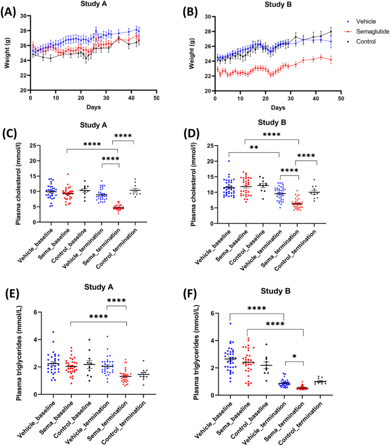
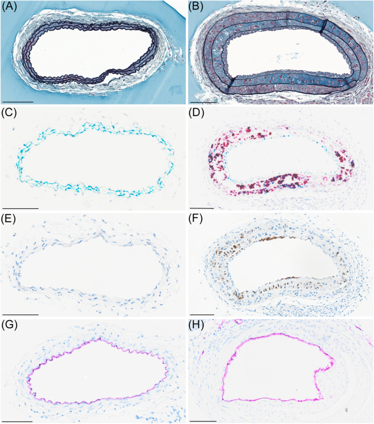
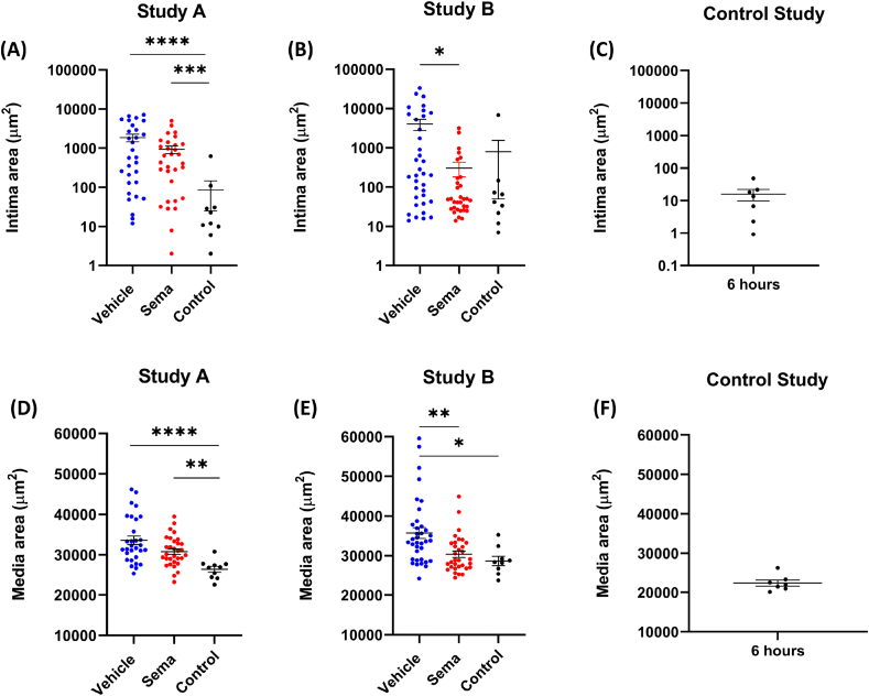
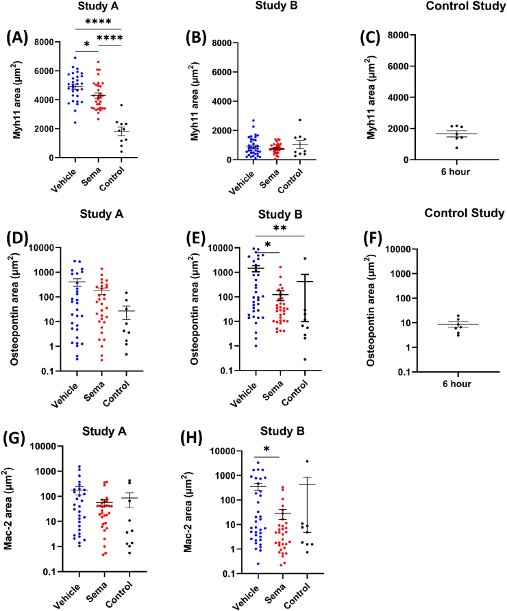
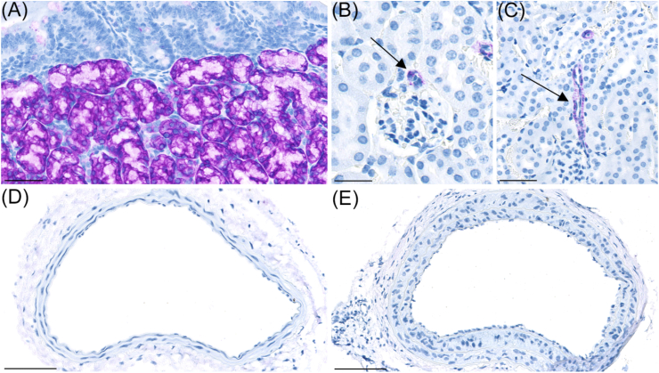
Results: Compared to vehicle, semaglutide treatment reduced the intimal and medial area by ∼66% (p = 0.007) and ∼11% (p = 0.0002), respectively. Following cuff placement, expression of the pro-inflammatory marker osteopontin and macrophage marker Mac-2 was reduced (p < 0.05) in the semaglutide-treated group compared to vehicle. GLP-1R were not expressed in murine carotid artery and human coronary vessels with and without atherosclerotic plaques, and semaglutide treatment did not affect proliferation of cultured primary human VSMCs.

Conclusions: Semaglutide treatment reduced vessel remodelling following electrical injury and blood flow perturbation in an atheroprone mouse model. This effect appears to be driven by anti-inflammatory and -proliferative mechanisms independent of GLP-1 receptor-mediated signalling in the resident vascular cells. This mechanism of action may be important for cardiovascular protection.

Keywords: ApoE−/−, Apolipoprotein E knock-out; Atherosclerosis; GLP 1RA, Glucagon-like peptide 1 receptor agonist; Glucagon-like peptide 1 receptor agonists; IHC, Immunohistochemistry; ISH, In situ hybridisation; LCCA, Left common carotid artery; Phenotypic switching; Plaque erosion; SSP1, Osteopontin; Semaglutide; SoC, Standard of care; VSMC, Vascular smooth muscle cells; Vascular injury; Vascular smooth muscle cells.


Conflict of interest statement

The authors declare the following financial interests/personal relationships which may be considered as potential competing interests: The authors BR, LMV, MKEO, JCB, AU, CP, RKK, HH, LBK and MN are employees at Novo Nordisk. Novo Nordisk markets semaglutide for the treatment of diabetes and obesity. DMJ, GFS, and JL are present or former employees at University of Copenhagen and have collaborated with Novo Nordisk on this project.

Figures

Fig. 1
Fig. 1
Body weight and lipid parameters. (A) Body weight at study start (baseline, day 0) for mice in Study A (electric injury) and Study B (plaque erosion)(B). Each data point is the average of animals in that group on the specific day. (C) Plasma cholesterol (mmol/L) at baseline and termination in mice from Study A (electric injury) and B (plaque erosion)(D). (E) Plasma triglyceride (mmol/L) at baseline and termination in mice from Study A (electric injury) and B (plaque erosion) (F). Statistical significance:∗p < 0.05, ∗∗p < 0.01, ∗∗∗p < 0.001, ∗∗∗∗p < 0.0001. Results are shown as mean ± SEM.
Fig. 2
Fig. 2
Histological depiction of vessels with (right panel) and without neointima (left panel). (A) Carotid artery without neointima (Movat's pentachrome stain). (B) Carotid artery with neointima showing increased tunica media and tunica intima area (Movat's pentachrome stain). (C) Carotid artery without neointima stained for Myh11 (teal) and osteopontin (red) (duplex ISH). (D) Carotid artery with neointima stained for Myh11 (teal) and osteopontin (red) (duplex ISH). (E) Carotid artery without neointima stained for Mac-2 (IHC). (F) Injured carotid artery with neointima stained for Mac-2 (IHC). (G) Carotid artery without plaque stained for CD31 (IHC). (H) Carotid artery with injury stained for CD31 (IHC). Scalebar is 100 μm in length.
Fig. 3
Fig. 3
Area of tunica intima and tunica media. (A) Intima area in mice from Study A (electric injury). (B) Intima area in mice from Study B (plaque erosion study). (C) Intima area in mice from the control study (constrictive cuff). (D) Tunica media area in mice from Study A (electric injury). (E) Tunica media area in mice from Study B (plaque erosion). (F) Tunica media area in mice from the control study (constrictive cuff). Intima and media area in Study A and Study B were quantified from approximately 40 slides in total per animal (every 10th pentachrome-stained slide starting from the bifurcation end). In the control study, intima and media area was quantified from approximately 20 slides in total per animal (every 10th pentachrome-stained slide starting from where the end of the cuff). Each data point represents one animal. Statistical significance:∗p < 0.05, ∗∗p < 0.01, ∗∗∗p < 0.001, ∗∗∗∗p < 0.0001. Results are shown as mean ± SEM. Note that the y-axis is log scale in (A), (B) and (C).
Fig. 4
Fig. 4
Area of Myh11, osteopontin and Mac-2. (A) Myh11 area in mice from Study A (electric injury). (B) Myh11 area in mice from Study B (plaque erosion study). (C) Myh11 area in mice from the control study (constrictive cuff). (D) Osteopontin area in mice from Study A (electric injury). (E) Osteopontin area in mice from Study B (plaque erosion). (F) Osteopontin area in mice from the control study (constrictive cuff). (G) Mac-2 area in mice from Study A (electric injury). (H) Mac-2 area in mice from Study B (plaque erosion study). Myh11 and osteopontin area was quantified by ISH duplex staining of approximately 10 slides in total per animal (every 50th stained slide). Mac-2 area was quantified by IHC staining of approximately 10 slides in total per animal (every 50th stained slide). Each data point represents one animal. Statistical significance:∗p < 0.05, ∗∗p < 0.01, ∗∗∗∗p < 0.0001. Results are shown as mean ± SEM. Note that the y-axis is log scale in (D), (E), (F), (G) and (H).
Fig. 5
Fig. 5
GLP1-R staining. (A) GLP1-R (purple) was detected at high levels in Brunner's gland in duodenum. Scalebar = 50 μm. (B) GLP-1R was also expressed in arterioles supplying glomeruli in mouse kidney (arrow). Scalebar = 50 μm. (C) GLP1-R was also expressed in larger arterioles of mouse kidney (arrow). Scalebar = 50 μm. GLP-1R expression was not observed in carotid artery without neointima (D) or in carotid artery with neointima formation (E). Scalebar in panel (D) and (E) = 100 μm.

References

    1. Pasterkamp G., Den Ruijter H.M., Libby P. Temporal shifts in clinical presentation and underlying mechanisms of atherosclerotic disease. Nat Rev Cardiol. 2016;14:21–29. doi: 10.1038/nrcardio.2016.166. - DOI - PubMed
    1. Basatemur G.L., Jørgensen H.F., Clarke M.C.H., Bennett M.R., Mallat Z. Vascular smooth muscle cells in atherosclerosis. Nat Rev Cardiol. 2019;16:727–744. doi: 10.1038/s41569-019-0227-9. - DOI - PubMed
    1. Wirka R.C., Wagh D., Paik D.T., Pjanic M., Nguyen T., Miller C.L., Kundu R., Nagao M., Coller J., Koyano T.K., Fong R., Woo Y.J., Liu B., Montgomery S.B., Wu J.C., Zhu K., Chang R., Alamprese M., Tallquist M.D., Kim J.B., Quertermous T. Atheroprotective roles of smooth muscle cell phenotypic modulation and the TCF21 disease gene as revealed by single-cell analysis. Nat. Med. 2019;25:1280–1289. doi: 10.1038/s41591-019-0512-5. - DOI - PMC - PubMed
    1. Aherrahrou R., Guo L., Nagraj V.P., Aguhob A., Hinkle J., Chen L., Yuhl Soh J., Lue Di, Alencar G.F., Boltjes A., Van Der Laan S.W., Farber E., Fuller D., Anane-Wae R., Akingbesote N., Manichaikul A.W., Ma L., Kaikkonen M.U., Björkegren J.L.M., Önengüt-Gümüşcü S., Pasterkamp G., Miller C.L., Owens G.K., Finn A., Navab M., Fogelman A.M., Berliner J.A., Civelek M. Genetic regulation of atherosclerosis-relevant phenotypes in human vascular smooth muscle cells. Circ. Res. 2020:1552–1565. doi: 10.1161/CIRCRESAHA.120.317415. - DOI - PMC - PubMed
    1. Wong D., Turner A.W., Miller C.L. Genetic insights into smooth muscle cell contributions to coronary artery disease. Arterioscler Thromb Vasc Biol. 2019;39:1006–1017. doi: 10.1161/ATVBAHA.119.312141. - DOI - PMC - PubMed