Case Reports
. 2023 Jan 12;14(1):23-28.
doi: 10.1159/000528408. eCollection 2023 Jan-Dec.

Late PAMM-Like Lesions in a Patient with HIV Retinopathy

Affiliations
Case Reports

Late PAMM-Like Lesions in a Patient with HIV Retinopathy

Menelaos Kanakis et al. Case Rep Ophthalmol. .

Abstract

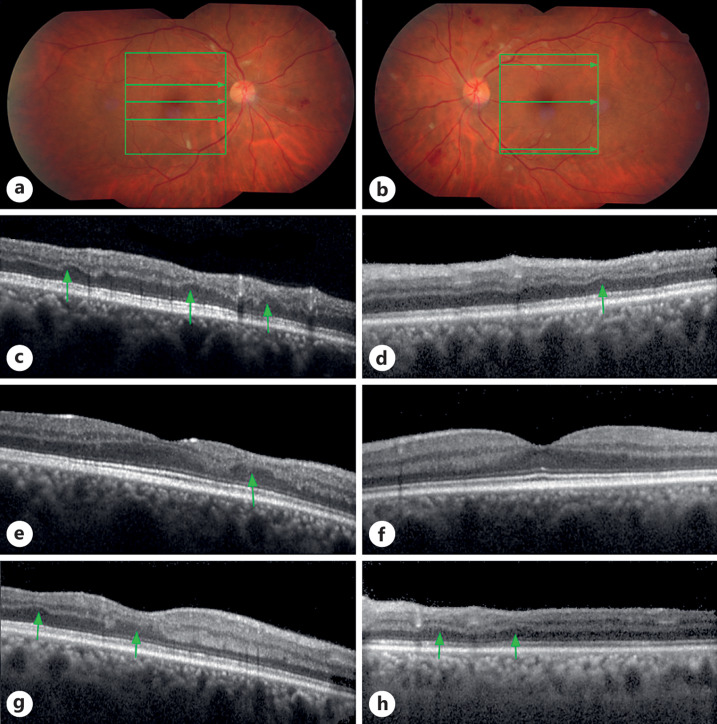
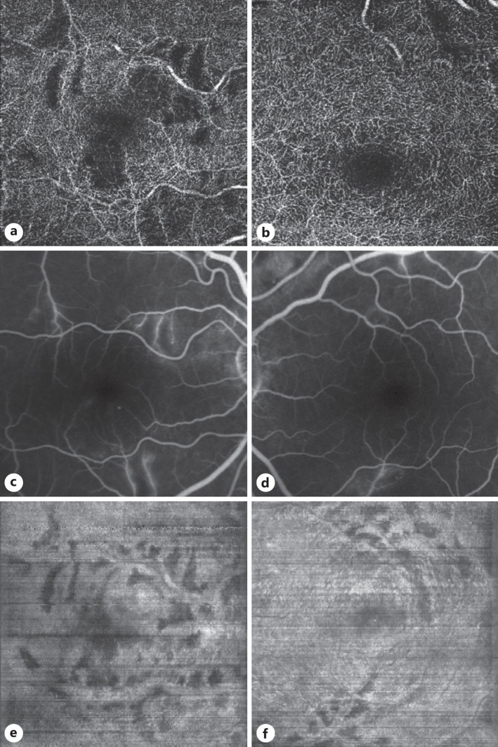
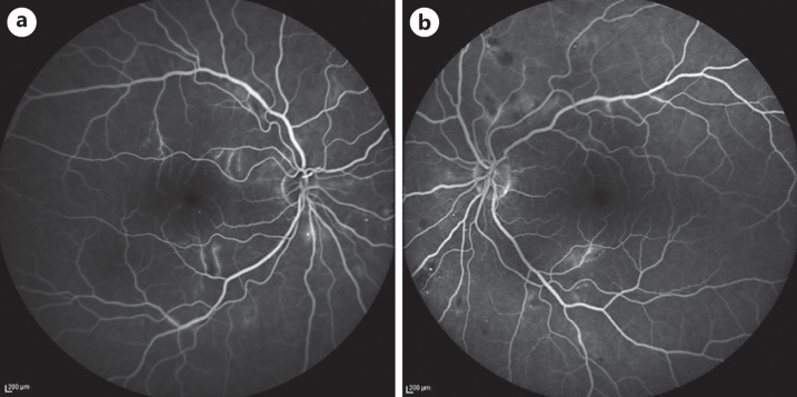
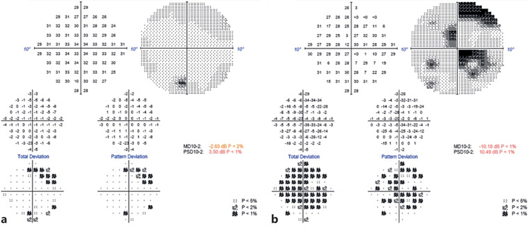
This report describes a case of a newly diagnosed 49-year-old HIV patient, who presented with decreased visual acuity and retinal lesions characterized by ischemia at the level of the deep retinal capillary plexus, documented with optical coherence tomography (OCT), OCT angiography, fluorescein angiography, and visual fields testing. These lesions closely resembled the morphologic and clinical characteristics of late paracentral acute middle maculopathy. The presence of these lesions suggests that HIV microangiopathy can potentially affect both superficial and deep retinal capillary plexuses.

Keywords: HIV; HIV Retinopathy; PAMM; Paracentral acute middle maculopathy.


Conflict of interest statement

The authors report no conflicts of interest to declare.

Figures

Fig. 1.
Fig. 1.
Color fundus and OCT imaging: HIV retinopathy and INL thinning. a Right eye at presentation: cotton wool spots, pre-, and intraretinal hemorrhages. OCT scan frame and scan lines are presented as green square and arrows. b Left eye at presentation: cotton wool spots, pre-, and intraretinal hemorrhages, extending in a larger region than in the right eye but respecting the macula. c, e, g OCT scans of the right eye, revealing areas with INL thinning (arrows), including the macula. Scan lines are presented in A above as green arrows. d, f, h OCT scans of the left eye, revealing areas with INL thinning (arrows), respecting the macula. Scan lines are presented in B above as green arrows.
Fig. 2.
Fig. 2.
En face OCT reconstruction, OCT angiography, and FA. a, b OCT angiography at the level of the deep capillary plexus. The areas devoid of capillaries coincide with the areas of thinning in a and b above, indicating the conjunction of DCP ischemia with the late PAMM-like lesions. c, d Enlarged images of the FA, to facilitate the comparison with the en face reconstructed images and OCT angiography. e, f En face OCT reconstruction at the INL-OPL level with concurrent shadow removal revealing dark areas of INL thinning consistent with late PAMM lesions. FA, fluorescein angiography; OPL, outer plexiform layer.
Fig. 3.
Fig. 3.
Full (55°) view of late-phase FA. Evident microaneurysms and areas of late leakage in both eyes. a Right eye. b Left eye. FA, fluorescein angiography.
Fig. 4.
Fig. 4.
VFs (SITA Fast, 10–2). a Left eye. b Right eye.

References

    1. Rahimy E, Sarraf D. Paracentral acute middle maculopathy spectral-domain optical coherence tomography feature of deep capillary ischemia. Curr Opin Ophthalmol. 2014 May;25(3):207–12. 10.1097/ICU.0000000000000045. - DOI - PubMed
    1. Rahimy E, Kuehlewein L, Sadda SR, Sarraf D. Paracentral acute middle maculopathy: what we knew then and what we know now. Retina. 2015 Oct;35. (10); p. 1921–30. 10.1097/IAE.0000000000000785. - DOI - PubMed
    1. Chen X, Rahimy E, Sergott RC, Nunes RP, Souza EC, Choudhry N, et al. . Spectrum of retinal vascular diseases associated with paracentral acute middle maculopathy. Am J Ophthalmol. 2015 Jul;160(1):26–34.e1. 10.1016/j.ajo.2015.04.004. - DOI - PubMed
    1. Ilginis T, Keane PA, Tufail A. Paracentral acute middle maculopathy in sickle cell disease. JAMA Ophthalmol. 2015 May;133(5):614–6. 10.1001/jamaophthalmol.2014.6098. - DOI - PubMed
    1. Castro CS, Ferreira AS, Silva NP, Lume MR, Furtado MJ. Paracentral acute middle maculopathy after COVID-19 disease: multimodal evaluation. Retin Cases Brief Rep. 2022 Jul 15. 10.1097/ICB.0000000000001301. - DOI - PubMed

Publication types