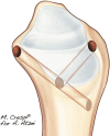
Allinside Anatomic Arthroscopic (3A) Reconstruction of Irreparable TFCC Tear
- PMID: 36644726
- PMCID: PMC9836779
- DOI: 10.1055/s-0041-1735981
Allinside Anatomic Arthroscopic (3A) Reconstruction of Irreparable TFCC Tear
Abstract
Background In recent years, new arthroscopic techniques have been introduced to address the irreparable tears of the triangular fibrocartilage complex (TFCC) (Palmer type 1B, Atzei class 4) by replicating the standard Adams-Berger procedure. These techniques, however, show the same limitations of the open procedure in relation to the anatomically defective location of the radial origins of the radioulnar ligaments (RUL) and the risk of neurovascular and/or tendon injury. Aiming to improve the quality of reconstruction and reduce surgical morbidity, a novel arthroscopic technique was developed, with the advantages of reproducing the anatomical origins of the RUL ligaments and providing all-inside tendon graft (TG) deployment and fixation. Description of Technique The Allinside anatomic arthroscopic (3A) technique is indicated for TG reconstruction of irreparable TFCC tears in the absence of distal radioulnar joint (DRUJ) arthritis. Standard wrist arthroscopy portals are used. A small incision in the radial metaphyseal area and arthroscopic control are required to set a Wrist Drill Guide and create two converging tunnels, whose openings are at the radial anatomical origins of the RUL. An ulnar tunnel is drilled at the fovea from inside-out via the 6U portal. A 3-mm tendon strip, from the palmaris longus or extensor carpi radialis brevis, is woven through the tunnels and then secured into the ulnar tunnel with an interference screw. Postoperative immobilization with restricted forearm rotation is discontinued at 5 weeks, and then postoperative rehabilitation is started. Patients and Methods The 3A technique was applied on 5 patients (2 females and 3 males), with an average age 42 years. DRUJ stability, range of motion (ROM), pain (0-10 visual analogue scale [VAS]), grip strength, modified Mayo wrist score (MMWS), and patient satisfaction were used for evaluation before surgery and at follow-up. Results No intraoperative or early complications were registered. At a mean follow-up of 26 months, DRUJ was stable in all patients, which recovered 99% ROM. Pain VAS decreased from 7 to 0.6. Grip strength increased from 38 to 48.8 Kgs. There were 4 excellent results and 1 good result on MMWS. All patient showed high satisfaction. Conclusions Although the 3A technique requires dedicated instrumentation and arthroscopic expertise, it takes advantage of improved intra-articular vision and minimized surgical trauma to reduce the risk of complications and obtain promising functional results.
Keywords: Arthroscopy; Chronic Tear; DRUJ; Instability; Reconstruction; TFCC; Tendon Graft; Wrist.
Thieme. All rights reserved.
Conflict of interest statement
Conflict of Interest None declared.
Figures


References
-
- Palmer A K. Triangular fibrocartilage complex lesions: a classification. J Hand Surg Am. 1989;14(04):594–606. - PubMed
-
- Atzei A, Luchetti R. Foveal TFCC tear classification and treatment. Hand Clin. 2011;27(03):263–272. - PubMed
-
- Adams B D, Berger R A. An anatomic reconstruction of the distal radioulnar ligaments for posttraumatic distal radioulnar joint instability. J Hand Surg Am. 2002;27(02):243–251. - PubMed
-
- Atzei A. New trends in arthroscopic management of type 1-B TFCC injuries with DRUJ instability. J Hand Surg Eur Vol. 2009;34(05):582–591. - PubMed
-
- Tse W L, Lau S W, Wong W Y. Arthroscopic reconstruction of triangular fibrocartilage complex (TFCC) with tendon graft for chronic DRUJ instability. Injury. 2013;44(03):386–390. - PubMed
Publication types
LinkOut - more resources
Full Text Sources
Miscellaneous

