. 2023 Jan;52(2):20220210.
doi: 10.1259/dmfr.20220210. Epub 2023 Jan 16.

A novel technique to quantify bone-to-implant contact of zygomatic implants: a radiographic analysis based on three-dimensional image registration and segmentation

Affiliations

A novel technique to quantify bone-to-implant contact of zygomatic implants: a radiographic analysis based on three-dimensional image registration and segmentation

Yuwei Gu et al. Dentomaxillofac Radiol. 2023 Jan.

Abstract

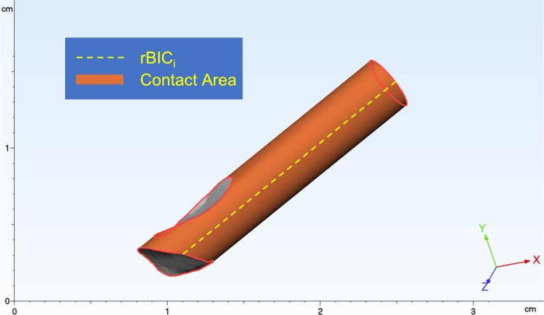
Objectives: The purpose of this study is to establish a novel, reproducible technique to obtain the BIC area (BICA) between zygomatic implants and zygomatic bone based on post-operative cone-beam computed tomography (CBCT) images. Three-dimensional (3D) image registration and segmentation were used to eliminate the effect of metal-induced artifacts of zygomatic implants.

Methods: An ex-vivo study was included to verify the feasibility of the new method. Then, the radiographic bone-to-implant contact (rBIC) of 143 implants was measured in a total of 50 patients. To obtain the BICA of zygomatic implants and the zygomatic bone, several steps were necessary, including image preprocessing of CBCT scans, identification of the position of zygomatic implants, registration, and segmentation of pre- and post-operative CBCT images, and 3D reconstruction of models. The conventional two-dimensional (2D) linear rBIC (rBICc) measurement method with post-operative CBCT images was chosen as a comparison.

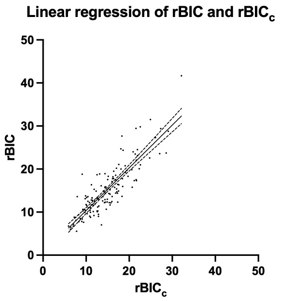
Results: The mean values of rBIC and rBICc were 15.08 ± 5.92 mm and 14.77 ± 5.14 mm, respectively. A statistically significant correlation was observed between rBIC and rBICc values ([Formula: see text]=0.86, p < 0.0001).

Conclusions: This study proposed a standardized, repeatable, noninvasive technique to quantify the rBIC of post-operative zygomatic implants in 3D terms. This technique is comparable to conventional 2D linear measurements and seems to be more reliable than these conventional measurements; thus, this method could serve as a valuable tool in the performance of clinical research protocols.

Keywords: Bone-to-implant contact; CBCT imaging; atrophic edentulous maxilla; post-operative radiographic bone-to-implant contact; zygomatic implants.


Figures

Figure 1.
Figure 1.
The contact margin was traced by a marker pen (red marker). Then, the zygomatic implant was extracted, and the contact margin curve was rubbed on a piece of grid paper.
Figure 2.
Figure 2.
The rubbing of the contact margin was traced by a marker pen on the grid paper.
Figure 3.
Figure 3.
This image depicts the interface between the zygomatic implant and the zygomatic bone (orange); this surface can be segmented into countless rBICi (yellow dashed line) regions. rBIC, radiographic BIC.
Figure 4.
Figure 4.
The green label shows the zygomatic bone once the threshold was set (A). The blue label indicates a cavity of cancellous bone filled semiautomatically (B).
Figure 5.
Figure 5.
The diameter was set as 3.75 mm, and the top & height was set as 52.5 mm using the tool “Implant Designer” in the coDiagnostiX implant planning software. Then, the new model was published to the implant database.
Figure 6.
Figure 6.
The yellow line shows the border line of the zygomatic bone. As shown above, the apex of the zygomatic implant was completely wrapped by the zygomatic bone but seemed to be partially wrapped on the cylinder model (the blue line) (A). That problem did not occur when zygomatic implants were punched through the zygomatic bone (B). To correct this error, the study divided post-operative implants into two types according to whether the apex was completely wrapped so that the error (the red part) could be compensated for in subsequent steps.
Figure 7.
Figure 7.
As shown in the image, the models (the 3D reconstruction model of the skull and virtual zygomatic implants) were imported into the 3-matic software. Every zygomatic implant was separated into two parts (red: contact area and green: non-contact area). 3D, three-dimensional.
Figure 8.
Figure 8.
Using the marking tool, the contact area (orange: marked area) was selected. If the zygomatic implant was the type completely wrapped by the zygomatic bone, the apex surface was selected together. The software was capable of computing the marked area (the highlighted line in the bottom of the right corner).
Figure 9.
Figure 9.
Using the function of the implant-centered tool, the section was adjusted to 0° (A) and 90° (B). The distance was measured on both the facial side and the temporal side.
Figure 10.
Figure 10.
This graph shows the linear regression of rBIC and rBICc, including 95% confidence bands of the best-fit line. Residual errors were distributed normally. rBIC, radiographic BIC.

References

    1. Sales P-H, Gomes M-V, Oliveira-Neto O-B, de Lima F-J, Leão J-C. Quality assessment of systematic reviews regarding the effectiveness of zygomatic implants: an overview of systematic reviews. Med Oral Patol Oral Cir Bucal 2020; 25: e541–48. doi: 10.4317/medoral.23569 - DOI - PMC - PubMed
    1. Stokholm R, Spin-Neto R, Nyengaard JR, Isidor F. Comparison of radiographic and histological assessment of peri-implant bone around oral implants. Clin Oral Implants Res 2016; 27: 782–86. doi: 10.1111/clr.12683 - DOI - PubMed
    1. Greenfield, EJ. Implantation of artificial crown and bridge abutments. Dent Cosmos 1913; 55: 364. - PubMed
    1. Davies JE. Understanding peri-implant endosseous healing. J Dent Educ 2003; 67: 932–49. doi: 10.1002/j.0022-0337.2003.67.8.tb03681.x - DOI - PubMed
    1. Puleo DA, Nanci A. Understanding and controlling the bone-implant interface. Biomaterials 1999; 20: 2311–21. doi: 10.1016/s0142-9612(99)00160-x - DOI - PubMed

Substances