

Single-cell RNA sequencing reveals transcriptional changes of human choroidal and retinal pigment epithelium cells during fetal development, in healthy adult and intermediate age-related macular degeneration
- PMID: 36645183
- PMCID: PMC10162434
- DOI: 10.1093/hmg/ddad007
Single-cell RNA sequencing reveals transcriptional changes of human choroidal and retinal pigment epithelium cells during fetal development, in healthy adult and intermediate age-related macular degeneration
Abstract
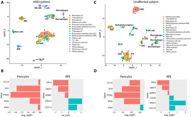
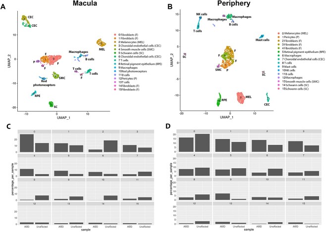
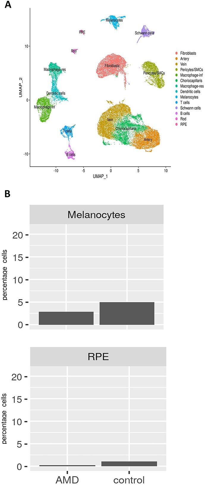
Age-related macular degeneration (AMD) is the most prevalent cause of blindness in the developed world. Vision loss in the advanced stages of the disease is caused by atrophy of retinal photoreceptors, overlying retinal pigment epithelium (RPE) and choroidal endothelial cells. The molecular events that underline the development of these cell types from in utero to adult as well as the progression to intermediate and advanced stages AMD are not yet fully understood. We performed single-cell RNA-sequencing (RNA-Seq) of human fetal and adult RPE-choroidal tissues, profiling in detail all the cell types and elucidating cell type-specific proliferation, differentiation and immunomodulation events that occur up to midgestation. Our data demonstrate that progression from the fetal to adult state is characterized by an increase in expression of genes involved in the oxidative stress response and detoxification from heavy metals, suggesting a better defence against oxidative stress in the adult RPE-choroid tissue. Single-cell comparative transcriptional analysis between a patient with intermediate AMD and an unaffected subject revealed a reduction in the number of RPE cells and melanocytes in the macular region of the AMD patient. Together these findings may suggest a macular loss of RPE cells and melanocytes in the AMD patients, but given the complex processing of tissues required for single-cell RNA-Seq that is prone to technical artefacts, these findings need to be validated by additional techniques in a larger number of AMD patients and controls.
Keywords: Schwann cells; age related macular degeneration (AMD); choroid; choroid endothelial cells; embryonic and fetal eye; melanocytes; pericytes; retinal pigment epithelium; single cell RNA-Seq; smooth muscle cells.
© The Author(s) 2023. Published by Oxford University Press.
Figures
References
-
- Mitchell, P., Liew, G., Gopinath, B. and Wong, T.Y. (2018) Age-related macular degeneration. Lancet, 392, 1147–1159. - PubMed
-
- Wong, W.L., Su, X., Li, X., Cheung, C.M., Klein, R., Cheng, C.Y. and Wong, T.Y. (2014) Global prevalence of age-related macular degeneration and disease burden projection for 2020 and 2040: a systematic review and meta-analysis. Lancet Glob. Health, 2, e106–e116. - PubMed
-
- Mrejen, S. and Spaide, R.F. (2013) Optical coherence tomography: imaging of the choroid and beyond. Surv. Ophthalmol., 58, 387–429. - PubMed
-
- Margolis, R. and Spaide, R.F. (2009) A pilot study of enhanced depth imaging optical coherence tomography of the choroid in normal eyes. Am J. Ophthalmol., 147, 811–815. - PubMed
-
- Provis, J.M., Penfold, P.L., Cornish, E.E., Sandercoe, T.M. and Madigan, M.C. (2005) Anatomy and development of the macula: specialisation and the vulnerability to macular degeneration. Clin. Exp. Optom., 88, 269–281. - PubMed
Publication types
MeSH terms
Grants and funding
- BB/T004460/1/BB_/Biotechnology and Biological Sciences Research Council/United Kingdom
- MR/R006237/1/MRC_/Medical Research Council/United Kingdom
- MR/S035826/1/MRC_/Medical Research Council/United Kingdom
- MR/X001687/1/MRC_/Medical Research Council/United Kingdom
- NC/C016106/1/NC3RS_/National Centre for the Replacement, Refinement and Reduction of Animals in Research/United Kingdom
LinkOut - more resources
Full Text Sources
Other Literature Sources
Medical
Molecular Biology Databases
