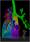
Preoperative visualization of congenital lung abnormalities: hybridizing artificial intelligence and virtual reality
- PMID: 36645240
- PMCID: PMC10481780
- DOI: 10.1093/ejcts/ezad014
Preoperative visualization of congenital lung abnormalities: hybridizing artificial intelligence and virtual reality
Abstract
Objectives: When surgical resection is indicated for a congenital lung abnormality (CLA), lobectomy is often preferred over segmentectomy, mostly because the latter is associated with more residual disease. Presumably, this occurs in children because sublobar surgery often does not adhere to anatomical borders (wedge resection instead of segmentectomy), thus increasing the risk of residual disease. This study investigated the feasibility of identifying eligible cases for anatomical segmentectomy by combining virtual reality (VR) and artificial intelligence (AI).
Methods: Semi-automated segmentation of bronchovascular structures and lesions were visualized with VR and AI technology. Two specialists independently evaluated via a questionnaire the informative value of regular computed tomography versus three-dimensional (3D) VR images.
Results: Five asymptomatic, non-operated cases were selected. Bronchovascular segmentation, volume calculation and image visualization in the VR environment were successful in all cases. Based on the computed tomography images, assignment of the CLA lesion to specific lung segments matched between the consulted specialists in only 1 out of the cases. Based on the three 3D VR images, however, the localization matched in 3 of the 5 cases. If the patients would have been operated, adding the 3D VR tool to the preoperative workup would have resulted in changing the surgical strategy (i.e. lobectomy versus segmentectomy) in 4 cases.
Conclusions: This study demonstrated the technical feasibility of a hybridized AI-VR visualization of segment-level lung anatomy in patients with CLA. Further exploration of the value of 3D VR in identifying eligible cases for anatomical segmentectomy is therefore warranted.
Keywords: Congenital lung abnormalities; Congenital pulmonary airway malformations; Paediatric lung surgery; Segmentectomy; Virtual reality.
© The Author(s) 2023. Published by Oxford University Press on behalf of the European Association for Cardio-Thoracic Surgery. All rights reserved.
Figures


References
-
- Andrade CF, da Costa Ferreira HP, Fischer GB.. Malformações pulmonares congênitas. J Bras Pneumol 2011;37:259–71. - PubMed
-
- Hermelijn SM, Elders BBLJ, Ciet P, Wijnen RMH, Tiddens HAWM, Schnater JM. et al. A clinical guideline for structured assessment of CT-imaging in congenital lung abnormalities. Paediatr Respir Rev 2021;37:80–8. - PubMed
-
- Stocker LJ, Wellesley DG, Stanton MP, Parasuraman R, Howe DT.. The increasing incidence of foetal echogenic congenital lung malformations: an observational study. Prenat Diagn 2015;35:148–53. - PubMed
-
- Stanton M, Njere I, Ade-Ajayi N, Patel S, Davenport M.. Systematic review and meta-analysis of the postnatal management of congenital cystic lung lesions. J Pediatr Surg 2009;44:1027–33. - PubMed
MeSH terms
Grants and funding
LinkOut - more resources
Full Text Sources
Medical

