

What's new in the WHO 2022 classification of kidney tumours?
- PMID: 36645398
- PMCID: PMC10342217
- DOI: 10.32074/1591-951X-818
What's new in the WHO 2022 classification of kidney tumours?
Abstract
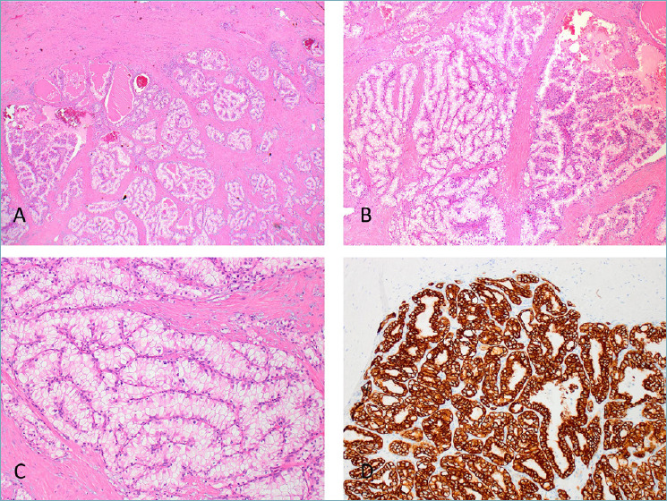
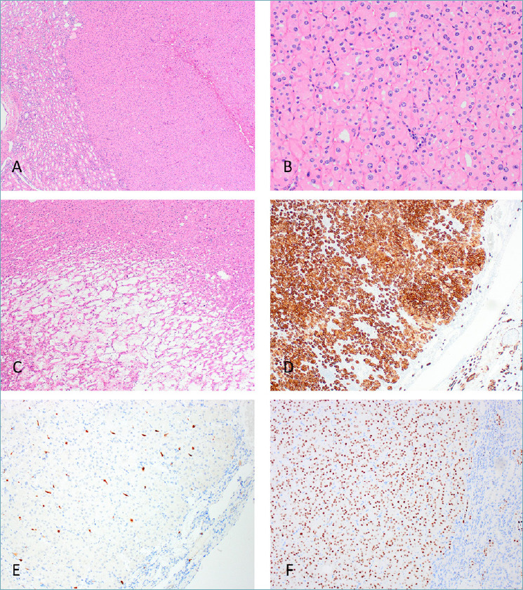
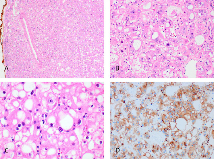
The World Health Organization (WHO) 2022 classification of urinary and male genital tumours (5th edition) has significantly improved our understanding of the morphologic, immunohistochemical, and molecular characteristics of renal tumours. The aim of this review is to outline the most important changes and diagnostic updates in the WHO 2022 classification of kidney tumours. A major change in this edition is the grouping of renal tumours into broader categories that include "clear cell renal tumours", "papillary renal tumours", "oncocytic and chromophobe renal tumours", "collecting duct tumours" as well as adding two categories of "other renal tumours" and "molecularly defined renal carcinomas". Novel entities included in the WHO 2022 classification are eosinophilic solid and cystic renal cell carcinoma (ESC RCC), anaplastic lymphoma kinase (ALK)-rearranged RCC and ELOC (formerly TCEB1)-mutated RCC. The category of "other renal tumours" includes a group of diverse, unrelated renal tumours that do not fit into other categories. The group of "molecularly defined renal carcinomas" reflects recent discoveries in the renal tumour genomics. These molecularly-defined renal entities demonstrate a set of morphologic features reflecting genotype-phenotype relationships. Final diagnosis of such entities rests on phenotypic and immunohistochemical (IHC) correlation, usually associated with IHC surrogate makers that reflect specific genetic abnormalities.
Keywords: WHO; classification; kidney; pathology; renal cell carcinoma.
Copyright © 2023 Società Italiana di Anatomia Patologica e Citopatologia Diagnostica, Divisione Italiana della International Academy of Pathology.
Conflict of interest statement
Authors declare no potential conflicts of interest with respect to the content, authorship, and/or publication of this article.
Figures
References
-
- Moch H, Humphrey PA, Ulbright TM, et al. WHO Classification of Tumours of the Urinary System and Male Genital Organs. 4th ed. Lyon (France): International Agency for Research on Cancer, 2016.
-
- Trpkov K, Williamson SR, Gill AJ, et al. Novel, emerging and provisional renal entities: The Genitourinary Pathology Society (GUPS) update on renal neoplasia. Mod Pathol 2021;34:1167-1184. https://doi.org/10.1038/s41379-021-00737-6 10.1038/s41379-021-00737-6 - DOI - PubMed
-
- Trpkov K, Hes O, Williamson SR, et al. New developments in existing WHO entities and evolving molecular concepts: The Genitourinary Pathology Society (GUPS) update on renal neoplasia. Mod Pathol 2021;34:1392-1424. https://doi.org/10.1038/s41379-021-00779-w 10.1038/s41379-021-00779-w - DOI - PubMed
-
- Board WCoTE. WHO Classification of Tumours Urinary and Male Genital Tumours. 5th ed. Lyon (France): International Agency for Research on Cancer, 2022.
-
- Choueiri TK, Cheville J, Palescandolo E, et al. BRAF mutations in metanephric adenoma of the kidney. Eur Urol 2012;62:917-922. https://doi.org/10.1016/j.eururo.2012.05.051 10.1016/j.eururo.2012.05.051 - DOI - PMC - PubMed
Publication types
MeSH terms
LinkOut - more resources
Full Text Sources
Medical
