. 2023 Jan 16;257(2):37.
doi: 10.1007/s00425-023-04072-9.

Steroidal glycoside profile differences among primary roots system and adventitious roots in Solanum dulcamara

Affiliations

Steroidal glycoside profile differences among primary roots system and adventitious roots in Solanum dulcamara

Ilaria Chiocchio et al. Planta. .

Abstract

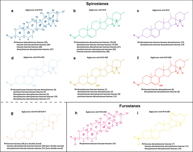
Solanum dulcamara primary and adventitious roots showed qualitative and quantitative differences in their steroidal glycosides profile. This opened new venues to evaluate the bioactivity of these molecules in belowground ecosystems. The Solanum genus is characterized by the presence of steroidal glycosides (SGs) that confer herbivore resistance and serve as drug precursors in the pharmaceutical industry. Solanum dulcamara is a self-compatible, sexually reproducing species that produces seeds after buzz-pollination. In addition, primordia on the stem facilitate clonal propagation via adventitious root (AR) formation. ARs contain aerenchyma being developmentally and morphologically different from primary roots (PRs). Therefore, we hypothesized that ARs and PRs have different SG profiles. Aiming to assess differences in SGs profiles in S. dulcamara roots in relation to their origins and morphologies, we used liquid chromatography coupled to electron spray ionization quadruple time of flight mass spectrometry (LC-ESI-qToF-MS) to profile SGs from PRs and ARs of seven S. dulcamara individuals. Mass fragmentation pattern analysis indicated the presence of 31 SG-type structures, including those with spirostans and furostans moieties. We assigned the 31 structures to 9 classes of steroidal aglycons (SAgls) that differ in hydroxylation and degree of unsaturation. We found that SAgls were conjugated with di-, tri- and tetra saccharides whereby one compound contained a malonylated sugar. Principle component analysis showed that SG profiles of PRs and ARs separated on the first principal component, supporting our hypothesis. Specifically, PRs contain higher number of SGs than ARs with some compounds exclusively present in PRs. Our results reveal a high level of novel chemodiversity in PRs and ARs of Solanum dulcamara. The knowledge gained will deepen our understanding of SGs biosynthesis and their functional role in plant-environment interactions.

Keywords: Chemodiversity; Glycosides; LC–MS; Mass spectrometry; Solanaceae; Steroids.


Conflict of interest statement

The authors declare that they have no conflicts of interest.

Figures

Fig. 1
Fig. 1
Proposed structures produced by interpreting the mass spectra of the 31 detected steroidal glycosides (SGs) in roots of Solanum dulcamara. Protonated adducts (X − H) are shown as deducted from LC–qToF mass spectra. SGs are grouped into 9 classes (a − i) according to the steroidal aglycone (SAgl) type. For simplicity, the SAgl m/z values are shown only as integers. A detailed description of the retention times and m/z values for all the SGs is shown in supplementary Table S1. SAgls a to g are interpreted as spirostanes whereas SAgls h and i are interpreted as furostanes. IUPAC carbon atom numbering (1 − 27) and ring lettering (a − f or a − e) are shown for spirostanes and furostanes. The mass spectra of SGs d − f do not allow the exact position of the hydroxyl group to be determined (thus the bonds are represented as dotted lines). Color codes correspond with colors shown in Figs. 3, 4
Fig. 2
Fig. 2
Mass spectrum of compound 15 (class H) showing an oxygen loss. This fragmentation supports the proposed SAgl structure consisting of a furostane bearing an aldehyde in the lateral chain
Fig. 3
Fig. 3
Principal component analysis (PCA) of the LC-qToF ion abundance (counts) as EIC integrals of the 31 glycosidic steroids (SGs) detected in roots of Solanum dulcamara. a Scores plot of primary (red squares -PRs-) and adventitious roots (blue circles -ARs-). Numbers 1 − 7 represent plant individuals, obtained from crossing or selfing of the two accessions TW12 and ZD04 (see Calf et al. 2018). b PC1 loadings for every EIC integral. Black bars represent SGs detected both in PRs and ARs, red bars represent SGs exclusively detected in PRs. Compound ID numbers (1 − 31) are shown in the color code for SAgl type (Figs. 1, 4)
Fig. 4
Fig. 4
Bubble plot representing the LC–qToF ion abundance. Each bubble is proportional to the area of an m/z value representing each SG (EIC integrals). The compound number ID (1 − 31) corresponds with the elution order. Steroidal aglycone (SAgl) class and color codes correspond with those shown in Figs. 1, 3. Plant number (1 − 7) corresponds with Fig. 3a. PRs primary roots, ARs adventitious roots

References

    1. Ahmed S, Shevlin P. The possible involvement of excited singlet methylene in the deoxygenation of formaldehyde by atomic carbon. J Am Chem Soc. 1983;105:6488–6490. doi: 10.1021/ja00359a021. - DOI
    1. Alaguero-Cordovilla A, Sánchez-García AB, Ibáñez S, Albacete A, Cano A, Acosta M, Pérez-Pérez JM. An auxin-mediated regulatory framework for wound-induced adventitious root formation in tomato shoot explants. Plant Cell Environ. 2021;44(5):1642–1662. doi: 10.1111/pce.14001. - DOI - PubMed
    1. Bailly C. The steroidal alkaloids α-tomatine and tomatidine: panorama of their mode of action and pharmacological properties. Steroids. 2021;176:108933. doi: 10.1016/j.steroids.2021.108933. - DOI - PubMed
    1. Bednarz H, Roloff N, Niehaus K. Mass spectrometry imaging of the spatial and temporal localization of alkaloids in nightshades. J Agric Food Chem. 2019;67:13470–13477. doi: 10.1021/acs.jafc.9b01155. - DOI - PubMed
    1. Bellini C, Pacurar D, Perrone I. Adventitious roots and lateral roots: similarities and differences. Annu Rev Plant Biol. 2014;65:639–666. doi: 10.1146/annurev-arplant-050213-035645. - DOI - PubMed