

Complications of cochlear implants with MRI scans in different body regions: type, frequency and impact
- PMID: 36645551
- PMCID: PMC9842833
- DOI: 10.1186/s13244-022-01353-x
Complications of cochlear implants with MRI scans in different body regions: type, frequency and impact
Abstract
Objectives: The aim was to assess the type, frequency and impact of MRI-related complications in patients with cochlear implants (CI) and MRI indications in different body regions.
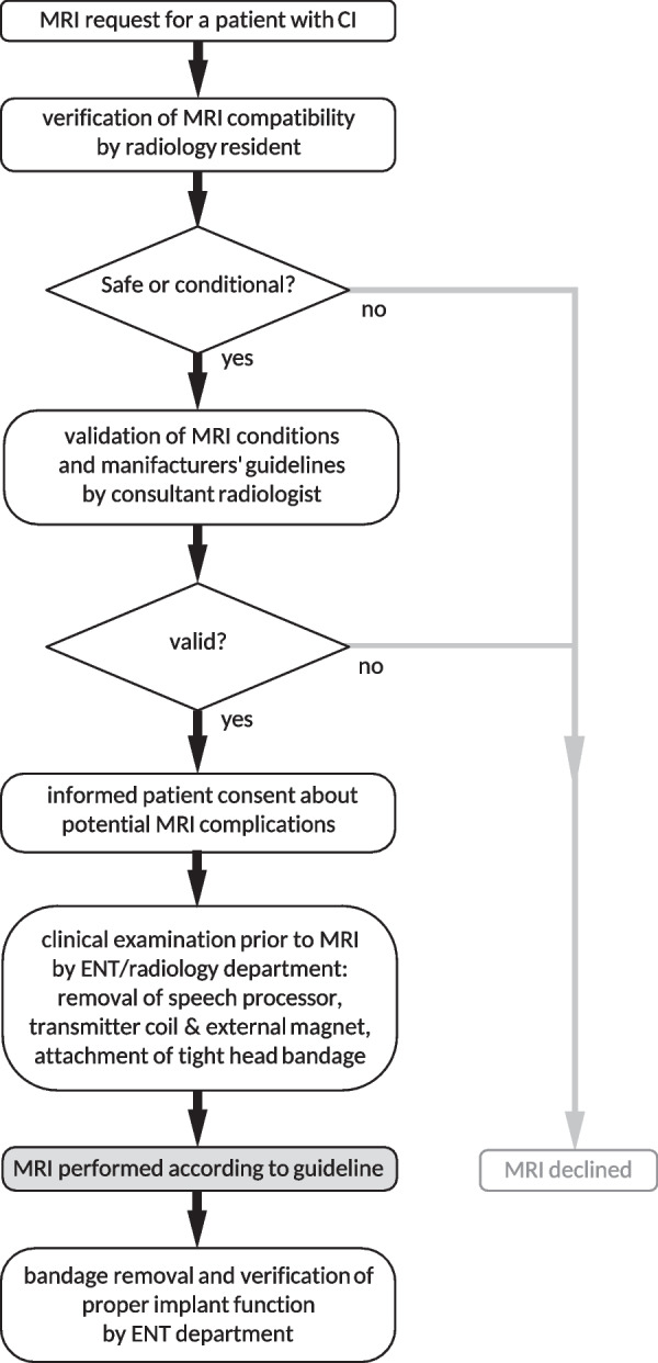
Methods: For that purpose, the institutional radiology database of a single tertiary hospital was searched for patients with a CI who underwent MRI between 2001 and 2018. The number of MRI examinations and complications were retrieved from the patient record. Examinations were categorized into five distinct body regions or combinations thereof. Records of CI artifacts in the head also included basic information on diagnostic image quality.
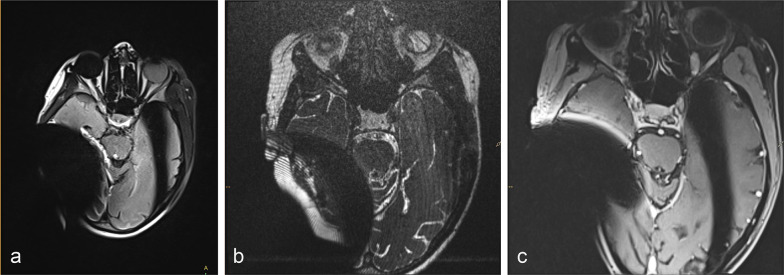
Results: Out of 1017 MRI database entries (examinations) of patients with a CI, 91 records were after implantation (71 patients) and 66 were attempted (no contraindications, 49 patients). In four cases (4/66, 6.1%), the magnet was dislocated and had to be replaced surgically. Three out of four severe complications occurred for examination regions outside the head. Thirteen MRI examinations were aborted due to pain (19.7%) and one because of artifacts-resulting in 48 scans (72.7%) completed successfully (36 patients). All cranial scans featured device artifacts in all sequences, but the majority of them did not affect proper imaging diagnostics in the respective region.
Conclusion: This retrospective, single-center analysis of patients with MRI-conditional cochlear implants shows that MRI-related complications were common, at least in models with a fixed magnet, despite appropriate precautions and compliance with the manufacturers' guidelines. MRI examinations of CI patients should therefore be indicated strictly until the exact causes have been clarified.
Keywords: Cochlea; Implant; MR complication; MR safety; MRI.
© 2023. The Author(s).
Conflict of interest statement
TD has received travel grants and honoraria for speaking at scientific meetings from Siemens Healthcare and Canon Medical as well as research funding from Siemens Healthcare. All other authors declare no relationships with any companies whose products or services may be related to the subject matter of the article.
Figures


References
LinkOut - more resources
Full Text Sources
