. 2023 Mar;10(7):e2205997.
doi: 10.1002/advs.202205997. Epub 2023 Jan 16.

Multifunctional Integrated Nanozymes Facilitate Spinal Cord Regeneration by Remodeling the Extrinsic Neural Environment

Affiliations

Multifunctional Integrated Nanozymes Facilitate Spinal Cord Regeneration by Remodeling the Extrinsic Neural Environment

Tiandi Xiong et al. Adv Sci (Weinh). 2023 Mar.

Abstract

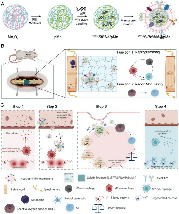
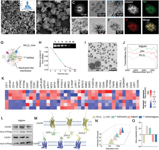
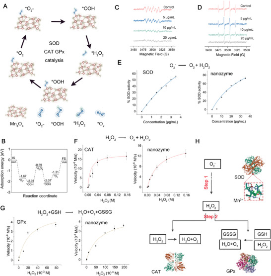
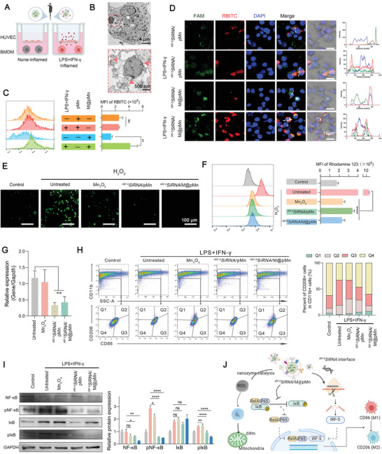
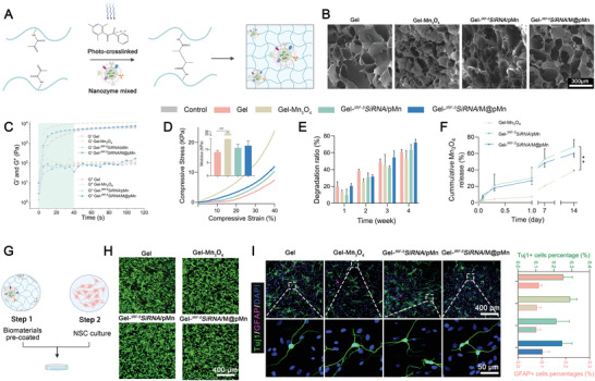
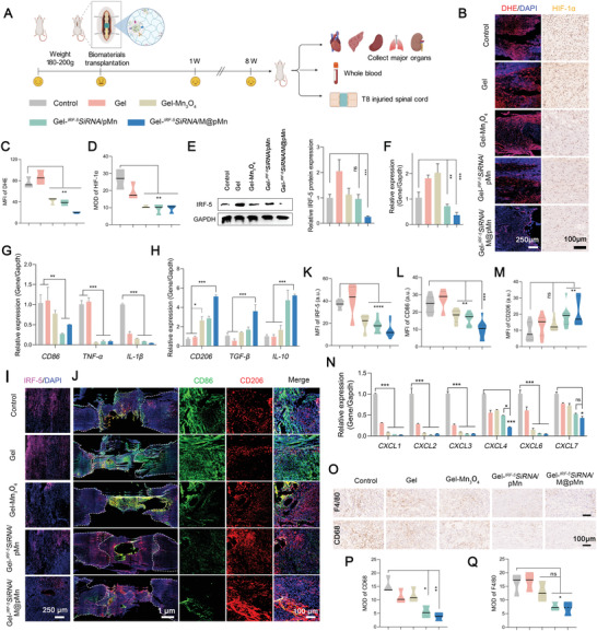
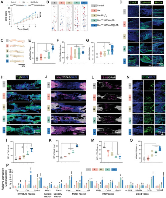
High levels of reactive oxygen species (ROS) and inflammation create a complicated extrinsic neural environment that dominates the initial post-injury period after spinal cord injury (SCI). The compensatory pathways between ROS and inflammation limited the efficacy of modulating the above single treatment regimen after SCI. Here, novel "nanoflower" Mn3 O4 integrated with "pollen" IRF-5 SiRNA was designed as a combination antioxidant and anti-inflammatory treatment after SCI. The "nanoflower" and "pollen" structure was encapsulated with a neutrophil membrane for protective and targeted delivery. Furthermore, valence-engineered nanozyme Mn3 O4 imitated the cascade response of antioxidant enzymes with a higher substrate affinity compared to natural antioxidant enzymes. Nanozymes effectively catalyzed ROS to generate O2 , which is advantageous for reducing oxidative stress and promoting angiogenesis. The screened "pollen" IRF-5 SiRNA could reverse the inflammatory phenotype by reducing interferon regulatory factors-5 (IRF-5) expression (protein level: 73.08% and mRNA level: 63.10%). The decreased expression of pro-inflammatory factors reduced the infiltration of inflammatory cells, resulting in less neural scarring. In SCI rats, multifunctional nanozymes enhanced the proliferation of various neuronal subtypes (motor neurons, interneurons, and sensory neurons) and the recovery of locomotor function, demonstrating that the remodeling of the extrinsic neural environment is a promising strategy to facilitate nerve regeneration.

Keywords: anti-inflammation; antioxidation; catalytic cascade reaction; spinal cord injury.


Conflict of interest statement

The authors declare no conflict of interest.

Figures

Scheme 1
Scheme 1
A) Schematic representation of integrated IRF‐5SiRNA/M@pMn nanozymes. Polyethyleneimine (PEI) was used to modify Mn3O4 to form pMn. The siRNA was then loaded into the pMn, generating IRF‐5SiRNA/pMn. The surface of pMn was coated with a neutrophil‐like membrane ( IRF‐5SiRNA/M@pMn). B) Schematic diagram of the multifunctional therapeutic ability of the prepared nanozymes that play the main function; macrophages reprogramming and redox modulatory. The multifunctional nanozyme was blended into a gelatin‐based hydrogel for T9 spinal cord transplantation. C) Diagram showing the recruitment of IRF‐5SiRNA/M@pMn to inflammatory macrophages.
Figure 1
Figure 1
Preparation and characterization of IRF‐5SiRNA/M@pMn. A,B) SEM, C) SAED patterns, D) TEM, and E,F) HAADF‐STEM images with the corresponding elemental mappings of Mn3O4. G) Schematic illustration of IRF‐5SiRNA/M@pMn particles. H) Characterization of pMn/SiRNA nanoparticles using agarose gel electrophoresis at the different volumes of siRNA (0, 4, 8, 12, 16, and 20 µL of 20 µM siRNA) with 2 µL of 1 mg mL−1 pMn. I) TEM images of IRF‐5SiRNA/M@pMn. J) FTIR spectra of Mn3O4, pMn, and M@pMn nanoparticles. K) Heatmap of membrane protein expression in HL‐60 and neutrophil‐like cells. L) Western blots of CXCR1/2 protein levels in M@pMn. Three lanes in this western blot image reflect triplicate tests. M) Computer simulation image of protein docking between CXCR1/2 and CXCL1/2/3. N) Diameter and O) zeta potential of the different nanoparticles (n = 3). Data are presented as mean ± SD. Statistical analysis was performed using a one‐way ANOVA followed by Tukey's post hoc test, *p < 0.05; **p < 0.01; ***p < 0.001; ns, no statistical significance.
Figure 2
Figure 2
Theoretical investigation and characterization of the multienzyme cascade activities of M@pMn. A) Proposed reaction routes of O2 reduction to H2O with optimum adsorption configurations. B) Free energy diagram for the O2 reduction reaction. EPR for the elimination of C) O2 and D) •OH. The spectra were tested at 5 min after mixing in PBS (pH 7.4). E–G) The SOD, CAT, GPx‐like activity of the nanozyme compared to natural SOD, CAT, GPx tested in the same condition (n = 3). H) Schematic illustration showing the theoretical nanozyme‐catalyzed cascade reactions. Insert images are the protein structures of the native enzymes. Data are presented as mean ± SD. Statistical analysis was performed using a one‐way ANOVA followed by Tukey's post hoc test, *p < 0.05; **p < 0.01; ***p < 0.001; ns, no statistical significance.
Figure 3
Figure 3
The biological function of IRF‐5SiRNA/M@pMn on BMDMs includes targeting inflamed macrophages, ROS scavenging, and reducing inflammation. A) Transwell schematic diagram to measure the targeting ability of the nanoparticles. B) Bio‐TEM images of BMDMs cultured with M@pMn (12.5 µg mL−1). C) Fluorescence of pMn and M@pMn under non‐inflammatory and inflammatory BMDMs as measured using FACS (n = 3). D) Representative confocal images of BMDM incubated with FAM‐IRF‐5SiRNA/RBITCpMn and M@ FAM‐IRF‐5SiRNA/RBITCpMn with or without stimulation using LPS and IFN‐γ (Blue, nuclei; red, RBITCpMn; green, FAMsiRNA). E) Fluorescence imaging of BMDMs stained with the DCFH‐DA probe. F) MMP quantification and analysis using flow cytometry (n = 3). G) mRNA expression levels of IRF‐5 as detected via qPCR (n = 5). H) Flow cytometry assay and quantification of BMDM polarization (n = 3). M1 and M2 subtypes are distinguished by the presence of CD86 (mostly in Q3) and CD206 (primarily in Q1). I) Western blotting and quantitative analysis of NF‐κB, pNF‐κB, IκB, and pIκB expression in BMDMs (n = 3). J) Illustration of antioxidant and anti‐inflammatory processes of IRF‐5SiRNA/M@pMn treatment in vitro. Data are presented as mean ± SD. Statistical analysis was performed using a one‐way ANOVA followed by Tukey's post hoc test, *p < 0.05; **p < 0.01; ***p < 0.001; ns, no statistical significance.
Figure 4
Figure 4
Characterization and biocompatibility analysis of gelatin hydrogels matched to native spinal cord healing processes. A) Schematic diagram of the prepared nanozymes mixed with hydrogels. B) SEM images of the different hydrogels. C) Cross‐linking of the prepolymer solution was monitored via rheological analyses. D) Representative stress−strain curves of the different hydrogels. Insert images represent the compressive modulus (n = 3). E) Degradation ratio of the hydrogel after incubation in saline for 1, 2, 3, and 4 weeks (n = 3). F) Mn3O4 sustained release behavior of the hydrogels (n = 4). G) Cell adhesion on the surface of the gelatin hydrogel. H) NSCs showed high cell viability even after 5 d of culture (green; live cells, red; dead cells). I) Representative images and quantification analysis (n = 6) of Tuj‐1 (for neurons) and GFAP (for astrocytes) expression, showing the growth of NSCs on the gelatin hydrogel after 7 d of culture. Data are presented as mean ± SD. Statistical analysis was performed using a one‐way ANOVA followed by Tukey's post hoc test, *p < 0.05; **p < 0.01; ***p < 0.001; ns, no statistical significance.
Figure 5
Figure 5
Evaluation of short‐term treatment effect in animal models. A) Treatment regime in rats. B) Representative fluorescence staining images of DHE and immunohistochemical images of HIF‐1α in the injured spinal cord tissues and quantitative analysis of C) DHE and D) HIF‐1α levels (n = 6). E) Western blot (n = 3) and quantification, and F) qPCR of IRF‐5 expression levels (n = 5). qPCR analysis of the expression levels of G) M1 markers (CD86, TNF‐α, IL‐1β) and H) M2 markers (CD206, TGF‐β, IL‐10) (n = 5). I) IRF‐5‐, J) CD86‐ (M1, green), and CD206‐ (M2, red) positive cells at the lesion site of SCI model rats. Quantification of K) IRF‐5, L) CD86, and M) CD206 expression (n = 6). N) qPCR to detect CXCL1, 2, 3, 4, 6, and 7 expression levels (n = 5). O) Representative immunohistochemical images and quantification analysis of F4/80 and CD68‐positive cells at the spinal cord injury site (n = 6). Data are presented as mean ± SD. Statistical analysis was performed using a one‐way ANOVA followed by Tukey's post hoc test, *p < 0.05; **p < 0.01; ***p < 0.001; ns, no statistical significance.
Figure 6
Figure 6
The functional hydrogel induced excellent recovery in long‐term animal experiments. A) BBB scores of rats from surgery to eight weeks post‐injury (n = 8). B) Examination of hind limb motor function recovery in rats using typical footprints (forepaw, blue ink; hind paw, red ink.) C) Representative spinal cord images after SCI repair. D) Representative immunofluorescence images after staining for ChAT, Calbindin, and Brn3a and quantification of E) ChAT, F) Calbindin, and G) Brn3a staining (n = 6). The neuronal differentiation profiles of H) Tuj1, I) Map2, L) CS56, and N) CD31, and their corresponding quantitative analyses J), K), M), O), respectively in the lesion area of SCI rats (n = 3). P) qPCR analysis of the expression of neuron and blood vessel markers (n = 5). Data are presented as mean ± SD. Statistical analysis was performed using a one‐way ANOVA followed by Tukey's post hoc test, *p < 0.05; **p < 0.01; ***p < 0.001; ns, no statistical significance.

References

    1. a) Ahuja C. S., Wilson J. R., Nori S., Kotter M. R. N., Druschel C., Curt A., Fehlings M. G., Nat. Rev. Dis. Primers 2017, 3, 17018; - PubMed
    2. b) Warner F. M., Cragg J. J., Jutzeler C. R., Finnerup N. B., Werhagen L., Weidner N., Maier D., Kalke Y. B., Curt A., Kramer J. L. K., J Neurotrauma 2019, 36, 1461; - PubMed
    3. c) Zipser C. M., Cragg J. J., Guest J. D., Fehlings M. G., Jutzeler C. R., Anderson A. J., Curt A. J. T. L. N., Lancet Neurol. 2022. - PubMed
    1. Orr M. B., Gensel J. C., Neurotherapeutics 2018, 15, 541. - PMC - PubMed
    1. a) Bains M., Hall E. D., Biochim. Biophys. Acta 2012, 1822, 675; - PMC - PubMed
    2. b) Fatima G., Sharma V. P., Das S. K., Mahdi A. A., Spinal Cord 2015, 53, 3; - PubMed
    3. c) Eli I., Lerner D. P., Ghogawala Z., Neurol Clin 2021, 39, 471; - PubMed
    4. d) Hall E. D., J. E. J. N., NeuroRx 2004, 80, 100; - PMC - PubMed
    5. e) Springer J. E., Rao R. R., Lim H. R., Cho S. I., Moon G. J., Lee H. Y., Park E. J., Noh J. S., Gwag B. J. J. J. o. n., J Neurotrauma 2010, 27, 139. - PMC - PubMed
    1. a) Knock G. A., Free Radic Biol Med 2019, 145, 385; - PubMed
    2. b) Murphy M. P., Biochem. J. 2009, 417, 10.1042/BJ20081386; - DOI - PubMed
    3. c) Go Y. M., Chandler J. D., Jones D. P., Free Radic Biol Med 2015, 84, 227. - PMC - PubMed
    1. a) Hall E., Yonkers P., Andrus P., Cox J., Anderson D. J. J. o. n., J Neurotrauma 1992, 9, 173; - PubMed
    2. b) Hamann K., Durkes A., Ouyang H., Uchida K., Pond A., Shi R. J. J. o. n., J. Neurochem. 2008, 107, 712. - PMC - PubMed

Publication types

LinkOut - more resources