Review
. 2023 Jan 16;8(1):32.
doi: 10.1038/s41392-022-01300-8.

Epigenetic and post-translational modifications in autophagy: biological functions and therapeutic targets

Affiliations
Review

Epigenetic and post-translational modifications in autophagy: biological functions and therapeutic targets

Feng Shu et al. Signal Transduct Target Ther. .

Abstract

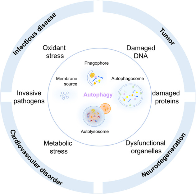
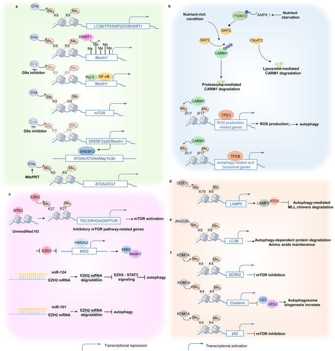
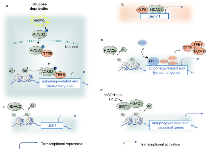
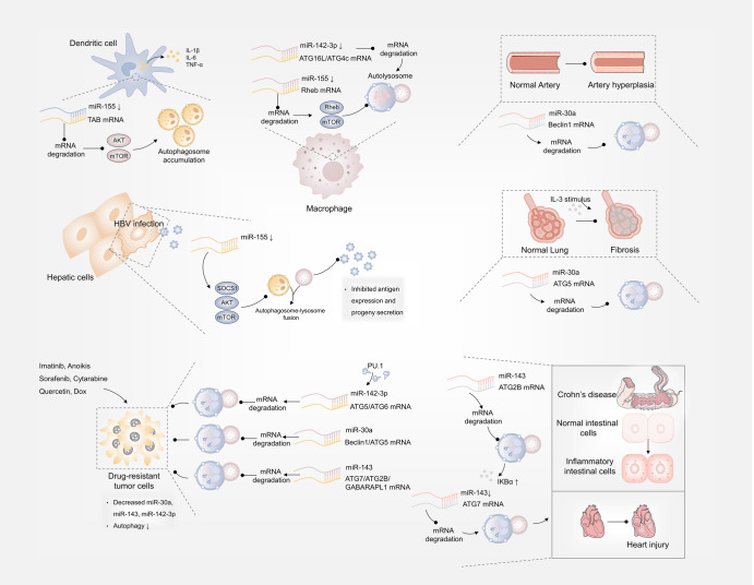
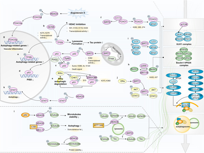
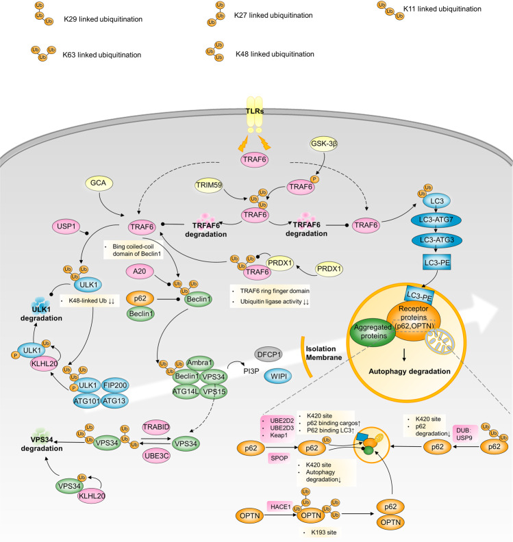
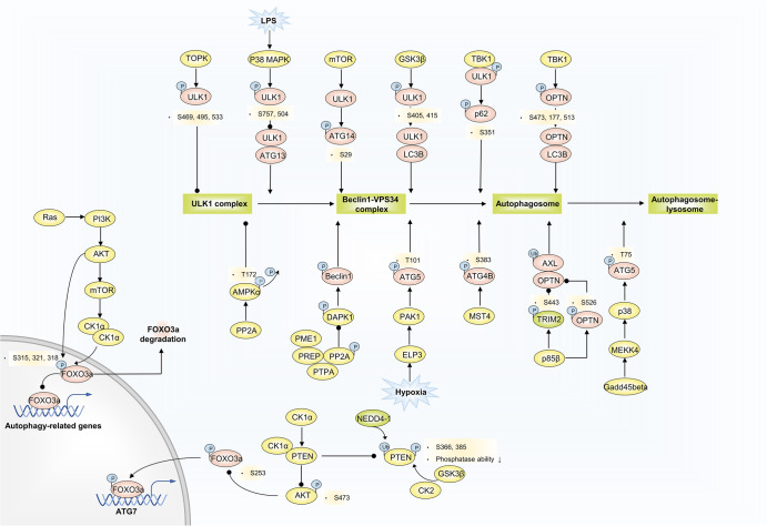
Autophagy is a conserved lysosomal degradation pathway where cellular components are dynamically degraded and re-processed to maintain physical homeostasis. However, the physiological effect of autophagy appears to be multifaced. On the one hand, autophagy functions as a cytoprotective mechanism, protecting against multiple diseases, especially tumor, cardiovascular disorders, and neurodegenerative and infectious disease. Conversely, autophagy may also play a detrimental role via pro-survival effects on cancer cells or cell-killing effects on normal body cells. During disorder onset and progression, the expression levels of autophagy-related regulators and proteins encoded by autophagy-related genes (ATGs) are abnormally regulated, giving rise to imbalanced autophagy flux. However, the detailed mechanisms and molecular events of this process are quite complex. Epigenetic, including DNA methylation, histone modifications and miRNAs, and post-translational modifications, including ubiquitination, phosphorylation and acetylation, precisely manipulate gene expression and protein function, and are strongly correlated with the occurrence and development of multiple diseases. There is substantial evidence that autophagy-relevant regulators and machineries are subjected to epigenetic and post-translational modulation, resulting in alterations in autophagy levels, which subsequently induces disease or affects the therapeutic effectiveness to agents. In this review, we focus on the regulatory mechanisms mediated by epigenetic and post-translational modifications in disease-related autophagy to unveil potential therapeutic targets. In addition, the effect of autophagy on the therapeutic effectiveness of epigenetic drugs or drugs targeting post-translational modification have also been discussed, providing insights into the combination with autophagy activators or inhibitors in the treatment of clinical diseases.


Conflict of interest statement

The authors declare no competing interests.

Figures

Fig. 1
Fig. 1
Autophagy and diseases. Autophagy dysregulation and tumor, inflammatory disease, neurodegenerative disease, cardiovascular disease
Fig. 2
Fig. 2
Histone methylation and autophagy regulation. a G9a negatively regulates core autophagy effectors and upstream autophagic regulators to influence autophagy level indirectly. b Under a nutrient-rich environment, SKP2 mediates CARM1 protein degradation, nutrient starvation activates AMPK-dependent FOXO3 phosphorylation, which transcriptionally represses SKP2, resulting in CARM1-mediated transcriptional activation of autophagy-related and lysosomal genes. c EZH2 repressively regulates autophagy via mTOR activation. EZH2 negatively controls TSC2/RHOA/DEPTOR gene transcription to elicit mTOR pathway, leading to autophagy inhibition. Additionally, EZH2 represses HMGA2 expression. HMGA2 can directly activate the MSI2 promoter region, which triggers autophagy via Beclin1 interactions. d DOT1L elevates LAMP5 expression via H3K79 methylation modification enhancements. LAMP5 directly interacts with ATG5 to interrupt an autophagy flux, protecting MLL chimeras from autophagy degradation. e JMJD2B promotes autophagy occurrence via histone demethylation at LC3B promoters, assisting in intracellular amino acid maintenance. f KDM1A negatively manipulates autophagy flux through SESN2- and CLU-dependent pathways or directly targeting p62. SESN2 inhibits mTORC1 activity, and CLU increases autophagosome biogenesis through MAP1LC3/LC3-ATG3 heterodimer stabilization
Fig. 3
Fig. 3
Histone acetylation and autophagy regulation. a Glucose deprivation assists AMPK-mediated ACSS2 phosphorylation and promotes its nuclear localization. In the nucleus, ACSS2 binds to TFEB and translocates to lysosomal and autophagy gene promoters, where ACSS2 generates acetyl-CoA for histone H3 acetylation and gene expression. b KLF5 protein and HDAC3 bind to the Beclin1 gene promoter and suppress its transcription, leading to autophagy suppression. c Coupled with MYC, HDACs, especially class I HDACs, epigenetically negatively regulate autophagic and lysosomal function. d HIST1H1C/H1.2 upregulates SIRT1 and HDAC1 to maintain the deacetylation of H4K16, leads to ATG proteins expression and then promotes autophagy, inflammation and cell toxicity. e Small-chain fatty acids treatment-reduced HDAC2 upregulates H3K27ac on ULK1 promoter, reduces ULK1 expression and autophagy flux and thus alleviate diabetic renal fibrosis
Fig. 4
Fig. 4
MicroRNA and autophagy regulation. In Behcet’s disease, HBV and mycobacterial infection, decreased miR-155 respectively inhibited TAB-AKT/mTOR-, SOCS1/Akt/mTOR- and Rheb/mTOR-dependent autophagy. miR-30a, miR-143 and miR-142-3p respectively targeted Beclin1/ATG5, ATG7/ATG2B and ATG5/ATG6 mRNA for degradation to inhibit autophagy and increase chemotherapy sensitiveness. miR-30a mediated autophagy suppression via targeting Beclin1 or ATG5 and alleviated arterial injury and airway fibrosis. Downregulated miR-142-3p could target ATG16L1, ATG4c to trigger autophagy and eliminate mycobacteria. miR-143 inhibited autophagy via targeting ATG7 or ATG2B, which induced Crohn’s disease or in other case, alleviated cardiac injury
Fig. 5
Fig. 5
Acetylation modification and autophagy regulation. a TFEB acetylation at K91, K103, K116, and K430 markedly activates the expression of its target genes related to autophagy and lysosomal biogenesis and induces cell death. b GCN5-catalyzed K274 and K279 acetylation of TFEB impedes lysosome formation and the clearance of Tau protein aggreagates. c Tip60-mediated p53 acetylation at K120 and K386 sumoylation function as a death signal to promote p53 cellular accumulation and autophagy. d P53 acetylation at K382 sites affects autophagy levels via the transcriptional regulation of the PISD enhancer. e P53 is phosphorylated by CHK1 activation and undergoes p300/CEP-mediated acetylation at the K373 and/or K382 sites, which triggers autophagy and autophagy-mediated IKKα degradation. f Cytoplasmic FOXO1 is acetylated at K262, K265, and K274 via SIRT2 dissociation, facilitating the interaction between cytosolic FOXO1 and ATG7 and the autophagic process. g Upregulated HDAC4 deacetylates FOXO3a to transcriptionally activate autophagy. h P300 and SIRT1/6-regulated acetylation of Beclin1 at the K430 and K437 sites. i SIRT1 overexpression deacetylates LC3, ATG5 and ATG7 and activates autophagy. j ERα-induced SIRT1 expression deacetylates and activates AKT and STAT3, resulting in suppression of autophagy via mTOR-ULK1 and p55 cascade. k HDAC4-STAT1 signaling-regulating autophagy inhibition. (l) HDAC6-mediated cytoskeleton proteins acetylation regulation
Fig. 6
Fig. 6
Ubiquitination and autophagy regulation. GCA triggers TRAF6 activity to induce K63-linked ULK1 ubiquitination. ULK1 deubiquitination by USP1 is required after TRAF6-induced ULK1 ubiquitination. Upon autophagy induction, ULK1 autophosphorylation facilitates its recruitment to KLHL20 for ubiquitination and proteolysis, which restrains the amplitude and duration of autophagy. TLR-mediated signaling can induce autophagy via the association of TRAF6 with the coiled-coil domain of Beclin1 and Beclin1 ubiquitination. A20 reduces the extent of K63-linked Beclin1 ubiquitination. PRDX1 binds to TRAF6 to inhibit K63-linked-ubiquitination of TRAF6, leading to reduced TRAF6 ubiquitin-ligase activity, which fails to activate Beclin1. KLHL20, UBE3C and TRABID reciprocally regulate K29/48-branched ubiquitination of VPS34, mediating its proteasome degradation. TRAF6 polyubiquitinates LC3B and promotes LC3B-ATG7 complex formation to drive selective autophagic CTNNB1 degradation. HACE1 ubiquitinates OPTN with K27 and K48 ubiquitin linkages and on multiple lysine residues of OPTN containing K193. OPTN ubiquitination at K193 promotes the interaction with p62 to activate autophagy. USP8 deubiquitinates p62 by removing the K11-linked ubiquitin chains from p62, and the deubiquitinated site is principally located at K420 site, which leads to p62 reduced degradation and autophagy flux. p62 can be also ubiquitinated by Keap1/Cullin3 to promote p62’s association with LC3 and autophagy
Fig. 7
Fig. 7
Phosphorylation and autophagy regulation. TOPK phosphorylates ULK1 at Ser469, Ser495, and Ser533, decreasing ULK1 activity and stability. LPS-activated p38 MAPK phosphorylates ULK1 at S757 and S504, preventing it from binding to ATG13, and reduced autophagy in microglia. GS3Kβ phosphorylates ULK1 at S405 and S415 sites, and thus promotes ULK1 and LC3B interaction. Activated ULK1 phosphorylates p62 and ATG14 to accelerate autophagy flux. Gadd45beta-MEKK4 pathway specifically directs p38 to autophagosomes, and p38 catalyzes phosphorylation of ATG5 at T75, resulting in an accumulation of autophagosomes through inhibition of lysosome fusion. ELP3 enhances PAK1 activity, thereby leading to ATG5 phosphorylation at T101. ATG4B is a direct MST4 substrate. MST4 phosphorylates ATG4B at S383 and contributes to increased autophagy activity. TRIM2 selectively ubiquitinates AXL to promote its autophagic degradation due to OPTN binding to the polyubiquitin chain of AXL. p85β mediates the phosphorylation of TRIM2 at the S443 residue and OPTN at S526, contributing to inhibited AXL degradation. Ras/PI3K/AKT/mTOR/CK1α/FOXO3 pathway limits an autophagy flux. CK1α/PTEN/AKT/FOXO3/ATG7 route excites autophagy. Polyubiquitination catalyzed by NEDD4-1 and phosphorylation by GSK3β and CK2 at the PTEN C-terminal reduce PTEN protein stability and inactivate phosphatase activity, activated PTEN blocks AKT phosphorylation at S473, subsequently inducing activated FOXO3a phosphorylation at S253. PP2A-DAPK1-Beclin1 and PP2A-AMPKα phosphorylation signaling pathways are involved in disease-related autophagy regulation
Fig. 8
Fig. 8
Crosstalk between phosphorylation and ubiquitination in autophagy regulation. SMURF1 forms a complex with UVRAG by binding to the PPxf motif and catalyzes K29- and K33-linked polyubiquitination of UVRAG at the K517 and K599 residues. Ubiquitinated UVRAG blocks RUBCN’s approach to the UVRAG-containing PIK3C3 complex, inhibiting the negative effect of RUBCN and promoting autophagosome maturation. CSNK1A1-mediated UVRAG phosphorylation at S522 disrupts the binding of SMURF1 to UVRAG and reverses UVRAG ubiquitination-mediated autophagosome maturation. CaMKII phosphorylates Beclin1 at S90 and subsequently increases TRAF6-catalyzed Beclin1 K63-ubiquitination, collectively contributing to autophagy induction for K63-linked ubiquitylated Id-1/2 degradation

References

    1. Kim KH, Lee MS. Autophagy–a key player in cellular and body metabolism. Nat. Rev. Endocrinol. 2014;10:322–337. doi: 10.1038/nrendo.2014.35. - DOI - PubMed
    1. Levine B, Kroemer G. Biological functions of autophagy genes: a disease perspective. Cell. 2019;176:11–42. doi: 10.1016/j.cell.2018.09.048. - DOI - PMC - PubMed
    1. Amaravadi R, Kimmelman AC, White E. Recent insights into the function of autophagy in cancer. Genes Dev. 2016;30:1913–1930. doi: 10.1101/gad.287524.116. - DOI - PMC - PubMed
    1. Garcia-Martinez L, Zhang Y, Nakata Y, Chan HL, Morey L. Epigenetic mechanisms in breast cancer therapy and resistance. Nat. Commun. 2021;12:1786. doi: 10.1038/s41467-021-22024-3. - DOI - PMC - PubMed
    1. Ling C, Ronn T. Epigenetics in human obesity and type 2 diabetes. Cell Metab. 2019;29:1028–1044. doi: 10.1016/j.cmet.2019.03.009. - DOI - PMC - PubMed

Publication types

MeSH terms