

Characterizing macular edema in retinitis pigmentosa through a combined structural and microvascular optical coherence tomography investigation
- PMID: 36646739
- PMCID: PMC9842653
- DOI: 10.1038/s41598-023-27994-6
Characterizing macular edema in retinitis pigmentosa through a combined structural and microvascular optical coherence tomography investigation
Abstract
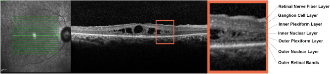
The aim of the study was to characterize macular edema (ME) in retinitis pigmentosa (RP) by means of quantitative optical coherence tomography (OCT)-based imaging. The study was designed as observational, prospective case series, with 1-year follow-up. All RP patients underwent complete ophthalmologic assessment, including structural OCT, OCT angiography, and microperimetry (MP). The primary outcome was the characterization through quantitative OCT-based imaging of RP eyes complicated by ME. A total of 68 RP patients' eyes (68 patients) and 68 eyes of 68 healthy controls were recruited. Mean BCVA was 0.14 ± 0.17 LogMAR at baseline and 0.18 ± 0.23 LogMAR at 1-year follow-up (p > 0.05). Thirty-four eyes (17 patients; 25%) showed ME, with a mean ME duration of 8 ± 2 months. Most of the eyes were characterized by recurrent ME. The ME was mainly localized in the inner nuclear layer in all eyes. LogMAR BCVA was similar in all RP eyes, whether with or without ME, although those with ME were associated with higher vessel density values, as well as thicker choroidal layers, than those without ME. In conclusion, the inner retina is closely involved in the pathogenesis of ME. The impairment of retinal-choroidal exchanges and Müller cell disruption might be a major pathogenic factor leading to the onset of ME in RP.
© 2023. The Author(s).
Conflict of interest statement
Francesco Bandello: Francesco Bandello consultant for Alcon (Fort Worth, Texas, USA), Alimera Sciences (Alpharetta, Georgia, USA), Allergan Inc (Irvine, California, USA), Farmila-Thea (Clermont-Ferrand, France), Bausch And Lomb (Rochester, New York, USA), Genentech (San Francisco, California, USA), Hoffmann-La-Roche (Basel, Switzerland), Novagali Pharma (Évry, France), Novartis (Basel, Switzerland), Bayer Shering-Pharma (Berlin, Germany), Sanofi-Aventis (Paris, France), Thrombogenics (Heverlee, Belgium), Zeiss (Dublin, USA), Pfizer (New York, USA), Santen (Osaka, Japan), Sifi (Aci Sant’Antonio, Italy). All the other authors have no financial disclosures. The authors declare no competing interests.
Figures

Similar articles
-
Morphological and functional involvement of the inner retina in retinitis pigmentosa.Eye (Lond). 2023 May;37(7):1424-1431. doi: 10.1038/s41433-022-02139-7. Epub 2022 Jun 29. Eye (Lond). 2023. PMID: 35768721 Free PMC article.
-
The impact of macular edema on microvascular and metabolic alterations in retinitis pigmentosa.Graefes Arch Clin Exp Ophthalmol. 2021 Mar;259(3):643-652. doi: 10.1007/s00417-020-04913-3. Epub 2020 Sep 10. Graefes Arch Clin Exp Ophthalmol. 2021. PMID: 32910309
-
Higher retinal vessel oxygen saturation: investigating its relationship with macular oedema in retinitis pigmentosa patients.Eye (Lond). 2018 Jul;32(7):1209-1219. doi: 10.1038/s41433-018-0043-1. Epub 2018 Mar 6. Eye (Lond). 2018. PMID: 29507331 Free PMC article.
-
Optical coherence tomography in the evaluation of retinitis pigmentosa.Ophthalmic Genet. 2020 Oct;41(5):413-419. doi: 10.1080/13816810.2020.1780619. Epub 2020 Jun 19. Ophthalmic Genet. 2020. PMID: 32552399 Free PMC article. Review.
-
Optical Coherence Tomographic Analysis of Retina in Retinitis Pigmentosa Patients.Ophthalmic Res. 2016;56(3):111-22. doi: 10.1159/000445063. Epub 2016 Jun 29. Ophthalmic Res. 2016. PMID: 27352292 Review.
Cited by
-
Evaluation of photoreceptor features in retinitis pigmentosa with cystoid macular edema by using an adaptive optics fundus camera.PLoS One. 2024 Jan 2;19(1):e0296493. doi: 10.1371/journal.pone.0296493. eCollection 2024. PLoS One. 2024. PMID: 38166083 Free PMC article.
-
Comparison of intravitreal anti-VEGF agents and oral carbonic anhydrase inhibitors in the treatment of cystoid macular edema secondary to retinitis pigmentosa.Front Pharmacol. 2024 Dec 10;15:1477889. doi: 10.3389/fphar.2024.1477889. eCollection 2024. Front Pharmacol. 2024. PMID: 39737070 Free PMC article.
-
Clinical Applications of Optical Coherence Tomography Angiography in Inherited Retinal Diseases: An Up-to-Date Review of the Literature.J Clin Med. 2023 Apr 28;12(9):3170. doi: 10.3390/jcm12093170. J Clin Med. 2023. PMID: 37176614 Free PMC article. Review.
-
Cystoid Macular Edema in Non-Syndromic Retinitis Pigmentosa: Associations With Causative Genes in a Large Cohort.Invest Ophthalmol Vis Sci. 2025 Sep 2;66(12):5. doi: 10.1167/iovs.66.12.5. Invest Ophthalmol Vis Sci. 2025. PMID: 40900079 Free PMC article.
-
Self-reported visual function and in-depth swept-source optical coherence tomography features of cystoid macular edema in retinitis pigmentosa.Int J Retina Vitreous. 2024 Nov 22;10(1):89. doi: 10.1186/s40942-024-00608-7. Int J Retina Vitreous. 2024. PMID: 39578919 Free PMC article.
References
Publication types
MeSH terms
LinkOut - more resources
Full Text Sources
