Clinical Trial
. 2023 Jan;29(1):190-202.
doi: 10.1038/s41591-022-02114-5. Epub 2023 Jan 16.

[11C]metomidate PET-CT versus adrenal vein sampling for diagnosing surgically curable primary aldosteronism: a prospective, within-patient trial

Affiliations
Clinical Trial

[11C]metomidate PET-CT versus adrenal vein sampling for diagnosing surgically curable primary aldosteronism: a prospective, within-patient trial

Xilin Wu et al. Nat Med. 2023 Jan.

Abstract

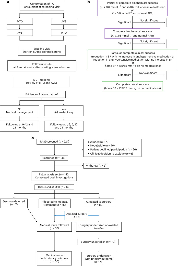
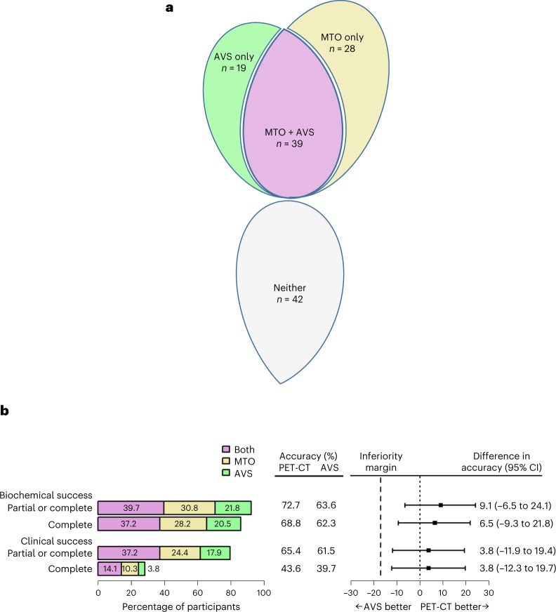
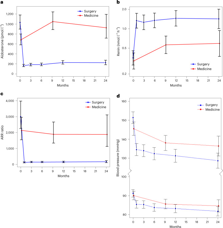
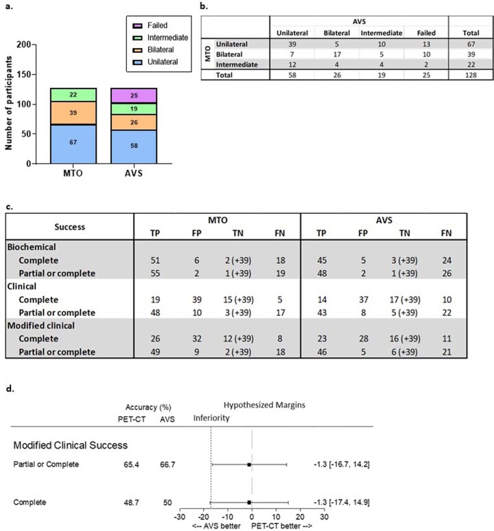
Primary aldosteronism (PA) due to a unilateral aldosterone-producing adenoma is a common cause of hypertension. This can be cured, or greatly improved, by adrenal surgery. However, the invasive nature of the standard pre-surgical investigation contributes to fewer than 1% of patients with PA being offered the chance of a cure. The primary objective of our prospective study of 143 patients with PA ( NCT02945904 ) was to compare the accuracy of a non-invasive test, [11C]metomidate positron emission tomography computed tomography (MTO) scanning, with adrenal vein sampling (AVS) in predicting the biochemical remission of PA and the resolution of hypertension after surgery. A total of 128 patients reached 6- to 9-month follow-up, with 78 (61%) treated surgically and 50 (39%) managed medically. Of the 78 patients receiving surgery, 77 achieved one or more PA surgical outcome criterion for success. The accuracies of MTO at predicting biochemical and clinical success following adrenalectomy were, respectively, 72.7 and 65.4%. For AVS, the accuracies were 63.6 and 61.5%. MTO was not significantly superior, but the differences of 9.1% (95% confidence interval = -6.5 to 24.1%) and 3.8% (95% confidence interval = -11.9 to 9.4) lay within the pre-specified -17% margin for non-inferiority (P = 0.00055 and P = 0.0077, respectively). Of 24 serious adverse events, none was considered related to either investigation and 22 were fully resolved. MTO enables non-invasive diagnosis of unilateral PA.


Conflict of interest statement

The authors declare no competing interests.

Figures

Fig. 1
Fig. 1. Study outline, hierarchical co-primary endpoints and CONSORT diagram.
a, Study outline. Patients with confirmed PA according to the Endocrine Society consensus guidelines underwent both AVS and MTO in random order. All patients were subsequently reviewed at a baseline visit and started on 50 mg spironolactone, which was up-titrated to 100 mg after 2 weeks. The blood pressure response to spironolactone was recorded at 2 and 4 weeks from the initiation of spironolactone. Eplerenone was used in those previously intolerant of spironolactone. Concurrently, the results of both investigations were reviewed at multidisciplinary team (MDT) meetings where MTO was always reviewed first, followed by AVS. A score of high, intermediate or low probability of unilateral PA was assigned to each investigation. Recommendations for unilateral adrenalectomy were made if either investigation indicated a high probability of unilateral PA or, in a small number of patients, where both investigations indicated intermediate probability and there was a clinical indication for surgery (for example, because of drug intolerance or uncontrolled blood pressure). Note that the blood pressure response to spironolactone was not used by the MDT in the decision-making process for recommending adrenalectomy. b, Hierarchical co-primary endpoints. The table shows definitions of partial and complete biochemical or clinical success, as defined by the PASO consensus (Supplementary Table 1), and the hierarchical order in which each definition of cure post-adrenalectomy was assessed. On an individual basis, a successful outcome was not dependent on success at the previous limb of the hierarchy. BP, blood pressure; K+, potassium. c, CONSORT diagram showing the disposition of study patients. Adrenalectomy was undertaken in 79 patients, of whom 78 had evaluable primary outcome data (assessed at 6 months post-surgery or after 9–12 months of medical therapy with spironolactone). Note that both biochemical and clinical primary outcome data were available in 77/78 patients. Only clinical primary outcome data were available for the remaining participant.
Fig. 2
Fig. 2. Primary outcomes.
a, Allocations to adrenalectomy and concordance of MTO and AVS. The number of patients scored as having a high probability of unilateral PA is shown for each independently scored investigation. The Venn diagram shows whether a high-probability score was achieved by one or both tests and includes the concordance between the investigations. 'Neither' indicates the number of patients who did not score as having a high probability of unilateral PA by either investigation and who were therefore recommended for medical therapy (total n = 128). b, Comparison of the accuracy of MTO and AVS in predicting a successful outcome from adrenalectomy. The four hierarchically analyzed measures of success (as outlined by the PASO consensus) are shown. The horizontal bars in the left panel are color coded as in a, reporting the proportion of high-probability scores for MTO or AVS, together or alone, that correctly predicted success. The middle panel shows the accuracy of each investigation, expressed as a percentage. The accuracy for each investigation was calculated as the number of patients who were scored as having a high probability of unilateral PA by that investigation (purple and gold bars for MTO and purple and green bars for AVS) and who achieved success, minus the number of patients in whom the investigation failed to predict a cure. The percentages exclude from the denominator the three patients who underwent adrenalectomy based on two medium-probability scores, on the basis of clinical grounds. The right panel shows the difference between the accuracies of MTO and AVS and the 95% CI around the difference, plotted for each outcome. All four sets of CI intervals cross zero, indicating that neither investigation was superior to the other. None of the lower bounds of the CI intervals cross the pre-specified margin of −17%, indicating that MTO is not inferior to AVS. The center of each error bar is the estimated difference in accuracy (%). P values for non-inferiority, from top to bottom, were: P = 0.00055, P = 0.0024, P = 0.0077 and P = 0.0091. The BinomDiffCI() function from R was used to calculate the 95% CI and to estimate the P values using a two-sided test (n = 78 for clinical success and n = 77 for biochemical success as post-operative biochemical data were not available for one patient; complete clinical success was achieved in this patient).
Fig. 3
Fig. 3. Secondary (pre- and post-intervention) outcomes.
a, Change in plasma aldosterone levels. For the surgical group, n = 80 at 0 months, n = 66 at 6 months, n = 57 at 12 months and n = 28 at 24 months. For the medical group, n = 59 at 0 months, n = 50 at 9 months and n = 18 at 24 months. b, Change in plasma renin activity. For the surgical group, n = 74 at 0 months, n = 69 at 6 months, n = 57 at 12 months and n = 29 at 24 months. For the medical group, n = 60 at 0 months, n = 48 at 9 months and n = 17 at 24 months. c, Change in the ARR. For the surgical group, n = 74 at 0 months, n = 66 at 6 months, n = 55 at 12 months and n = 28 at 24 months. For the medical group, n = 58 at 0 months, n = 48 at 9 months and n = 16 at 24 months. Reduction in aldosterone, de-suppression of renin and consequent reduction in ARR were seen post-surgery. ARR remained unchanged with medical therapy. d, Reduction in systolic and diastolic home blood pressure post-treatment. All effects seen were sustained for at least 2 years. For the surgical group, n = 76 at 0 months, n = 67 at 6 months, n = 56 at 12 months and n = 33 at 24 months. For the medical group, n = 59 at 0 months, n = 41 at 9 months and n = 19 at 24 months. The data represent least-squares means from mixed-effects models, adjusted for baseline covariates. Error bars show 95% CIs. 0 months indicates the baseline visit. Reference ranges were 150–550 pmol l−1 for aldosterone, 0.5–3.0 nmol l−1 h−1 for renin activity and <1,000 pmol l−1 (nmol l−1 h−1) for ARR (activity).
Fig. 4
Fig. 4. Predictors of clinical outcome from surgery.
ad, Influence of age (a), sex (b), baseline SBP (c) and ethnicity (d) on clinical outcomes. As seen from a and b, age and female sex are associated with a higher likelihood of complete clinical success. For age treated as categorical groups, statistical significance was determined by Fisher’s exact test (P = 0.065). For age treated as continuous, in univariate proportional odds logistical regression, the odds ratio was 0.43 (95% CI = 0.27 to 0.67) for an improvement of one level of outcome per 10 years of age (P = 0.00022). For sex, statistical significance was determined by Fisher’s exact test (P = 0.000036). For female versus male in a univariate proportional odds model, the odds ratio of going up one level of outcome was 9.078 (95% CI = 3.07 to 26.81) (P = 0.000065). n = 78. In c, the baseline SBP was lower in patients achieving complete or absent clinical success than in those achieving partial success. Each dot represents a single patient and is color coded to indicate the individual’s ethnicity. The center line of the box represents the median, the upper and lower boundaries of the box represent the interquartile range and the upper and lower whiskers represent the maximum and minimum values in the range, respectively. n = 78. For d, the absence of clinical success (shown in blue) was significantly more frequent in patients who were Black compared with other ethnicities. In a univariate proportional odds model, the odds ratio of going up one level was 0.14 (95% CI = 0.042 to 0.49). Statistical significance was determined by two-sided t-test (P = 0.002). e, Correlation between the decrease in SBP after 4 weeks of spironolactone therapy and the decrease in SBP at 6 months post-surgery (r = 0.53 (95% CI = 0.32 to 0.69) and P = 0.000012). The P value comes from a correlation test whose test statistic follows a t distribution. SBP is shown as an adjusted percentage change, which was calculated with Oldham’s correction (corrected change in blood pressure (%) = 100× the actual change in blood pressure divided by the average of the baseline and post-treatment pressure). n = 67. f, SBP after 4 weeks of spironolactone predicts clinical success. All patients whose SBP dropped to ≤135 mmHg after 4 weeks of spironolactone achieved either partial or complete clinical success. SBP ≤ 135 mmHg after 1 month of spironolactone was the best univariate predictor of complete clinical success with an odds ratio of 13.0 (3.72 to 45.24). Statistical significance was determined by two-sided t-test (P = 0.000057). n = 67.
Extended Data Fig. 1
Extended Data Fig. 1. Results from MTO and AVS.
a. Number of patients in whom MTO or AVS identified unilateral or bilateral primary aldosteronism (PA). ‘Intermediate’ suggests some asymmetry in the adrenals but not meeting criteria for unilateral disease. ‘Failed’ indicates unsuccessful cannulation of adrenal vein(s) and therefore inability to interpret AVS result. n = 128, all patients who reached the primary endpoint time-point (6 or 9-12 months post surgery or medical therapy respectively). b. Cross tabulation table showing results from MTO and AVS, indicating relative contribution of each investigation. n = 128. c. Accuracy of each investigation in predicting biochemical and clinical outcomes post surgery. The table assumes the 39 patients found to have bilateral disease on both investigations and did not go for surgery, indicated by brackets, were true negatives. This assumption (both investigations were accurate) allows calculation of the false positive rates but does not differentiate between the relative performance of each investigation, which is determined by cases where the investigation outcomes disagreed. *n = 116 for biochemical success, n = 117 for clinical success, based on a total of 78 patients who went underwent adrenalectomy and 39 who were thought to have bilateral disease based on both investigations. TP, true positive; FP, false positive; TN, true negative; FN, false negative. d. Modified clinical success outcomes. Forest plot of modified clinical success outcomes, determined by change in number of classes of antihypertensive medications (for definitions please refer to Supplementary Table 3). Column 2 and 3 show percentage of success accurately predicted by MTO and AVS respectively. Right hand column show percentage difference. The centre for the error bars is the Estimated Difference in Accuracy in (%). It, along with the 95% CI (shown in square brackets), is given in the figure’s last 3 columns. Non-inferiority p values: partial or complete modified clinical success p = 0.045; complete modified clinical success p = 0.057. The BinomDiffCI() function from R was used to calculate the 95% CI and to estimate the p-values using a 2-sided test. n = 78.
Extended Data Fig. 2
Extended Data Fig. 2. Conversion between renin activity and mass.
Different assays were used for the measurement of renin at the three sites. CUH and GSTT measured renin mass (mU/L), while SBH measured renin activity (nmol/L/hr). A renin mass level (pg/ml) was also sent off at selected (baseline and 6-9 month) visits at the SBH site to allow within-patient comparisons between renin mass and activity. A conversion of 1.67 was used to convert between renin mass measured in pg/ml to mU/L. The scatter plot above shows correlation between renin activity and mass within the SBH cohort. Pearson’s correlation r = 0.7851 (95% CI 0.7107 – 0.8422), p < 0.0001 (two-tailed test). For the study a conversion factor of 10.989 was used for conversion from renin activity (nmol/L/hr) to mass (mU/L), taken from the ARR thresholds for diagnosing PA: ARRactivity 1000, ARRmass 91 (1000/91 = 10.989). n = 136. CUH, Cambridge University Hospital; SBH, St Bartholomew’s Hospital; GSTT, Guy’s and St Thomas’ Hospital; ARR, aldosterone renin ratio; PA primary aldosteronism.
Extended Data Fig. 3
Extended Data Fig. 3. Pre- and post-intervention outcomes.
Change in aldosterone, renin and ARR observed at baseline (time 0) and after surgery and medical therapy, divided into cohorts where a. Renin activity (SBH) and b. Renin mass was measured (GSTT and CUH). A similar trend is observed in both cohorts. Data are least squares means from mixed effects models, adjusted for baseline covariates. Error bars show 95% CI. 0 months indicate baseline visit. For aldosterone levels with renin mass: Surgical group: n = 64 at 0 months, n = 51 at 6 months, n = 27 at 12 months, n = 15 at 24 months. Medical group: n = 50 at 0 months, n = 32 at 9 months, n = 9 at 24 months. For renin as renin mass: Surgical group: n = 64 at 0 months, n = 53 at 6 months, n = 28 at 12 months, n = 16 at 24 months. Medical group: n = 51 at 0 months, n = 32 at 9 months, n = 10 at 24 months. For ARR calculated using renin mass: Surgical group: n = 64 at 0 months, n = 51 at 6 months, n = 27 at 12 months, n = 15 at 24 months. Medical group: n = 50 at 0 months, n = 32 at 9 months, n = 9 at 24 months. For aldosterone levels with renin activity: Surgical group: n = 41 at 0 months, n = 40 at 6 months, n = 35 at 12 months, n = 18 at 24 months. Medical group: n = 40 at 0 months, n = 40 at 9 months, n = 16 at 24 months. For renin as renin activity: Surgical group: n = 41 at 0 months, n = 41 at 6 months, n = 36 at 12 months, n = 18 at 24 months. Medical group: n = 42 at 0 months, n = 40 at 9 months, n = 16 at 24 months. For ARR calculated using renin activity: Surgical group: n = 41 at 0 months, n = 40 at 6 months, n = 35 at 12 months, n = 18 at 24 months. Medical group: n = 40 at 0 months, n = 40 at 9 months, n = 16 at 24 months. SBH, St Bartholomew’s Hospital; GSTT, Guy’s and St Thomas’ Hospital; CUH, Cambridge University Hospital.
Extended Data Fig. 4
Extended Data Fig. 4. Cardiovascular Outcomes.
a. Fold change in plasma BNP levels between baseline and 6 months post surgery or after 9-12 months of medical therapy. Surgery median = -0.69 (IQR -1.14,-0.06), Medical median = -0.10 (IQR -0.53,0.40), p = 0.00013. n = 68 surgical group, n = 40 medical group. The centre line of the box represents the median, the upper and lower boundaries of the box represent the inter-quartile range and the upper and lower whiskers represent the maximum and minimum values in the range respectively. b. Correlation between fold change in plasma aldosterone levels and plasma BNP levels, pre and post-treatment. c. Correlation between fold change in systolic BP and plasma BNP levels pre and post treatment. For b. and c., the p-value for the vertical location comes from the t value for the treatment variable in a linear model where BNP fold change is the dependent variable and treatment and b. aldosterone fold change or c. SBP are explanatory variables. A two-sided T-test was used. The p-value for the difference in slope comes from the t value for the treatment: b. aldosterone fold change or c. SBP change interaction variable in a linear model where BNP fold change is the dependent variable and treatment, b. aldosterone fold change and treatment: aldosterone fold change interaction or c. SBP change and treatment: SBP change interaction are explanatory variables. A two sided t-test was used. d. Left ventricular volumes, ejection fraction and mass were measured using CMR at baseline and at 6 months post surgery, or after 9-12 months of medical therapy. Y axis shows volume in mililiters for LV EDV, ESV and SV; % for LV EF and grams for LVM. Pre and post-intervention measurements are shown for each treatment group. 15.1% reduction in LV EDV and 15.8% reduction in LV SV is seen post surgery, while 5.6% reduction in LV EDV and 5.0% reduction in LV SV is seen after medical therapy. Two-sided Mann-Whitney-Wilcoxon tests: **p = 0.00186 ***p = 0.00037. n = 25. The bars represent the mean and error bars represent + /- 1 standard error from that mean. IQR, interquartile range; BNP, N-terminal pro-B-type natriuretic peptide. LV, left ventricular; EDV, end diastolic volume; ESV, end systolic volume; SV, stroke volume; EF, ejection fraction; LVM, left ventricular mass.
Extended Data Fig. 5
Extended Data Fig. 5. Sub-types of APA, phenotype-genotype correlations and immunohistochemistry (IHC).
a. Frequency of APAs harbouring each of the known somatic gene mutations. [i] Each genotype was sub-divided to show variation in ethnicity, demonstrating significant differences between genotypes. n = 60. Fisher’s Exact test p = 0.0312. Genotyping by sex is shown in [ii] male, n = 39 and [iii] female, n = 21 patients. A correlation between ethnicity and genotype was seen in female (p = 0.0014) but not male patients (p = 0.3261). b. CYP11B1 and CYP11B2 mRNA expression by APA genotype. mRNA expression of APAs is the normalised read count on RNA sequencing. The bar represents mean and error bars show +/− 1 standard deviation from mean. For CYP11B2 expression, KCNJ5 v other genotypes, Kruskal-Wallis test, p = 2.95×10⁻⁷; For CYP11B1 expression, KCNJ5 v other genotypes, Kruskal-Wallis test, p = 2.0×10⁻⁵. Total with genotype and gene expression, n = 51. Numbers of each genotype are smaller than in Extended Data Fig. 5a, because not all adrenals had an RNAlater sample collected at the time of surgery for RNA sequencing. c. Intensity of CYP11B1 and CYP11B2 expression of APAs on IHC staining was quantified using the H-score system. The average H-score for each genotype showed differences in CYP11B1 expression between KCNJ5-mutant and other APAs. ANOVA p = 0.0002; KCNJ5 v CACNA1D p = 0.0009, KCNJ5 v ATP1A1 p = 0.0128, KCNJ5 v ATP2B3 p = 0.0022. No differences in CYP11B2 expression on IHC was detected between genotypes. ANOVA p = 0.937. Paired comparisons were made using Tukey’s honestly significant difference test. Note small sample size within each group in above analysis (n = 3-9), although these did pass the Shapiro-Wilk test for normality. H-scoring only performed on APAs of known gene mutations from Cambridge University Hospital, total. n = 30. d. Two illustrative cases demonstrating selective [11C]-metomidate uptake in patients with two or more adrenal adenomas. [i] Patient 1 has two adenomas on their adrenal, shown in separate axial MTO images. The first nodule is [11C]-metomidate avid while the second is ‘cold’. IHC staining of the two nodules show strong positive staining for CYP11B2 in nodule 1 while nodule 2 is negative for CYP11B2. IHC staining for CYP11B1 is positive in both nodules. [ii] Patient 2 had multiple adrenal nodules, only one of which (nodule 1) demonstrated high [11C]-metomidate uptake on PET-CT (left hand panels). Top middle and top right hand panels show high power views of patient 2’s adrenal, with CYP11B2 and CYP11B1 IHC staining respectively. Bottom middle panel: RNA sequencing results showing increased mRNA expression for CYP11B1 (compared to AAG) in both nodules, and increase mRNA expression for CYP11B2 in nodule 1 only. Scale bar represents 5 mm. APA, aldosterone producing adenoma; AAG, adjacent adrenal gland; IHC, immunohistochemistry; NFA, non-functioning adenoma.
Extended Data Fig. 6
Extended Data Fig. 6. Gene expression profiles in aldosterone producing adenomas (APAs) and non-functioning adenomas (NFAs).
Heat map representation of 200 most differentially expressed genes between adrenal adenomas and adjacent adrenal. Each column represents the expression profile of the APA or NFA. The unsupervised cluster analysis of samples separated the expression profile of APAs from NFAs, and between different genotypes. Red represents upregulation and blue represents downregulation of genes. Arrow shows the gene nephronectin, NPNT (All text is legible by increasing zoom/magnification of file).
Extended Data Fig. 7
Extended Data Fig. 7. Heat map representation of differentially expressed genes between APAs and NFAs, and between genotypes.
Each column represents the expression profile of the APA or NFA. Both genes and individual APAs are hierarchically clustered. Three nodes of interest are shown: a. node containing CYP11B2; b. node showing most upregulated genes in CACNA1D-mutant APAs. c. node showing most upregulated genes in KCNJ5-mutant APAs. The CYP11B2 nodes includes several genes previously noted to be upregulated in APAs, for example VSNL1, CALN1. Genes which are significantly upregulated in APMs are also seen in this node: TMEM266 (TMEM266 (C15ORF27), PPP1R16B, SEMA3D, KIAA1549L (C11ORF41), CFAP221 (PCDP1). Red represents upregulation and blue represents downregulation of genes. APA, aldosterone producing adenoma; AAG, adjacent adrenal gland; IHC, immunohistochemistry; NFA, non-functioning adenoma; APM, aldosterone producing micronodule (previously known and aldosterone producing cell clusters, APCCs).
Extended Data Fig. 8
Extended Data Fig. 8. Predictions of clinical success.
a. Genotypes and clinical success associations. Number of patients achieving each definition of clinical success by genotype. n = 58. Fisher Exact test p = 0.00021. KCNJ5 v Other: Univariate Proportional Odds Model, Odds Ratio of going up 1 level 10.37 (95% CI (2.50, 42.99)) p = 0.00123. CACNA1D v Other: Univariate Proportional Odds Model, Odds Ratio of going up 1 level 0.38 (95% CI (0.11, 1.36)) p = 0.13703. b. Predictors of complete clinical success. Results from logistic regression analysis of variables predicting complete clinical success. Note the two patients with double GNAQ/CTNNB1 mutations were excluded from this analysis since 16/16 patients with this double mutation were previously reported to be clinically cured. p-values based on the t values from the proportional odds logistic regression c.w., compared with. c. Urine steroid profile and genotype associations. 18-hydroxycortisol/cortisol ratio collected at baseline are shown by genotype. A difference in 18-hydroxycortisol/cortisol ratio was seen between APA genotypes: Kruskal Wallis test p < 0.0001; KCNJ5 v ATP2B3 p = 0.071, KCNJ5 v ATPA1A p = 0.003, CACNA1D p < 0.0001. Paired comparisons were made using Bonferroni p-value correction. The center line of the box represents the median, the upper and lower boundaries of the box represent the inter-quartile range, and the upper and lower whiskers represent the maximum and minimum values in the range respectively. Due to small sample size GNAQ and CLCN2 were excluded from the above analysis. n = 37.

Comment in

References

    1. Rossi, G. P. et al. A prospective study of the prevalence of primary aldosteronism in 1,125 hypertensive patients. J. Am. Coll. Cardiol.48, 2293–2300 (2006). - PubMed
    1. Monticone, S. et al. Prevalence and clinical manifestations of primary aldosteronism encountered in primary care practice. J. Am. Coll. Cardiol.69, 1811–1820 (2017). - PubMed
    1. Käyser, S. C. et al. Study heterogeneity and estimation of prevalence of primary aldosteronism: a systematic review and meta-regression analysis. J. Clin. Endocrinol. Metab.101, 2826–2835 (2016). - PubMed
    1. Libianto, R. et al. Detecting primary aldosteronism in Australian primary care: a prospective study. Med. J. Aust.216, 408–412 (2022). - PubMed
    1. Monticone, S. et al. Cardiovascular events and target organ damage in primary aldosteronism compared with essential hypertension: a systematic review and meta-analysis. Lancet Diabetes Endocrinol.6, 41–50 (2018). - PubMed

Publication types

MeSH terms

Associated data