. 2023 May;7(5):754-764.
doi: 10.1038/s41562-022-01502-8. Epub 2023 Jan 16.

Neuronal activity in the human amygdala and hippocampus enhances emotional memory encoding

Affiliations

Neuronal activity in the human amygdala and hippocampus enhances emotional memory encoding

Salman E Qasim et al. Nat Hum Behav. 2023 May.

Abstract

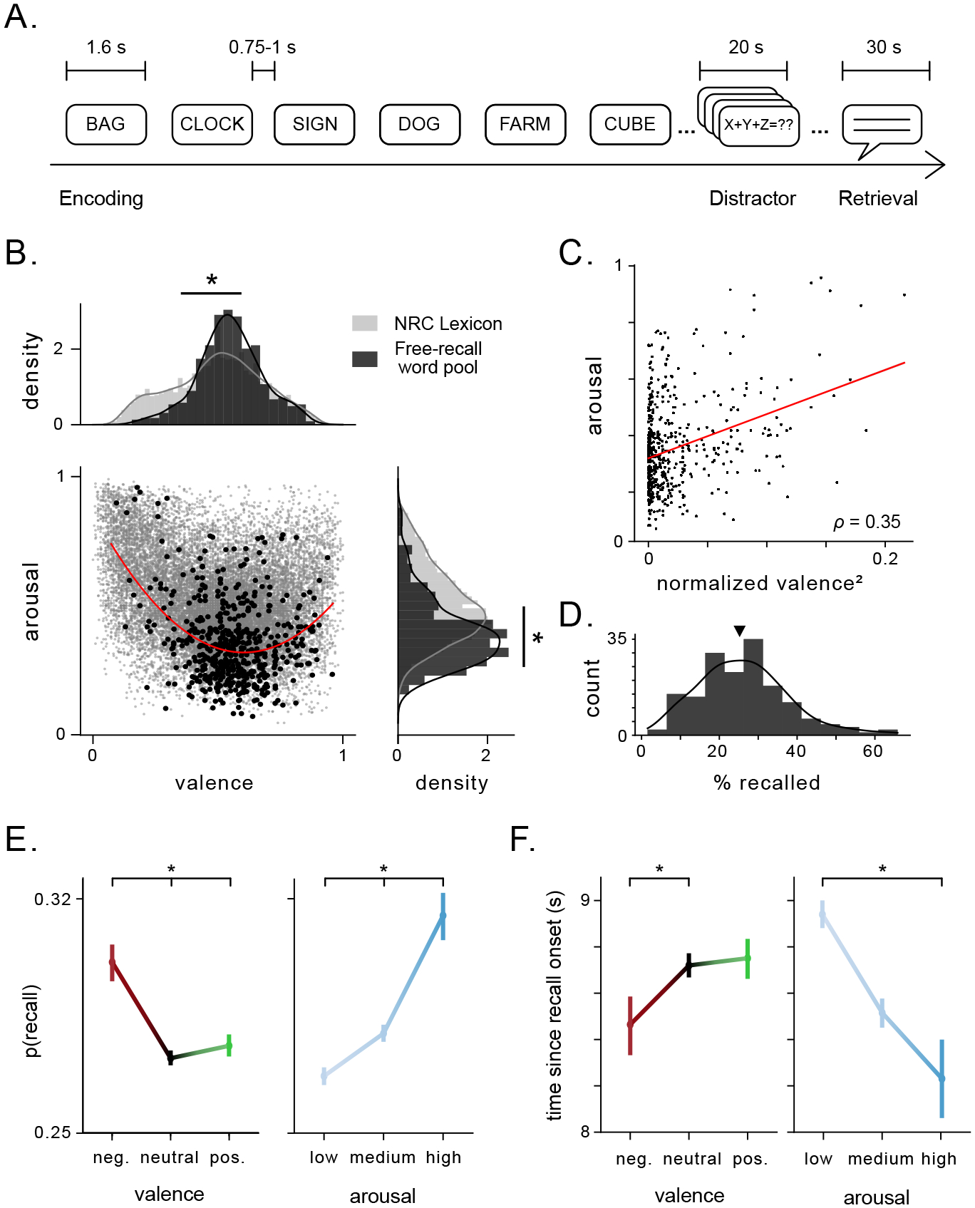
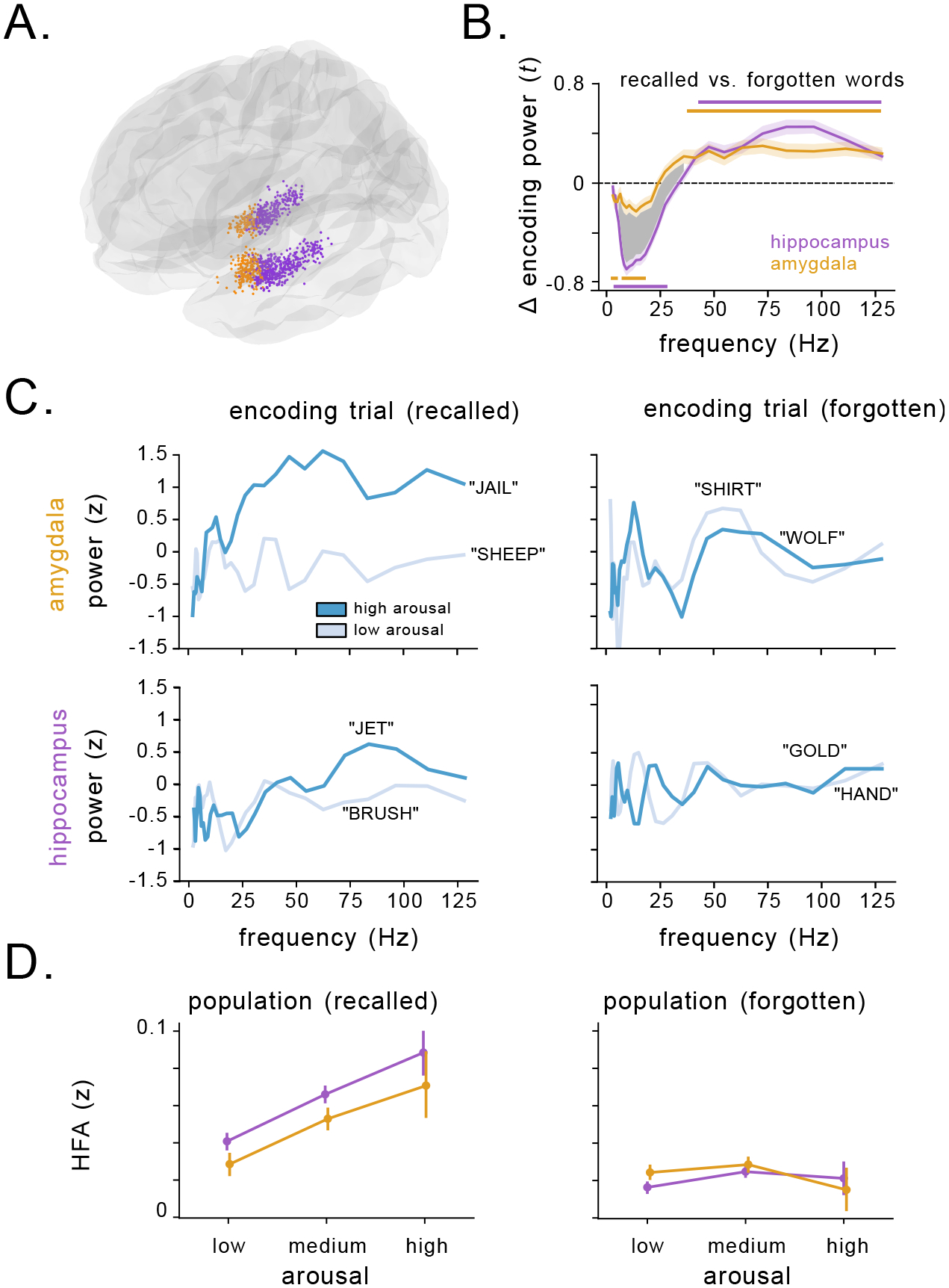
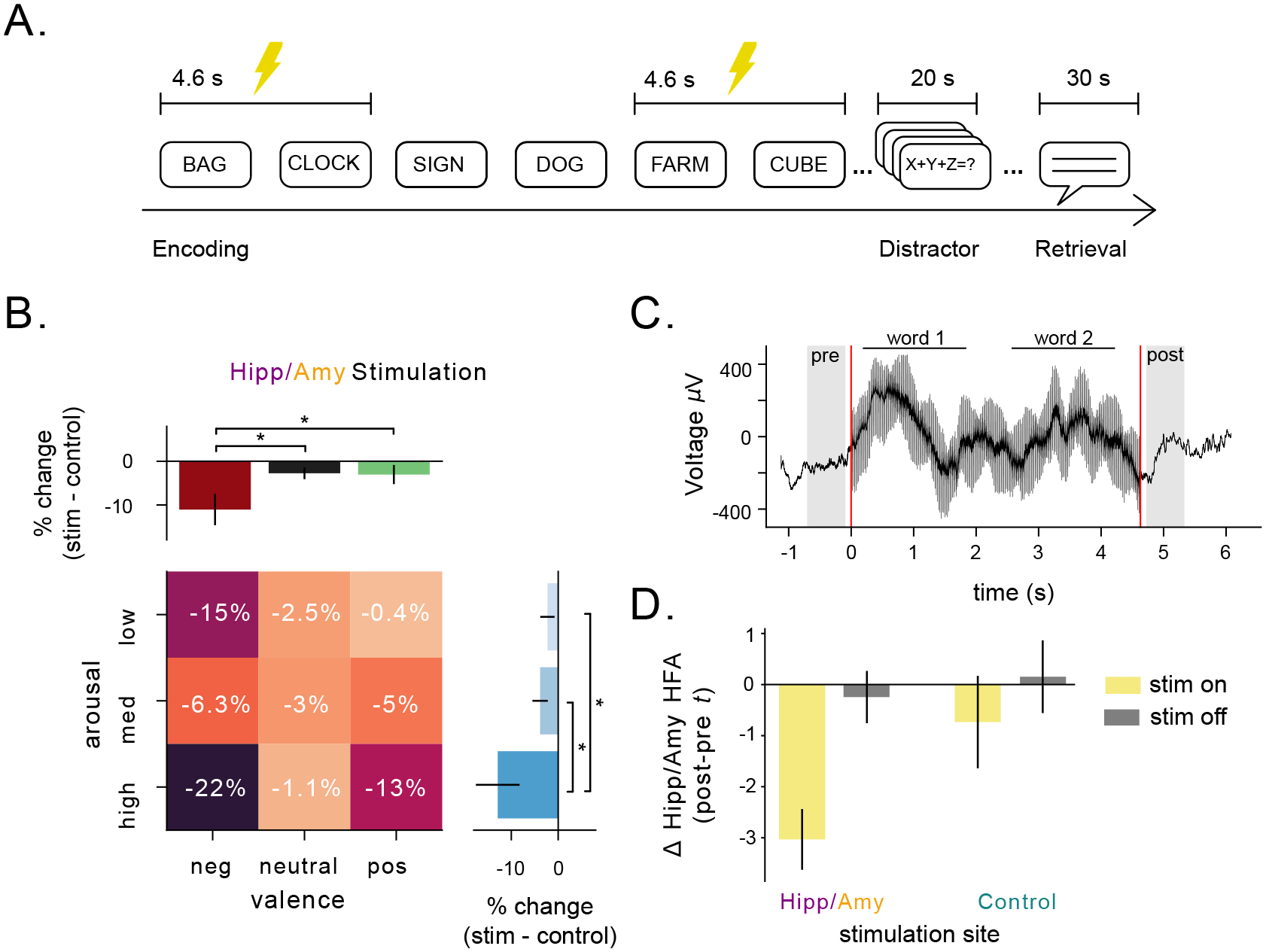
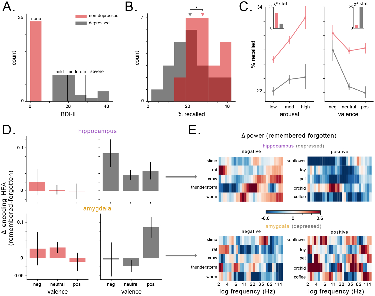
Emotional events comprise our strongest and most valuable memories. Here we examined how the brain prioritizes emotional information for storage using direct brain recording and deep brain stimulation. First, 148 participants undergoing intracranial electroencephalographic (iEEG) recording performed an episodic memory task. Participants were most successful at remembering emotionally arousing stimuli. High-frequency activity (HFA), a correlate of neuronal spiking activity, increased in both the hippocampus and the amygdala when participants successfully encoded emotional stimuli. Next, in a subset of participants (N = 19), we show that applying high-frequency electrical stimulation to the hippocampus selectively diminished memory for emotional stimuli and specifically decreased HFA. Finally, we show that individuals with depression (N = 19) also exhibit diminished emotion-mediated memory and HFA. By demonstrating how direct stimulation and symptoms of depression unlink HFA, emotion and memory, we show the causal and translational potential of neural activity in the amygdalohippocampal circuit for prioritizing emotionally arousing memories.


Conflict of interest statement

Declaration of Interests

The authors declare no competing interests.

Figures

Figure 1:
Figure 1:. Emotional features of stimuli in a verbal free recall task influence recall performance.
A) Schematic of task design showing the time intervals during and between task stages. Subject encoded 12 words per list. B) Joint scatter plot and marginal distributions of valence and arousal ratings in the general rating lexicon (gray) and the word pool for the free recall tasks performed by subjects (black). Asterisks indicate significant difference between the mean ratings for valence and arousal between the word pool and the general lexicon. Red line denotes polynomial fit to valence and arousal ratings of free-recall word pool. C) Arousal plotted as a function of mean-normalized, squared valence. Red line denotes linear fit. Pearson’s correlation coefficient is indicated. D) Distribution of recall performance across all subjects (mean = 26%, black arrow). E) Probability of recall significantly differed as a function of valence (p< 5 × 10⁻⁸, χ2(2) = 33.8) and arousal (p< 2 × 10⁻¹⁵, χ2(2) = 68.6). Vertical bars denote standard error. Asterisks denote significant difference in proportions across categories. F) Time since recall onset tended to be shorter for negative vs. neutral words (p= 0.05, t = −1.94), and was significantly lower for high vs. low arousal words (p< 4 × 10⁻⁵, t = −4.16). Asterisks denote significant difference in recall times across categories.
Figure 2:
Figure 2:. Arousal modulates high-frequency activity related to successful memory encoding in the hippocampus and amygdala.
A) Location of all 1,376 electrodes recorded across all subjects. Purple circles indicate electrodes localized to hippocampus, orange circles indicate electrodes localized to the amygdala. B) Comparison of SME (t-statistic) averaged over hippocampal (purple) and amygdala (orange) electrodes. Horizontal lines denote SMEs that significantly deviate from 0 for electrodes in the hippocampus (p’s<= 0.001, cluster-permutation test), and the amygdala (p’s<= 0.02, cluster-permutation test). The shaded region denotes a significant difference between the magnitude of the SME for hippocampus vs. amygdala (p’s<= 0.001, cluster-permutation test). C) Within-list z-scored power during example encoding trials from a single amygdala (top) and hippocampal (bottom) electrode in two subjects during memory encoding. Power during encoding of a high arousal word from the list is depicted in dark blue, while power for a low arousal word from the list is depicted in light blue. HFA increases during successful encoding (left) of high arousal words vs. low arousal words, but not during failed encoding (right). D) Z-scored HFA in the amygdala (orange) and hippocampus (purple) during the encoding phase as a function of word arousal for recalled (left) and forgotten (right) words. Circles represent mean of binned arousal, with vertical lines denoting the standard error of the bin.
Figure 3:
Figure 3:. Direct stimulation of hippocampus and amygdala during encoding impairs emotion-mediated memory and decreases HFA.
A) Schematic of task design showing the time intervals during direct brain stimulation. Stimulation was applied to alternating pairs of words in a list. B) Effect of stimulation administered to electrodes located either in the hippocampus or the amygdala, split by arousal and valence. Asterisks indicate significant differences in the effect of stimulation on recall as a function of emotional features of words (p’s< 0.05, likelihood-ratio test). Heatmap numbers indicate percentage of change in recall performance during stimulation. C) Single-trial example local-field potential during stimulation of a word-pair. Shaded regions indicate pre and post-periods used for analysis. Word presentation is indicated by horizontal lines. Red lines indicate onset and offset of stimulation. D) The difference in HFA in the hippocampus and amygdala (z-scored) before and after stimulation, measured by a paired t-test. The yellow bars denote words with stimulation turned on, while gray bars denote words with stimulation turned off. Bars on the left side correspond to stimulation applied to the hippocampus or amygdala, while bars on the right side correspond to stimulation applied to control regions in the MTL.
Figure 4:
Figure 4:. Valence, rather than arousal, modulates memory performance and HFA in subjects with depression.
A) Histogram of BDI-II scores for patients, split by depressive characterization. B) Distribution of recall performance across subjects as a function of depression rating. Depressed patients exhibit significantly worse recall performance (t = −2.4, p = 0.02). C) Recall percentage expressed as a function of arousal (left) and valence (right) for both depressed and non-depressed subjects. Inset depicts the χ2-statistic assessing the affect of arousal and valence on recall performance in both groups of subjects. D) The difference in HFA (z-scored) during the encoding period for remembered and forgotten items, for subjects with and without depression in the hippocampus (top) and amygdala (bottom). E) Heatmaps of hippocampal (top) and amygdalar (bottom) power (z-scored within session) for specific words from the task wordpool, averaged across sessions and subjects. Words were selected from the 30 words with the lowest valence ratings (left) or highest valence ratings (right). Warm colors indicate higher values while cool colors indicate lower values.

References

    1. Bradley MM, Greenwald MK, Petry MC & Lang PJ Remembering pictures: pleasure and arousal in memory. Journal of Experimental Psychology: Learning, Memory, and Cognition 18, 379 (1992). - PubMed
    1. Kensinger EA & Corkin S Memory enhancement for emotional words: are emotional words more vividly remembered than neutral words? Mem Cognit 31, 1169–80 (2003). - PubMed
    1. Reisberg D & Hertel P Memory and emotion (Oxford University Press, Oxford, 2004). URL http://www.loc.gov/catdir/toc/ecip041/2003006595.html.
    1. Klein-Koerkamp Y, Baciu M & Hot P Preserved and impaired emotional memory in alzheimer’s disease. Front Psychol 3, 331 (2012). - PMC - PubMed
    1. Klüver H & Bucy PC Preliminary analysis of functions of the temporal lobes in monkeys. J Neuropsychiatry Clin Neurosci 9, 606–20 (1939). - PubMed

Publication types

LinkOut - more resources