. 2023 May;19(5):607-613.
doi: 10.1038/s41589-022-01229-7. Epub 2023 Jan 16.

A structural basis for prion strain diversity

Affiliations

A structural basis for prion strain diversity

Szymon W Manka et al. Nat Chem Biol. 2023 May.

Abstract

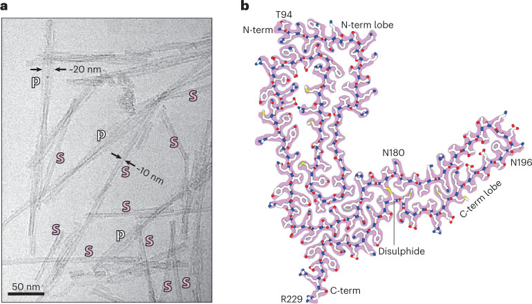
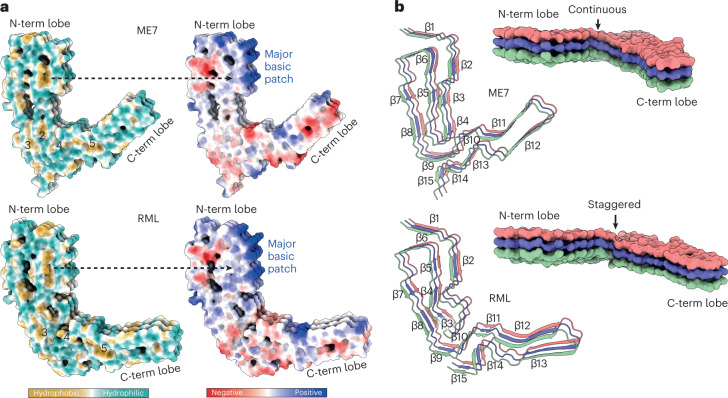
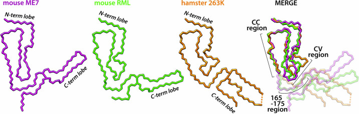
Recent cryogenic electron microscopy (cryo-EM) studies of infectious, ex vivo, prion fibrils from hamster 263K and mouse RML prion strains revealed a similar, parallel in-register intermolecular β-sheet (PIRIBS) amyloid architecture. Rungs of the fibrils are composed of individual prion protein (PrP) monomers that fold to create distinct N-terminal and C-terminal lobes. However, disparity in the hamster/mouse PrP sequence precludes understanding of how divergent prion strains emerge from an identical PrP substrate. In this study, we determined the near-atomic resolution cryo-EM structure of infectious, ex vivo mouse prion fibrils from the ME7 prion strain and compared this with the RML fibril structure. This structural comparison of two biologically distinct mouse-adapted prion strains suggests defined folding subdomains of PrP rungs and the way in which they are interrelated, providing a structural definition of intra-species prion strain-specific conformations.


Conflict of interest statement

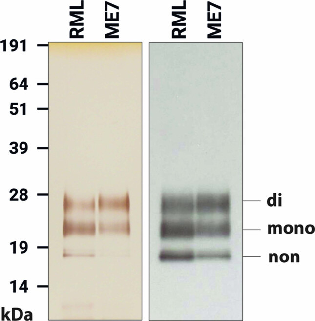
J.C. is a Director and J.C. and J.D.F.W. are shareholders of D-Gen Ltd., an academic spin-out company working in the field of prion disease diagnosis, decontamination and therapeutics. D-Gen supplied the ICSM35 and ICSM18 antibodies used for western blot and ELISA performed in this study. The other authors declare no potential conflicts of interest.

Figures

Fig. 1
Fig. 1. ME7 fibril morphologies and the atomic structure of their constituent PrP subunit.
a, Representative cryo-EM image from a dataset comprising 6,370 multi-frame movies (300-kV FEI Krios G3i, K3 camera) showing examples of single ME7 protofilaments (s) alongside their paired assemblies (p) with their approximate diameters. b, Protein-only density of a single amyloid rung (pink) with the fitted atomic model of the mouse PrP chain shown with sticks colored by heteroatom: C, white; N, blue; O, red; S, yellow. The start (T94) and end (R229) residues of the fitted polypeptide and both N-glycosylation sites are indicated.
Fig. 2
Fig. 2. Comparison of ME7 and RML protofibrils and assignment of folding subdomains in their core.
a, Rendered density views of the helix crossover or half-pitch (180° helical turn) distance, with annotated locations of PTA polyanions (semi-transparent white), N196 (yellow) and GPI anchor (blue). b, Density cross-sections with overlaid PrP backbone models colored by relative deviation based on global 3D alignment (UCSF Chimera; d), with annotations corresponding to a. C-α atoms of both N-glycosylation sites (N180 and N196) are marked with yellow circles. c, Diagrams of the PrP subunits with approximate dimensions of the inter-lobe grooves and the longest C-α distances in each model, measured between the indicated C-α atoms (dotted lines). Positions of amino acid side chains are shown with circles (positively charged, blue; negatively charged, red; neutral, green; hydrophobic, white; aromatic, gray) on either side of the backbone (black line). β-strands are indicated with thick black arrowheaded lines. d, Top, superposition of PrP backbones colored as in b and shaded according to the folding subdomain assignment. N-glycosylation sites are marked with yellow circles. PK-resistant core starts with residue 89. Bottom, mouse PrP sequence from the start of the amyloid core (T94) to the C-terminus (S230-GPI anchor), with color-coded folding subdomain assignment. Secondary structure annotation for PrPC (gray), ME7 fibril (magenta) and RML fibril (green) is included below the sequence (α-helix, zig-zag; β-sheet, arrow; disordered, undulated dashed line). Start and end residues are numbered, and the β2-α2 region of PrPC is indicated. N-glycosylation sites (N180 and N196) are marked with yellow circles.
Fig. 3
Fig. 3. Hydrophobic and polar domains of mouse prion protofibrils and details of their PIRIBS arrangements.
Models of three consecutive PrP rungs shown as: a, solvent-excluded surface colored by hydrophobicity and by electrostatic charge distribution (ChimeraX), and hydrophobic clusters are labeled 1–5; b, ribbons with secondary structures and solvent-excluded surface models colored by chain.
Fig. 4
Fig. 4. Lateral interactions stabilizing mouse prion fibrils.
Depiction of polar and non-polar lateral contacts. Protein backbone is shown with cartoon (licorice) representation, amino acid side chains as white sticks colored by heteroatom (O, red; N, blue; S, yellow) and selected hydrogen bonds as black lines. The hydrophobic patches are numbered 1–5.
Fig. 5
Fig. 5. Summary of structural differences between ME7 and RML prion protofibril structures.
a, Licorice backbone models (ChimeraX) of ME7 and RML protofibrils colored according to PrPSc folding subdomains (blue, CC region; red, CV region; yellow, DS hairpin; white, intervening regions). Positive charge of the major basic patch (transparent surface) is indicated with + signs, and the sialoglycan occupancy corresponds to the intensity of red shading. The N-glycosylation sites (N180 and N196 side chains) and the disulphide bond are shown as sticks and colored by heteroatom (C, yellow; N, blue; O, red; S, yellow). b, Alignment of PrP sequences from different mammals focused on the PrPSc folding subdomains. Human residues 127 and 129 in the CV region (highlighted with dark red) are polymorphic (G/V and M/V, respectively).
Extended Data Fig. 1
Extended Data Fig. 1. Silver-stained SDS-PAGE (left) and western blot (right) of purified ME7 and RML rods.
Labelling shows the migration positions of di-, mono- and non-glycosylated PrP. The samples were prepared as described in Methods. Uncropped and unprocessed SDS-PAGE and western blot data are provided in a Source Data file. Findings are representative of ~30 purifications. Source data
Extended Data Fig. 2
Extended Data Fig. 2. Comparisons between cryo-EM maps and atomic models of mouse prion fibrils.
a Density cross-sections (pixel size: RML, 1.067 Å; ME7, 0.828 Å) showing the protein core and the non-protein extra densities in the final cryo-EM map. b Top views of rendered reconstructions with indicated common folding sub-domains. c backbone alignments of the common folding sub-domains and the root mean square deviations (RMSD) between all of their respective atom pairs, as calculated using UCSF Chimera. CC, conformationally conserved; CV, conformationally variable; DS, disulphide-stapled.
Extended Data Fig. 3
Extended Data Fig. 3. Comparison of rodent prion structures.
Backbones of the three structures are aligned on the conformationally conserved (CC) region in the N-terminal lobe (MERGE, dimmed beyond the CC region). The C-terminal lobe of mouse prion strains has divergent orientations in line with differences in the N-terminal lobe’s conformationally variable (CV) region that interfaces with the C-terminal lobe. The C-terminal lobe of the hamster strain diverges further from the mouse strains due to additional differences in its primary structure (PrP sequence). The 165-176 region, which corresponds to the β2-α2 loop in PrPC, is part of the inter-lobe interface in all three rodent prion fibril structures.

References

    1. Prusiner SB. Prions. Proc. Natl Acad. Sci. USA. 1998;95:13363–13383. doi: 10.1073/pnas.95.23.13363. - DOI - PMC - PubMed
    1. Collinge J, Clarke A. A general model of prion strains and their pathogenicity. Science. 2007;318:930–936. doi: 10.1126/science.1138718. - DOI - PubMed
    1. Collinge J. Mammalian prions and their wider relevance in neurodegenerative diseases. Nature. 2016;539:217–226. doi: 10.1038/nature20415. - DOI - PubMed
    1. Collinge J. Variant Creutzfeldt–Jakob disease. Lancet. 1999;354:317–323. doi: 10.1016/S0140-6736(99)05128-4. - DOI - PubMed
    1. Wadsworth JD, Asante EA, Collinge J. Contribution of transgenic models to understanding human prion disease. Neuropathol. Appl. Neurobiol. 2010;36:576–597. doi: 10.1111/j.1365-2990.2010.01129.x. - DOI - PMC - PubMed

Publication types