

Placental DNA methylation profile as predicting marker for autism spectrum disorder (ASD)
- PMID: 36647002
- PMCID: PMC9843962
- DOI: 10.1186/s10020-022-00593-3
Placental DNA methylation profile as predicting marker for autism spectrum disorder (ASD)
Abstract
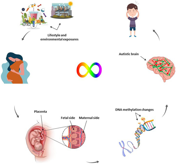
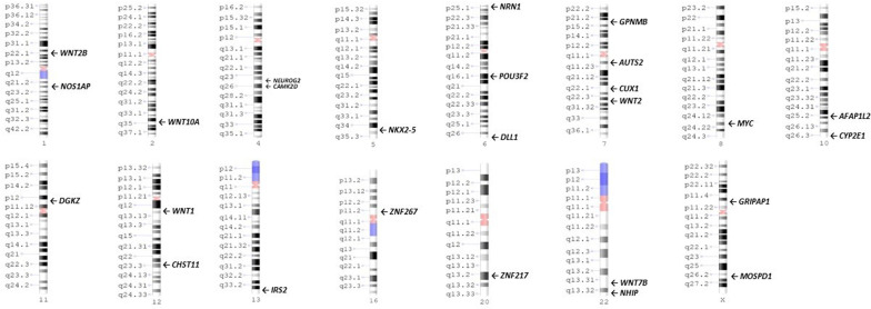
Autism spectrum disorder (ASD) is a neurodevelopmental disorder that impairs normal brain development and socio-cognitive abilities. The pathogenesis of this condition points out the involvement of genetic and environmental factors during in-utero life. Placenta, as an interface tissue between mother and fetus, provides developing fetus requirements and exposes it to maternal environment as well. Therefore, the alteration of DNA methylation as epigenetic consequence of gene-environmental interaction in the placenta could shed light on ASD pathogenesis. In this study, we reviewed the current findings on placental methylation status and its association with ASD. Differentially methylated regions (DMRs) in ASD-developing placenta were found to be mainly enriched in ASD gene loci affecting synaptogenesis, microtubule dynamics, neurogenesis and neuritogenesis. In addition, non-genic DMRs in ASD-placenta proposes an alternative contributing mechanism for ASD development. Our study highlights the importance of placental DNA methylation signature as a biomarker for ASD prediction.
Keywords: Autism; Brain; DNA methylation; Epigenetic; Placenta.
© 2023. The Author(s).
Conflict of interest statement
The authors declare that they have no competing interests.
Figures
References
-
- Ali F, Thaver I, Khan SA. Assessment of dietary diversity and nutritional status of pregnant women in Islamabad, Pakistan. J Ayub Med Coll Abbottabad. 2014;26(4):506–509. - PubMed
-
- American Psychiatric Association. DSM 5. Diagnostic and statistical manual of mental disorders (5th ed.). 2013.
-
- Autism Spectrum Disorders Working Group of The Psychiatric Genomics Consortium Meta-analysis of GWAS of over 16,000 individuals with autism spectrum disorder highlights a novel locus at 10q2432 and a significant overlap with schizophrenia. Mol Autism. 2017;8:21. doi: 10.1186/s13229-017-0137-9. - DOI - PMC - PubMed
Publication types
MeSH terms
Substances
LinkOut - more resources
Full Text Sources
Medical
