. 2023 Jan 16;11(1):14.
doi: 10.1186/s40478-023-01506-z.

Clinico-pathological and epigenetic heterogeneity of diffuse gliomas with FGFR3::TACC3 fusion

Collaborators, Affiliations

Clinico-pathological and epigenetic heterogeneity of diffuse gliomas with FGFR3::TACC3 fusion

Alice Métais et al. Acta Neuropathol Commun. .

Abstract

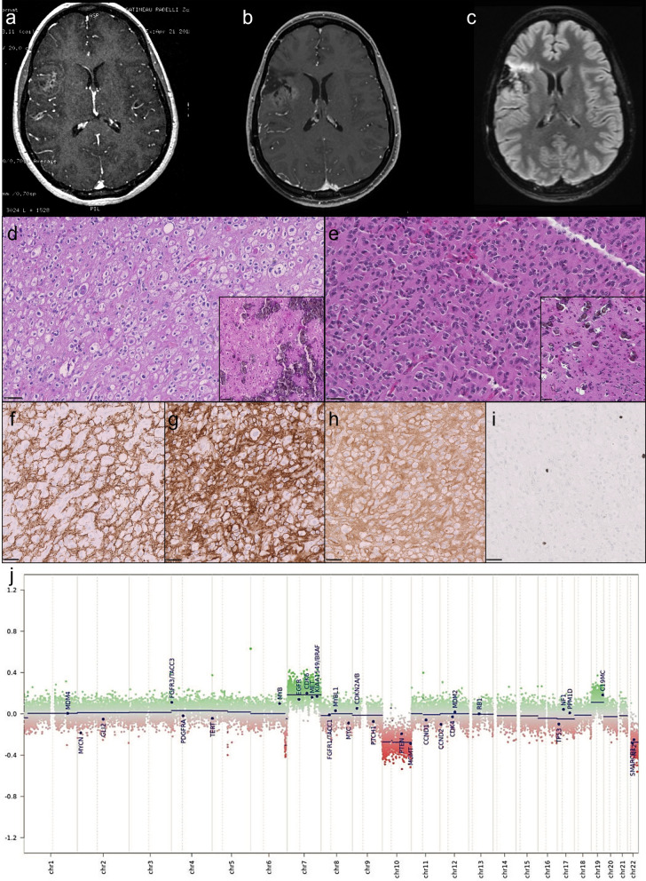
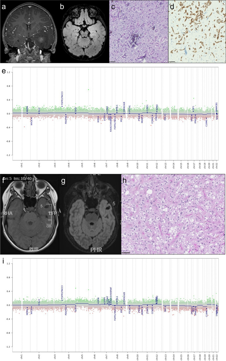
Background: Gliomas with FGFR3::TACC3 fusion mainly occur in adults, display pathological features of glioblastomas (GB) and are usually classified as glioblastoma, IDH-wildtype. However, cases demonstrating pathological features of low-grade glioma (LGG) lead to difficulties in classification and clinical management. We report a series of 8 GB and 14 LGG with FGFR3:TACC3 fusion in order to better characterize them.

Methods: Centralized pathological examination, search for TERT promoter mutation and DNA-methylation profiling were performed in all cases. Search for prognostic factors was done by the Kaplan-Meir method.

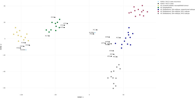
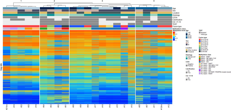
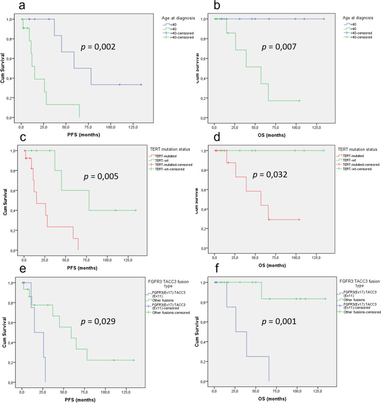
Results: TERT promoter mutation was recorded in all GB and 6/14 LGG. Among the 7 cases with a methylation score > 0.9 in the classifier (v12.5), 2 were classified as glioblastoma, 4 as ganglioglioma (GG) and 1 as dysembryoplastic neuroepithelial tumor (DNET). t-SNE analysis showed that the 22 cases clustered into three groups: one included 12 cases close to glioblastoma, IDH-wildtype methylation class (MC), 5 cases each clustered with GG or DNET MC but none with PLNTY MC. Unsupervised clustering analysis revealed four groups, two of them being clearly distinct: 5 cases shared age (< 40), pathological features of LGG, lack of TERT promoter mutation, FGFR3(Exon 17)::TACC3(Exon 10) fusion type and LGG MC. In contrast, 4 cases shared age (> 40), pathological features of glioblastoma, and were TERT-mutated. Relevant factors associated with a better prognosis were age < 40 and lack of TERT promoter mutation.

Conclusion: Among gliomas with FGFR3::TACC3 fusion, age, TERT promoter mutation, pathological features, DNA-methylation profiling and fusion subtype are of interest to determine patients' risk.

Keywords: 2021 WHO classification of CNS tumours; DNA-methylation profiling; FGFR3:TACC3 fusion; Glioblastoma; Pediatric low grade glioma.


Conflict of interest statement

The authors declare that they have no competing interests.

Figures

Fig. 1
Fig. 1
Illustration of an adult case presenting histologically as a low grade glioma (#12). This is an adult case of glioma (24 years at diagnosis) with FGFR3:TACC3 fusion with a low grade glioma (LGG) histological appearance initially and at recurrence. MRI at initial diagnosis showed a right frontal lesion with calcification and contrast enhancement (a). MRI at recurrence (b, c), showed an anterior high signal on FLAIR (c) without contrast enhancement (b). Histology at initial diagnosis (d, hematoxylin–phloxin–safron) and recurrence (e hematoxylin–phloxin–safron) showed an infiltrative and circumscribed growth pattern (f anti-neurofilamnet immunostaining), of an oligodendroglioma-like tumor with profuse microcalcifications (boxes) and without microvascular proliferation or tumoral necrosis. There was a strong and diffuse CD34 extra-vascular expression (g anti-CD34 immunostaining) and a strong and diffuse FGFR3 expression (h anti-FGFR3 immunostaining). No mitotic activity was observed and proliferation index was low (i anti-MIB1 immunostaining). Patient was simply monitored after surgeries and is still alive to date. Methylation class with v12.5 was ganglioglioma at both initial and recurrence with calibrated scores (CS) of respectively 0.41 and 0.61. Copy number variation (CNV) plots generated by the molecularneuropathology.org platform were the same between initial and recurrence (j). Gain of chromosome 7/loss of chromosome 10 was observed and there was mutation of TERT promotor. Scale bars: D, E: 50µm; F, G, H, I: 100 µm
Fig. 2
Fig. 2
Illustration of a pediatric case of FGFR3::TACC3 low grade glioma (#22 & #14). Upper panel ae illustrates the case of a 1-year-old at diagnosis child (#22). Clinical presentation was a West syndrome starting at 6 months of life. MRI revealed a left temporal infiltrative tumor (a T1 with gadolinium injection; b FLAIR sequence). Surgical resection was performed at 1 year old. Histological examination c showed a diffuse glial tumor with calcifications, there was no microvascular proliferation, no necrosis, nor mitotic activity. There was no CD34 extravascular expression (d). Methylation class with v12.5 was ganglioglioma with a calibrated score of 0.39, and the case clustered with ganglioglioma cluster on t-SNE, it fell within cluster 1 with other low-grade tumors on hierarchical clustering. Copy number plot generated by the platform did not show copy number variation (e). Lower panel fi illustrates the case of a 12-year-old child (#14) with chronic epilepsy. MRI show a left temporal without contrast enhancement (f T1 with gadolinium injection, g FLAIR sequence), surgical resection at 12 years old and revealed a diffuse oligodendroglioma-like tumor with calcification and strong and diffuse CD34 expression and FGFR3(ex17)::TACC3(ex13) fusion, very suggestive of a polymorphous low grade neuroepithelial tumor of the young (PLNTY) in the absence of ganglion cells (h hematoxylin–phloxin–saffron). There was no TERT promoter mutation and no chromosome + 7/− 10 (i CNV plots from DNA methylation profiling). Methylation class was ganglioglioma with 0.99 calibrated score. Scale bars: 50 µm
Fig. 3
Fig. 3
t-distributed stochastic neighbor embedding DNA-methylation profiling data analysis. t-SNE analysis of 23 glioma samples with FGFR3::TACC3 fusion (black triangles and blue triangles for recurrences) compared to reference cohort samples retrieved from our DNA-methylation profiling database that were classified by the v12.5 version of the Heidelberg classifier with calibrated scores (CS) > 0.9 as follows: glioblastoma, IDH-wildtype mesenchymal subtype methylation class (MC) (12 cases, blue dots); glioblastoma, IDH-wildtype RTK1 subtype MC (12 cases, purple dots); glioblastoma, IDH-wildtype RTK2 subtype MC (12 cases, grey dots); LGG DNET MC (11 cases, yellow dots); and LGG GG MC (10 cases, green dots)
Fig. 4
Fig. 4
Unsupervised hierarchical clustering of DNA-methylation profiling data. Unsupervised hierarchical clustering of 23 glioma samples with FGFR3:TACC3 fusion based on the 10,000 most variably methylated probes. Samples with CS > 0.9 were considered as matching with the MC proposed by the v12.5 version of the DKFZ classifier, CS ≤ 0.9 were considered not matching. Chr chromosome, DNET dysembryoplastic neuroepithelial tumor, GG ganglioglioma, IHG infantile hemispheric glioma, LGG low grade glioma, GB IDH-WT glioblastoma IDH-wildtype, GB_MES_TYP glioblastoma mesenchymal subtype, GB_RTK2 glioblastoma RTK2 subtype, MYXGNT, PDGFRA-mutant (novel) Myxoid glioneuronal tumor, PDGFRA-mutant (novel), RGNT rosette forming glioneuronal tumor
Fig. 5
Fig. 5
Survival analysis according to WHO CNS 5 diagnosis and age at diagnosis. Kaplan-Meier curves for progression-free (a) and overall (b) survival for gliomas with FGFR3:TACC3 fusion according to age at initial diagnosis (> 40 vs. < 40). Kaplan–Meier curves for progression-free (c) and overall (d) survival for gliomas with FGFR3:TACC3 fusion according to TERT promotor mutation status (mutated vs. wild-type). Kaplan–Meier curves for progression-free (e) and overall (f) survival for gliomas with FGFR3:TACC3 fusion according to FGFR3:TACC3 fusion type (FGFR3(Ex17)::TACC3(Ex11) vs. other fusions). OS overall survival, PFS progression-free survival

References

    1. Ahrendsen JT, Sinai C, Meredith DM, Malinowski SW, Cooney TM, Bandopadhayay P, Ligon KL, Alexandrescu S. Molecular alterations in Pediatric Low-Grade Gliomas that led to death. J Neuropathol Exp Neurol. 2021 doi: 10.1093/jnen/nlab097. - DOI - PubMed
    1. Appay R, Bielle F, Sievers P, Barets D, Fina F, Boutonnat J, Adam C, Gauchotte G, Godfraind C, Lhermitte B, Maurage C-A, Meyronet D, Mokhtari K, Rousseau A, Tauziède-Espariat A, Tortel M-C, Uro-Coste E, Burel-Vandenbos F, Chotard G, Pesce F, Varlet P, Colin C, Figarella-Branger D. Rosette-forming glioneuronal tumours are midline, FGFR1-mutated tumours. Neuropathol Appl Neurobiol. 2022;48:e12813. doi: 10.1111/nan.12813. - DOI - PubMed
    1. Appay R, Fina F, Barets D, Gallardo C, Nanni-Metellus I, Scavarda D, Henaff D, Vincent J, Grewis L, Pourquier P, Colin C, Figarella-Branger D. Multiplexed droplet digital PCR assays for the simultaneous screening of major genetic alterations in tumors of the central nervous system. Front Oncol. 2020;10:579762. doi: 10.3389/fonc.2020.579762. - DOI - PMC - PubMed
    1. Asif S, Fatima R, Krc R, Bennett J, Raza S. Comparative proteogenomic characterization of glioblastoma. CNS Oncol. 2019;8:CNS37. doi: 10.2217/cns-2019-0003. - DOI - PMC - PubMed
    1. Bale TA, Rosenblum MK. The 2021 WHO classification of tumors of the central nervous system: an update on pediatric low-grade gliomas and glioneuronal tumors. Brain Pathol Zurich Switz. 2022;32:e13060. doi: 10.1111/bpa.13060. - DOI - PMC - PubMed

Publication types