Optically driven microtools with an antibody-immobilised surface for on-site cell assembly
- PMID: 36647211
- PMCID: PMC10190638
- DOI: 10.1049/nbt2.12114
Optically driven microtools with an antibody-immobilised surface for on-site cell assembly
Abstract
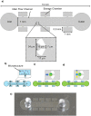
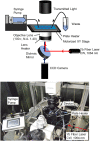
To enable the accurate reproduction of organs in vitro, and improve drug screening efficiency and regenerative medicine research, it is necessary to assemble cells with single-cell resolution to form cell clusters. However, a method to assemble such forms has not been developed. In this study, a platform for on-site cell assembly at the single-cell level using optically driven microtools in a microfluidic device is developed. The microtool was fabricated by SU-8 photolithography, and antibodies were immobilised on its surface. The cells were captured by the microtool through the bindings between the antibodies on the microtool and the antigens on the cell membrane. Transmembrane proteins, CD51/61 and CD44 that facilitate cell adhesion, commonly found on the surface of cancer cells were targeted. The microtool containing antibodies for CD51/61 and CD44 proteins was manipulated using optical tweezers to capture HeLa cells placed on a microfluidic device. A comparison of the adhesion rates of different surface treatments showed the superiority of the antibody-immobilised microtool. The assembly of multiple cells into a cluster by repeating the cell capture process is further demonstrated. The geometry and surface function of the microtool can be modified according to the cell assembly requirements. The platform can be used in regenerative medicine and drug screening to produce cell clusters that closely resemble tissues and organs in vivo.
Keywords: bioMEMS; laser beam applications; microchannel flow; microfabrication.
© 2023 The Authors. IET Nanobiotechnology published by John Wiley & Sons Ltd on behalf of The Institution of Engineering and Technology.
Conflict of interest statement
There are no conflicts to declare.
Figures


Similar articles
-
Non-destructive handling of individual chromatin fibers isolated from single cells in a microfluidic device utilizing an optically driven microtool.Lab Chip. 2014 Feb 21;14(4):696-704. doi: 10.1039/c3lc51111a. Lab Chip. 2014. PMID: 24356711
-
On-site processing of single chromosomal DNA molecules using optically driven microtools on a microfluidic workbench.Sci Rep. 2021 Apr 12;11(1):7961. doi: 10.1038/s41598-021-87238-3. Sci Rep. 2021. PMID: 33846479 Free PMC article.
-
Optically Manipulated Microtools to Measure Adhesion of the Nanoparticle-Targeting Ligand Glutathione to Brain Endothelial Cells.ACS Appl Mater Interfaces. 2021 Aug 25;13(33):39018-39029. doi: 10.1021/acsami.1c08454. Epub 2021 Aug 16. ACS Appl Mater Interfaces. 2021. PMID: 34397215
-
Single cell screening approaches for antibody discovery.Methods. 2017 Mar 1;116:34-42. doi: 10.1016/j.ymeth.2016.11.006. Epub 2016 Nov 15. Methods. 2017. PMID: 27864085 Review.
-
Microfluidic strategies for design and assembly of microfibers and nanofibers with tissue engineering and regenerative medicine applications.Adv Healthc Mater. 2015 Jan 7;4(1):11-28. doi: 10.1002/adhm.201400144. Epub 2014 May 23. Adv Healthc Mater. 2015. PMID: 24853649 Review.
Cited by
-
Optofluidic Tweezers: Efficient and Versatile Micro/Nano-Manipulation Tools.Micromachines (Basel). 2023 Jun 28;14(7):1326. doi: 10.3390/mi14071326. Micromachines (Basel). 2023. PMID: 37512637 Free PMC article. Review.
References
MeSH terms
Substances
Grants and funding
- 20H02118/Japan Society for the Promotion of Science
- JPMJFR212D/Japan Science and Technology Agency
- JPMJPR14FB/Precursory Research for Embryonic Science and Technology
- Ministry of Education, Culture, Sports, Science and Technology
- Nakatani Foundation for Advancement of Measuring Technologies in Biomedical Engineering
LinkOut - more resources
Full Text Sources
Miscellaneous

