. 2023 Feb 20;36(2):162-176.
doi: 10.1021/acs.chemrestox.2c00172. Epub 2023 Jan 16.

Characterization of a Novel Thermostable DNA Lyase Used To Prepare DNA for Next-Generation Sequencing

Affiliations

Characterization of a Novel Thermostable DNA Lyase Used To Prepare DNA for Next-Generation Sequencing

Tuvshintugs Baljinnyam et al. Chem Res Toxicol. .

Abstract

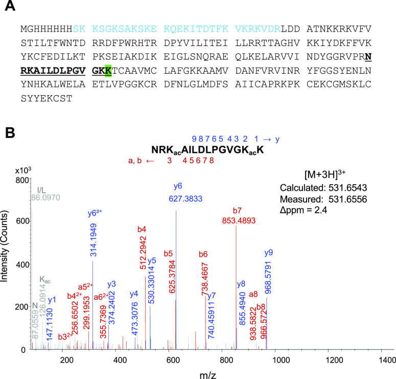
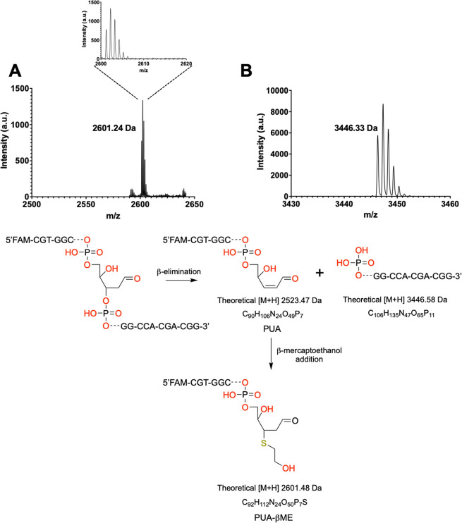
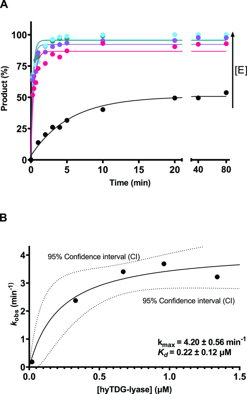
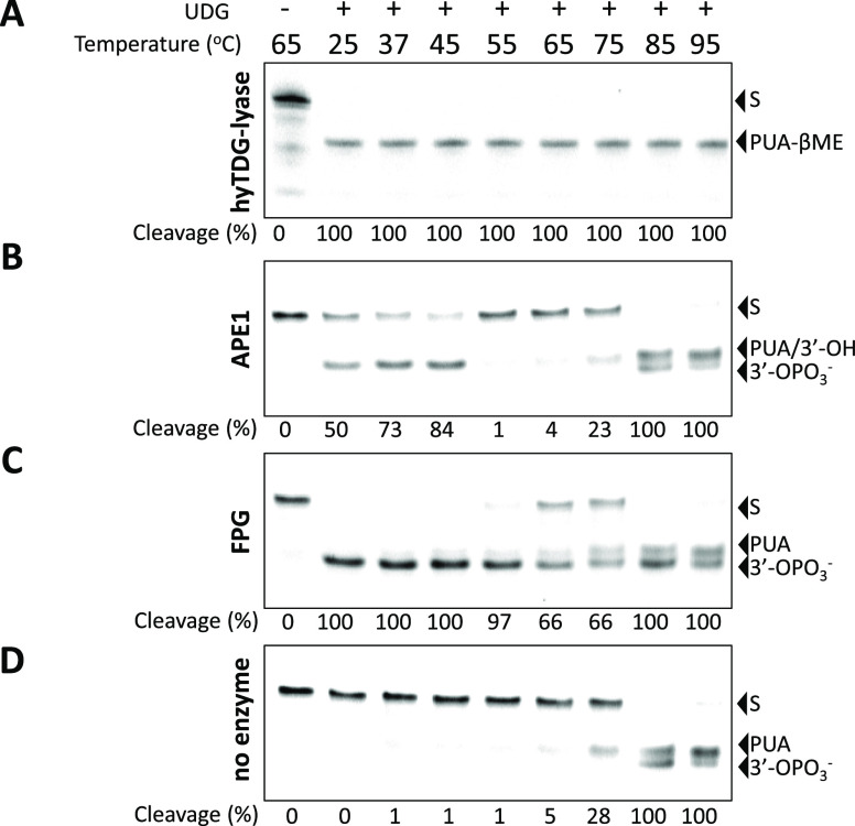
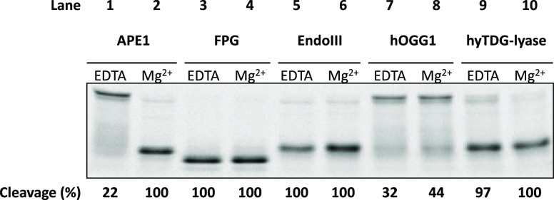
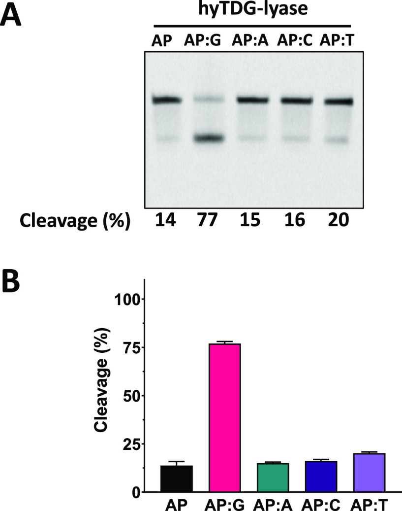
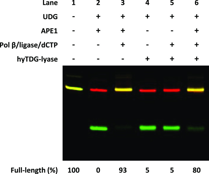
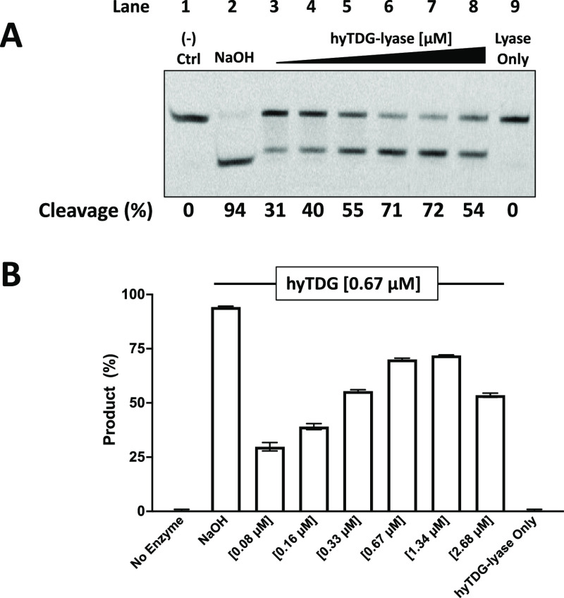
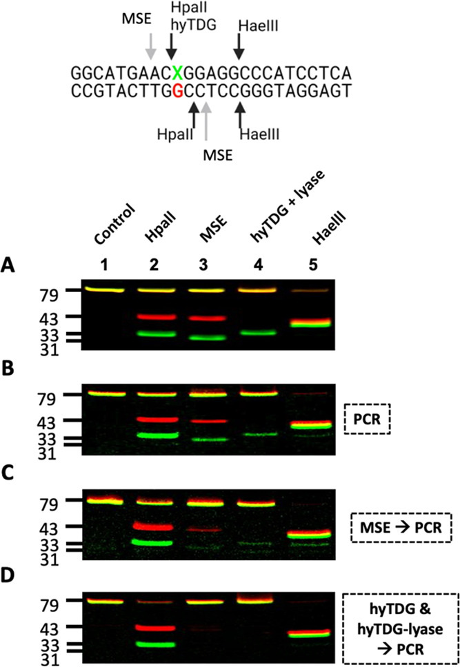
Recently, we constructed a hybrid thymine DNA glycosylase (hyTDG) by linking a 29-amino acid sequence from the human thymine DNA glycosylase with the catalytic domain of DNA mismatch glycosylase (MIG) from M. thermoautotrophicum, increasing the overall activity of the glycosylase. Previously, it was shown that a tyrosine to lysine (Y126K) mutation in the catalytic site of MIG could convert the glycosylase activity to a lyase activity. We made the corresponding mutation to our hyTDG to create a hyTDG-lyase (Y163K). Here, we report that the hybrid mutant has robust lyase activity, has activity over a broad temperature range, and is active under multiple buffer conditions. The hyTDG-lyase cleaves an abasic site similar to endonuclease III (Endo III). In the presence of β-mercaptoethanol (β-ME), the abasic site unsaturated aldehyde forms a β-ME adduct. The hyTDG-lyase maintains its preference for cleaving opposite G, as with the hyTDG glycosylase, and the hyTDG-lyase and hyTDG glycosylase can function in tandem to cleave T:G mismatches. The hyTDG-lyase described here should be a valuable tool in studies examining DNA damage and repair. Future studies will utilize these enzymes to quantify T:G mispairs in cells, tissues, and genomic DNA using next-generation sequencing.


Conflict of interest statement

The authors declare the following competing financial interest(s): The authors have submitted a patent application for hyTDG and hyTDG-lyase. Otherwise, the authors declare that they have no conflicts of interest with the contents of this article.

Figures

Figure 1
Figure 1
Confirmation of the primary amino acid sequence of hyTDG-lyase. (A) Amino acid sequence of hyTDG-lyase. Underlined and in bold is the peptide that contains the Y163K amino acid substitution (highlighted). The protein has a his-tag followed by a 29 amino acid sequence from human thymine DNA glycosylase on the N-terminus (blue). (B) Mass spectrum of the NRKAILDLPGVGKK peptide containing the Y163K substitution obtained by nLC-MS/MS. The fragmentation pattern confirms the predicted sequence. To obtain high sequence coverage and high-quality peptides, lysines were acetylated to prevent trypsin over digestion. Table S1 contains additional peptides that were detected.
Figure 2
Figure 2
hyTDG-lyase generates a β-elimination product that undergoes a Michael addition with β-mercaptoethanol. An 18-base oligonucleotide duplex with a 5′-FAM label and a U:G mispair (100 pmol, 4 μM) was incubated with hyTDG (25 pmol, 1 μM) and hyTDG-lyase (12.5 pmol, 0.5 μM) for 2 h at 65 °C. The resulting fragments were examined by MALDI-TOF-MS. (A) The fragment that contains the 5′-FAM label and a 3′-terminus had a measured m/z of 2601.24. The observed mass is consistent with the formation of a β-elimination product, a PUA, that forms an adduct with β-ME (PUA-βME) (theoretical m/z 2601.48 Da). (B) Corresponding 11-base fragment formed from the cleavage of the 3′-end of the abasic site had a measured m/z of 3446.33 (theoretical m/z 3446.58 Da) consistent with a 5′-phosphate. The schematic demonstrates the possible structure of the proposed fragment consistent with the measured mass.
Figure 3
Figure 3
hyTDG-lyase kinetics. (A) To estimate the kmax and Kd of hyTDG-lyase, we incubated various concentrations of hyTDG-lyase: 0.02, 0.33, 0.67, 0.96, and 1.34 μM, at 65 °C with a FAM-labeled duplex oligonucleotide (0.05 μM) containing an AP:G site. This was done by first incubating a U:G oligonucleotide with UDG for 1 h at 37 °C to generate an abasic site. Product formation was monitored as a function of time using a gel and fit a single exponential (smooth curves) to individual data points (dots) to obtain the kobs for each concentration of hyTDG-lyase. The arrow represents increasing hyTDG-lyase concentration [E]. (B) We then fit our kobs data as a function of hyTDG-lyase concentration to determine kmax and Kd using a nonlinear hyperbolic fit (solid curve). Errors for kmax and Kd are reported as the standard error of the mean associated with the fit of the curve.
Figure 4
Figure 4
hyTDG-lyase is thermostable. A FAM-labeled 18-base oligonucleotide containing a U:G mispair (2.5 pmol, 0.2 μM) was incubated with UDG (2.5 U, 0.84 pmol, 0.07 μM) at 37 °C for 1 h in TDG buffer. Then hyTDG-lyase (16.8 pmol, 1.34 μM), APE1 (5 U, 0.18 pmol, 0.01 μM), FPG (4 U, 6.44 pmol, 0.52 μM), or a no lyase control was added at the indicated temperatures for 1 h. (A) hyTDG-lyase cleaves oligonucleotides (S) at an abasic site generated by UDG at all temperatures tested and produces a PUA-βME adduct. (B) APE1 cleaves an abasic site from 25 to 45 °C to form a free 3′-OH but was inactive at higher temperatures (55–95 °C). Spontaneous β and β,δ-elimination occurred at the abasic site at higher temperatures (65–95 °C), resulting in both formation of a PUA and 3′-OPO3, respectively. (C) FPG was highly active from 25 to 55 °C and produces a 3′-OPO3. Its activity was greatly reduced at higher temperatures (65–95 °C). (D) As a control, we heated the abasic site containing oligo for the same duration at the indicated temperatures. Heating an abasic site for 1 h at 65–75 °C produced a β-elimination product. At higher temperatures, all the abasic sites were cleaved and we observed a mixture of both β and β,δ-elimination, upper and lower product bands respectively, seen in panels B–D.
Figure 5
Figure 5
AP endonucleases but not AP lyases require magnesium. APE1 requires the presence of Mg2+ for strand cleavage, while FPG, Endo III, hOGG1, and hyTDG-lyase do not. This suggests that these bifunctional glycosylases and our hyTDG-lyase are AP lyases and not endonucleases. An abasic site was generated by treating an 18-base oligonucleotide containing uracil with UDG and subsequently adding the indicated enzyme. 2.5 units of UDG (0.84 pmol, 0.07 μM) for 1 h at 37 °C. This substrate was then treated with 5 units of APE1 (0.18 pmol, 0.01 μM), 4 units of FPG (6.44 pmol, 0.52 μM), 5 units of Endo III (0.13 pmol, 0.01 μM), 0.25 μg of hOGG1 (6.2 pmol, 0.5 μM), or 0.5 μg of hyTDG-lyase (16.8 pmol, 1.34 μM) for an addition 1 h. The reaction with hyTDG-lyase was incubated at 65 °C, while the others were held at 37 °C. Reactions were prepared in TDG buffer (10 mM K2HPO4, 30 mM NaCl, 40 mM KCl, pH 7.9) supplemented with either 2 mM EDTA or 10 mM Mg-Ac, as indicated. Each reaction had 2.5 pmol (0.2 μM) of oligo.
Figure 6
Figure 6
hyTDG-lyase prefers cleaving an abasic site opposite G. hyTDG-lyase retains a preference for activity opposite G but can cleave abasic sites in single-stranded DNA and all other base-pairing contexts. (A) Representative gel of a 30 s reaction of hyTDG-lyase with an abasic site containing oligonucleotides in different base-pairing contexts. (B) Quantification of gel pictures (n = 3). Error bars represent the standard deviation. A 5′-FAM-labeled single-stranded oligonucleotide containing U was used or was annealed to a complementary strand containing either a G, A, C, or T opposite U. DNA substrates (2.5 pmol, 0.2 μM), were incubated with 2.5 units of UDG (0.84 pmol, 0.07 μM) in buffer at 37 °C for 1 h to generate an abasic site. hyTDG-lyase was then added, and cleavage of the abasic site was quantified after incubating at 65 °C for 30 s.
Figure 7
Figure 7
3′-terminus produced by hyTDG-lyase cannot be extended by DNA Pol β but is resolved by APE1. In lane 1, A 5′-FAM-labeled 79 base oligonucleotide containing a U in a U:G mispair (2.5 pmol, 0.2 μM). In lane 2, the oligonucleotide was incubated with UDG (2.5 U, 0.84 pmol, 0.07 μM) for 1 h at 37 °C in CutSmart buffer, and subsequently, the abasic site was cleaved by incubating with APE1 (5 U, 0.18 pmol, 0.01 μM) for an additional 30 min. In lane 3, Pol β (6.2 pmol, 0.5 μM) and E. coli ligase (5 U, 4 pmol, 0.32 μM), dCTP (250 pmol, 20 μM), and NAD+ (325 pmol, 26 μM) were added for an additional 1 h to simulate short-patch BER. Lane 4 was otherwise identical to lane 2, but hyTDG-lyase was used instead of APE1 at 37 °C for 30 min. Lane 5 was otherwise identical to lane 3, except hyTDG-lyase (26.9 pmol, 2.15 μM) was used instead of APE1 at 37 °C. In lane 6, we similarly generated an abasic site that was then cleaved by hyTDG-lyase for 30 min at 37 °C. Then APE1, Pol β, dCTP, and E. coli ligase were added and incubated for an additional 1 h. Lane 6 demonstrates that the AP endonuclease activity of APE1 can clean up the PUA-βME 3′-end produced by the hyTDG-lyase. The complementary strand is labeled with a 5′-Cy5 fluorophore (red). The overlap between the full-length 5′-FAM-labeled oligo and its 5′-Cy5 complement is depicted as yellow.
Figure 8
Figure 8
hyTDG-lyase competitively inhibits hyTDG glycosylase. (A) 5′-FAM-labeled 18-baseoligonucleotides containing a U:G mispair (0.6 pmol, 0.05 μM) was treated with hyTDG (8.4 pmol, 0.67 μM) alone or simultaneously incubated with increasing concentrations of hyTDG-lyase (1.05, 2.1, 4.2, 8.4, 16.8, or 33.6 pmol) (0.08–2.68 μM) or hyTDG-lyase alone (33.6 pmol, 2.68 μM) in TDG buffer. To confirm removal of U following incubation with hyTDG, one sample was treated with NaOH. To confirm that hyTDG-lyase has no glycosylase activity, we had a hyTDG-lyase only control lane. All samples were incubated for 1 h at 65 °C. (B) Quantification of three independent experiments. Error bars represent the standard deviation.
Figure 9
Figure 9
hyTDG and hyTDG-lyase are more thorough than MSE at removing T:G mismatches in DNA prior to PCR amplification for NGS applications. (A) 79/77 base oligonucleotide duplexes containing a C:G or T:G mismatch were prepared by ligation and mixed in a 0.9:1 ratio, respectively (X = C or T). This mixture was then treated with HpaII, MSE, hyTDG and hyTDG-lyase, or HaeIII (lanes 1–5). HpaII cleaves both strands of only a C:G oligo. MSE cleaves both strands of a T:G mispaired oligo with a two-nucleotide overhang 5′ to the T:G mismatch. hyTDG and hyTDG-lyase remove the T and cleave the T-containing strand. HaeIII cleaves only duplex DNA. (B) PCR amplification: Unlabeled C:G and T:G oligos were mixed in a 1:1 ratio with a 10% excess of the complementary G strand and then used as PCR template to amplify the DNA and attach fluorescent labels for visualization purposes. PCR products were column purified and treated with enzymes, as in A. (C) MSE then PCR: The same oligo mixture in B was first treated with MSE and then used as a PCR template. MSE does not remove the T and leaves overhangs, which can provide a template during PCR, resulting in amplification and T:A mutations. In lane 2, HpaII digestion shows a significant amount of full-length T:A oligo. To confirm this, in lanes 3 and 4, we see only minor cleavage with MSE or hyTDG and hyTDG-lyase. (D) hyTDG and hyTDG-lyase then PCR: Same as C except DNA was first treated with hyTDG for 1 h followed by the addition of hyTDG-lyase for an additional 1 h, at 65 °C. Quantification is found in Table S6.

Similar articles

References

    1. Friedberg E. C. A History of the DNA Repair and Mutagenesis Field. The Discovery of Base Excision Repair. DNA Repair 2016, 37, A35–A39. 10.1016/j.dnarep.2015.12.003. - DOI - PubMed
    1. Howard M. J.; Wilson S. H. DNA Scanning by Base Excision Repair Enzymes and Implications for Pathway Coordination. DNA Repair 2018, 71, 101–107. 10.1016/j.dnarep.2018.08.013. - DOI - PMC - PubMed
    1. Mullins E. A.; Rodriguez A. A.; Bradley N. P.; Eichman B. F. Emerging Roles of DNA Glycosylases and the Base Excision Repair Pathway. Trends Biochem. Sci. 2019, 44, 765–781. 10.1016/j.tibs.2019.04.006. - DOI - PMC - PubMed
    1. Zhao S.; Tadesse S.; Kidane D.. Significance of Base Excision Repair to Human Health, 1st ed.; Elsevier Inc., 2021; Vol. 364. - PubMed
    1. Bordin D. L.; Lirussi L.; Nilsen H. Cellular Response to Endogenous DNA Damage: DNA Base Modifications in Gene Expression Regulation. DNA Repair 2021, 99, 10305110.1016/j.dnarep.2021.103051. - DOI - PubMed

Publication types