

[Prognostic Value of the Expression of Myeloid Leukemia Factor 1-Interacting Protein in Gastric Cancer and Its Regulatory Role in Tumor Progression]
- PMID: 36647653
- PMCID: PMC10409039
- DOI: 10.12182/20230160103
[Prognostic Value of the Expression of Myeloid Leukemia Factor 1-Interacting Protein in Gastric Cancer and Its Regulatory Role in Tumor Progression]
Abstract
Objective: To investigate the prognostic value of the expression of myeloid leukemia factor 1-interacting protein (MLF1IP) in gastric cancer tissue and its regulatory role in tumor progression.
Methods: Gene Expression Omnibus (GEO) database was used to analyze the expression level of MLF1IP in tumor tissues of gastric cancer patients. Kaplan-Meier Plotter database was used to analyze the relationship between MLF1IP expression level and patient prognosis. We conducted a retrospective analysis of 108 gastric cancer patients who had undergone radical surgery at our hospital between January 2015 and December 2015. The expression of MLF1IP in gastric cancer tissue and adjacent tissues was examined. We analyzed the relationship between MLF1IP and the clinicopathological parameters of gastric cancer patients and its impact on the long-term prognosis of gastric cancer patients. Univariate and multivariate regression analyses were done to identify the risk factors affecting the long-term prognosis of gastric cancer patients. The assessment value of MLF1IP for long-term prognosis of gastric cancer was analyzed with ROC curve. The effects of MLF1IP on the proliferation, migration, and invasion of gastric cancer cells were analyzed in vitro with gastric cancer cell line (MGC803). A xenograft tumor model was established with nude mice to analyze in vivo the effect of MLF1IP on tumor growth.
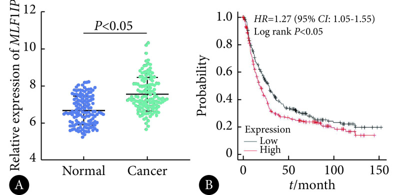
Results: The results of the gastric cancer cohort GSE29272 of GEO database showed that the expression level of MLF1IP in gastric cancer tissues was significantly higher than that in normal tissues ( P<0.05). Analysis with Kaplan-Meier Plotter database indicated that high MLF1IP expression was correlated with poor prognosis in gastric cancer patients. Immunohistochemical analysis showed that the expression level of MLF1IP in gastric cancer tissues was higher than that in adjacent tissues ( P<0.05). Correlation analysis showed that the MLF1IP level in gastric cancer tissue was positively correlated with Ki67 ( r=0.609, P<0.01), peripheral blood carcinoembryonic antigen (CEA) ( r=0.572, P<0.01) and carbohydrate antigen 19-9 (CA19-9) ( r=0.623, P<0.01). Kaplan-Meier (K-M) survival analysis showed that the 5-year survival rate of patients in the MLF1IP high expression group was significantly lower than that in the MLF1IP low expression group ( P<0.01). Cox regression analysis showed that independent risk factors for 5-year survival after radical gastrectomy for gastric cancer included the expression of MLF1IP ( HR=2.508, 95% CI: 1.259-4.999), CEA≥5 μg/L ( HR=2.171, 95% CI: 1.152-4.092), CA19-9≥37 kU/L ( HR=2.401, 95% CI: 1.094-5.269), and T3-T4 stages ( HR=2.779, 95% CI: 1.049-7.358) and N2-N3 stages ( HR=2.072, 95% CI: 1.100-3.904). ROC analysis showed that the sensitivity, specificity, and accuracy of MLF1IP (the cut-off value was 3.00 relative protein expression level) in assessing the 5-year survival rate after radical gastrectomy for gastric cancer was 75.00%, 76.92%, and 76.2%, respectively ( P<0.05). CCK-8, Transwell assay, and scratch assays showed that in vitro knocking down of MLF1 IP gene expression significantly inhibited the proliferation, migration and invasion of gastric cancer cells. Subcutaneous tumor xenograft experiment in nude mice showed that knocking down MLF1 IP gene significantly inhibited tumor growth.
Conclusion: Increased expression of MLF1IP in gastric cancer tissue, which may be involved in the malignant activities of proliferation, migration, and invasion of gastric cancer cells, has a certain predictive value for poor prognosis.
目的: 探讨胃癌组织中骨髓增生异常白血病因子1相互作用蛋白(MLF1IP)表达的预后价值及其在肿瘤进展中的调控作用。
方法: 采用GEO、Kaplan-Meier Plotter数据库分析胃癌肿瘤组织中MLF1IP的表达水平及其与患者预后的关系;回顾性分析2015年1–12月在我院行胃癌根治术的患者108例,检测胃癌及癌旁组织中MLF1IP的表达情况;分析MLF1IP与胃癌患者临床病理参数间的关系及其对患者远期预后的影响;进一步采用单因素与多因素回归分析胃癌患者远期预后的危险因素;ROC曲线分析MLF1IP对胃癌远期预后的评估价值。选择胃癌细胞系MGC803体外分析MLF1IP对癌细胞增殖、迁移与侵袭能力的影响。建立裸鼠移植瘤模型分析在体环境下MLF1IP对肿瘤生长的影响。
结果: GEO数据库胃癌队列GSE29272结果显示胃癌组织中MLF1IP表达水平高于正常组织(P<0.05),Kaplan-Meier Plotter数据库提示MLF1IP高表达患者预后不良。免疫组化分析显示,胃癌组织中MLF1IP表达水平高于癌旁组织(P<0.05);相关性分析显示MLF1IP水平与胃癌组织中Ki67(r=0.609,P<0.01)、外周血癌胚抗原(carcinoembryonic antigen, CEA)(r=0.572,P<0.01)及糖类抗原19-9(carbohydrate antigen 19-9, CA19-9)(r=0.623,P<0.01)均呈正相关;Kaplan-Meier(K-M)生存分析显示MLF1IP高表达组患者术后5年生存率低于MLF1IP低表达组(P<0.01);Cox回归分析显示,MLF1IP表达量(HR=2.508,95%CI:1.259~4.999)、CEA≥5 μg/L(HR=2.171,95%CI:1.152~4.092)、CA19-9≥37 kU/L(HR=2.401,95%CI:1.094~5.269)、T分期为3~4期(HR=2.779,95%CI:1.049~7.358)及N分期为2~3期(HR=2.072,95%CI:1.100~3.904)均是影响胃癌根治术后5年生存率的独立危险因素;ROC分析表明MLF1IP(以蛋白相对表达量3.00为截点值)评估胃癌患者根治术后5年生存率的敏感性为75.00%,特异性为76.92%,准确度为76.2%(P<0.05)。CCK-8、Transwell及划痕实验证明,体外干扰MLF1IP基因表达可显著抑制胃癌细胞的增殖、迁移和侵袭。裸鼠皮下移植瘤实验显示,干扰MLF1IP基因可显著抑制肿瘤的生长。
结论: MLF1IP在胃癌组织中表达升高并对患者预后不良有一定的预测价值,其可能参与了胃癌细胞增殖、迁移和侵袭等恶性行为。
Keywords: Gastric cancer; Invasion; MLF1IP; Migration; Prognosis; Proliferation.
Copyright© by Editorial Board of Journal of Sichuan University (Medical Sciences).
Conflict of interest statement
Figures


References
Publication types
MeSH terms
Substances
LinkOut - more resources
Full Text Sources
Medical