

[Extraction of Extracelluar Vesicles Derived from Mycobacterium tuberculosis and Their Effect on the Production of Reactive Oxygen Species and Expression of Inflammatory Factors in Mouse Bone Marrow-Derived Dendritic Cells]
- PMID: 36647654
- PMCID: PMC10409048
- DOI: 10.12182/20230160201
[Extraction of Extracelluar Vesicles Derived from Mycobacterium tuberculosis and Their Effect on the Production of Reactive Oxygen Species and Expression of Inflammatory Factors in Mouse Bone Marrow-Derived Dendritic Cells]
Abstract
Objective: To isolate extracellular vesicles (EVs) from Mycobacterium tuberculosis ( Mtb), to examine their morphology, particle size, and distribution, to study the effect of EVs derived from Mtb ( Mtb-EVs) on intracellular reactive oxygen species (ROS) production and cytokine secretion in dendritic cells (DCs), and to make preliminary exploration of Mtb-EVs' effect on the immune regulation of DCs.
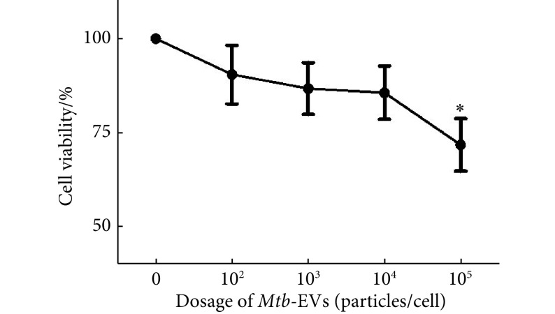
Methods: Mtb-EVs were obtained by ultrafiltration concentration and the protein concentration was determined by BCA assay. The morphology of Mtb-EVs was observed through negative staining electron microscopy (EM). The particle size distribution and concentration of Mtb-EVs were determined by nanoparticle tracking analysis (NTA). Mouse bone marrow was isolated through sterile procedures and mice myeloid DCs were induced and amplified by the combined use of recombinant mouse granulocyte-macrophage colony-stimulating factor (rm GM-CSF) and recombinant mouse interleukin-4 (rm IL-4). Then, morphological and immunophenotypic characterization was performed. After that, the DCs were treated with Mtb-EVs at different concentrations and CCK-8 assay was done to measure their effect on the survival rate of DCs and to identify the appropriate stimulation concentration for subsequent experimental procedures. The intracellular ROS levels of DCs were evaluated with DCFH-DA fluorescence probe and the cytokine secretion of DCs was determined by ELISA.
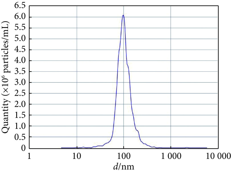
Results: EM observation showed that Mtb-EVs isolated by ultrafiltration concentration were spherical vesicles of varied sizes, all being approximately 100 nm in diameter and displaying typical morphology. NTA results from NanoSight nanoparticle tracker showed that the peak particle size was 98.5 nm, that the average particle size was 110.2 nm, and that the particle size was mainly distributed between 68.4-155.7 nm. Mtb-EVs that were smaller than 250 nm accounted for 98.39% of the total. Mouse myeloid DCs directionally induced and amplified in vitro displayed typical DC phenotype and morphology, and the purity exceeded 85%. EM verified the abundance of microvilli and radial protuberance on the surface of DCs, which had uniform cytoplasm and clear nuclear membrane. Loaded with Mtb-EVs at different concentrations, including 10 2, 10 3, and 10 4 particles/cell, the DCs had significantly upregulated levels of intracellular ROS ( P<0.05). In addition, Mtb-EVs induced the release of IL-1β and IL-6 in a dose-dependent manner ( P<0.05).
Conclusion: We established in the study a technical process for the extraction of Mtb-EVs by ultrafiltration concentration and obtained Mtb-EVs with sound morphology, high purity, and concentrated particle size distribution. Furthermore, Mtb-EVs can upregulate the intracellular ROS level in DCs and induce the release of IL-1β and IL-6 in a dose-dependent manner.
目的: 提取结核分枝杆菌(M. tuberculosis, Mtb)胞外囊泡(extracellular vesicles, EVs),检测其形态、粒径大小及分布,研究Mtb-EVs对树突状细胞(dendritic cell, DC)胞内活性氧(ROS)和细胞因子水平的影响,初步探讨其对DC的免疫调节作用。
方法: 超滤浓缩法分离获取Mtb-EVs,BCA法检测蛋白浓度,负染电镜检测Mtb-EVs形态,纳米颗粒跟踪分析技术检测其粒径大小分布和浓度;无菌分离获取小鼠骨髓,经重组小鼠粒细胞-巨噬细胞集落刺激因子(rm GM-CSF)和重组小鼠白细胞介素-4(rm IL-4)联合诱导扩增出DC,并进行形态学及免疫表型鉴定;用不同剂量Mtb-EVs作用于DC,DCFH-DA荧光探针法检测DC胞内ROS水平,ELISA法检测DC细胞IL-1β和IL-6分泌。
结果: 超滤浓缩法提取的Mtb-EVs为大小不等的球状囊泡结构,形态典型,直径约100 nm;NanoSight纳米颗粒追踪仪检测结果显示,粒径峰值98.5 nm,平均粒径110.2 nm,主要分布在68.4~155.7 nm之间,小于250 nm囊泡数量占总量98.39%;体外定向诱导扩增的细胞具有典型DC的形态特征,纯度可达85%以上,透射电镜可见DC表面有丰富微绒毛及放射状突起,胞浆均匀,核膜清晰;102、103、104 particles/cell Mtb-EVs处理DC后,ROS 水平与Mtb-EVs剂量呈正相关(r= 0.9694 , P<0.05),并以剂量依赖方式诱导细胞释放产生IL-1β和IL-6(P<0.05)。
结论: 本研究建立了超滤浓缩法分离提取Mtb-EVs的技术流程,可得到形态完整、纯度较高、粒径分布集中的细胞外囊泡。同时,Mtb-EVs可以诱发DC胞内ROS水平上调,并以剂量依赖方式诱导细胞因子IL-1β和IL-6的释放。
Keywords: Dendritic cells; Extracelluar vesicles; Mycobacterium tuberculosis.
Copyright© by Editorial Board of Journal of Sichuan University (Medical Sciences).
Conflict of interest statement
Figures


References
-
- 国家卫生健康委疾病预防控制局 2019年全国法定传染病疫情概况. 中国病毒病杂志. 2020;10(4):245.
Publication types
MeSH terms
Substances
LinkOut - more resources
Full Text Sources