

A new method for preparing a rat intracerebral hemorrhage model by combining focused ultrasound and microbubbles
- PMID: 36647712
- PMCID: PMC10158945
- DOI: 10.1002/ame2.12303
A new method for preparing a rat intracerebral hemorrhage model by combining focused ultrasound and microbubbles
Abstract
Background: We aimed to prepare a non-invasive, reproducible, and controllable rat model of intracerebral hemorrhage with focused ultrasound (FUS).
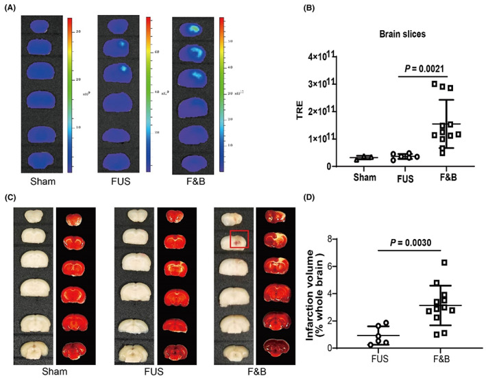
Methods: A rat intracerebral hemorrhage (ICH) model was established by combining FUS and microbubbles (μBs), and edaravone was used to verify whether the free radical scavenger had a protective effect on the model. The brain tissue of each group was sectioned to observe the gross histology, blood-brain barrier (BBB) permeability, cerebral infarction volume, and histopathological changes.
Results: Compared with the FUS group, the BBB permeability was significantly increased in the FUS + μBs (F&B) group (p = 0.0021). The second coronal slice in the F&B group had an obvious hemorrhage lesion, and the FUS + μBs + edaravone (F&B&E) group had smaller hemorrhage areas; however, ICH did not occur in the FUS group. The cerebral infarction volume in the F&B group was significantly larger than that in the FUS group (p = 0.0030) and F&B&E group (p = 0.0208). HE staining results showed that nerve fibrinolysis, neuronal necrosis, microglia production, and erythrocytes were found in both the F&B group and the F&B&E group, but the areas of the nerve fibrinolysis and neuronal necrosis in the F&B group were larger than the F&B&E group.
Conclusions: A rat ICH model was successfully prepared using the μBs assisted FUS treatment, and edaravone had a therapeutic effect on this model. This model can be used to study the pathophysiological mechanism of ICH-related diseases and in preclinical research on related new drugs.
Keywords: ICH model; focused ultrasound (FUS); microbubbles (μBs); rat.
© 2023 The Authors. Animal Models and Experimental Medicine published by John Wiley & Sons Australia, Ltd on behalf of The Chinese Association for Laboratory Animal Sciences.
Conflict of interest statement
The authors have no competing interests to declare. Zhihui Zhong is an Editorial Board member of AMEM and a co‐author of this article. To minimize bias, he was excluded from all editorial decision‐making related to the acceptance of this article for publication.
Figures


Similar articles
-
Stereotactic Administration of Edaravone Ameliorates Collagenase-Induced Intracerebral Hemorrhage in Rat.CNS Neurosci Ther. 2016 Oct;22(10):824-35. doi: 10.1111/cns.12584. Epub 2016 Jul 8. CNS Neurosci Ther. 2016. PMID: 27390192 Free PMC article.
-
Detection of intracerebral hemorrhage and transient blood-supply shortage in focused-ultrasound-induced blood-brain barrier disruption by ultrasound imaging.Ultrasound Med Biol. 2012 Aug;38(8):1372-82. doi: 10.1016/j.ultrasmedbio.2012.03.013. Epub 2012 May 12. Ultrasound Med Biol. 2012. PMID: 22579546
-
Drug-loaded bubbles with matched focused ultrasound excitation for concurrent blood-brain barrier opening and brain-tumor drug delivery.Acta Biomater. 2015 Mar;15:89-101. doi: 10.1016/j.actbio.2014.12.026. Epub 2015 Jan 7. Acta Biomater. 2015. PMID: 25575854
-
Evaluating the safety profile of focused ultrasound and microbubble-mediated treatments to increase blood-brain barrier permeability.Expert Opin Drug Deliv. 2019 Feb;16(2):129-142. doi: 10.1080/17425247.2019.1567490. Epub 2019 Jan 29. Expert Opin Drug Deliv. 2019. PMID: 30628455 Free PMC article. Review.
-
Focused ultrasound-mediated drug delivery through the blood-brain barrier.Expert Rev Neurother. 2015 May;15(5):477-91. doi: 10.1586/14737175.2015.1028369. Expert Rev Neurother. 2015. PMID: 25936845 Free PMC article. Review.
References
-
- Virani S, Alonso A, Benjamin EJ, et al. Heart disease and stroke statistics‐2020 update: a report from the American Heart Association. Circulation. 2020;141(9):e139‐e596. - PubMed
-
- Anderson C, Heeley E, Huang Y, et al. Rapid blood‐pressure lowering in patients with acute intracerebral hemorrhage. N Engl J Med. 2013;368(25):2355‐2365. - PubMed
-
- Schrag M, Kirshner H. Management of intracerebral hemorrhage: JACC focus seminar. J Am Coll Cardiol. 2020;75(15):1819‐1831. - PubMed
-
- Woo D, Broderick J. Spontaneous intracerebral hemorrhage: epidemiology and clinical presentation. Neurosurg Clin N Am. 2002;13(3):265‐279. - PubMed
Publication types
MeSH terms
Substances
LinkOut - more resources
Full Text Sources
