Review
. 2023 Jan 25;55(1):1-10.
doi: 10.3724/abbs.2022196.

Ironing out macrophages in atherosclerosis

Affiliations
Review

Ironing out macrophages in atherosclerosis

Lei Wang et al. Acta Biochim Biophys Sin (Shanghai). .

Abstract

The most common cause of death worldwide is atherosclerosis and related cardiovascular disorders. Macrophages are important players in the pathogenesis of atherosclerosis and perform critical functions in iron homeostasis due to recycling iron by phagocytosis of senescent red blood cells and regulating iron availability in the tissue microenvironment. With the growth of research on the "iron hypothesis" of atherosclerosis, macrophage iron has gradually become a hotspot in the refined iron hypothesis. Macrophages with the M1, M2, M(Hb), Mox, and other phenotypes have been defined with different iron-handling capabilities related to the immune function and immunometabolism of macrophages, which influence the progression of atherosclerosis. In this review, we focus on macrophage iron and its effects on the development of atherosclerosis. We also cover the contradictory discoveries and propose a possible explanation. Finally, pharmaceutical modulation of macrophage iron is discussed as a promising target for atherosclerosis therapy.

Keywords: atherosclerosis; iron; macrophage.


Conflict of interest statement

The authors declare that they have no conflict of interest.

Figures
None
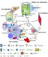
Figure 1
Regulation of systemic iron metabolism Organs and cell types involved in systemic iron homeostasis are shown. Dietary iron absorption: Duodenal enterocytes absorb dietary iron via divalent metal transporter 1 (DMT1) located on the apical surface upon reduction of Fe 3+ to Fe 2+ by Fe reductase (DcytB). At the basolateral membrane, ferroportin (FPN) cooperates with Fe oxidase (hephaestin) to convert Fe 2+ to Fe 3+. Iron utilization: plasma transferrin (Tf) captures and circulates iron in the body. By binding to transferrin receptor 1 (TfR1) and subsequent endocytosis, iron-loaded transferrin (Holo-Tf) provides iron to all types of cells. Iron recycling: iron is recycled by macrophages from senescent red blood cells. The uptake of Hb-Hp and Hx-heme is mediated by CD163 and LDL (low-density lipoprotein)-related receptor 1 (LRP1, also known as CD91). Heme oxygenase 1 (HO-1) catabolizes intracellular heme-Fe for inclusion into the cytosolic labile iron pool or cellular ferritin pool or trafficked into the mitochondria. Non-Tf-bound iron (NTBI) is imported via DMT1. Ceruloplasmin (Cp) facilitates iron export by FPN by oxidizing Fe 2+ to Fe 3+, allowing apo-Tf to sequester it. Iron regulation: the hepatic hormone hepcidin modulates the stability of FPN, which controls iron outflow from these cells.
None
Figure 2
Macrophage subpopulations and iron states in atherosclerotic plaques Interferon (IFN) and lipopolysaccharide (LPS), as well as oxidized low-density lipoprotein (oxLDL) and cholesterol crystals, are stimuli for M1 polarization; IL-4 is an inducer of M2 polarization, oxidized phospholipids (oxPL) for the Mox phenotype, and the Hb/Hp complex for the M(Hb) phenotype. M1 macrophages display proinflammatory and iron retention profiles. Iron activates the Toll-like receptor (TLR)/nuclear factor kappa-light-chain-enhancer of activated B cells (NF-κB) signaling pathway, which is in charge of macrophage inflammatory activity. OxLDL and iron stimulate the TLR4 pathway, resulting in the autocrine production of hepcidin. This results in the exacerbation of iron accumulation, ROS production, and the inflammatory response through a positive feedback loop. Furthermore, iron retention promotes foam cell formation by increasing CD36-mediated cholesterol absorption and decreasing ABC transporter ABCA1/ABCG1-mediated reverse cholesterol efflux via interference with CYP27A1 and liver X receptor (LXR) signaling, respectively. M (Hb) and M2 macrophages play a crucial role in iron handling and prevent foam cell formation. While these nonfoam M(Hb) macrophages are supposed to be antiatherogenic in theory, intracellular iron deficiency causes hypoxia-inducible factor 1α (HIF1α) stability and vascular endothelial growth factor (VEGF) release, which has proatherosclerotic effects by increasing vascular permeabilization and intraplaque neoangiogenesis. Mox macrophages exhibit reduced phagocytic capacity and express antioxidant genes. However, iron retention and lipid accumulation in Mox macrophages likely contribute to lesion development and plaque instability in atherosclerosis.

References

    1. Björkegren JLM, Lusis AJ. Atherosclerosis: recent developments. Cell. . 2022;185:1630–1645. doi: 10.1016/j.cell.2022.04.004. - DOI - PMC - PubMed
    1. Libby P, Buring JE, Badimon L, Hansson GK, Deanfield J, Bittencourt MS, Tokgözoğlu L, et al. Atherosclerosis. Nat Rev Dis Primers. . 2019;5:56. doi: 10.1038/s41572-019-0106-z. - DOI - PubMed
    1. Engelen SE, Robinson AJB, Zurke YX, Monaco C. Therapeutic strategies targeting inflammation and immunity in atherosclerosis: how to proceed? Nat Rev Cardiol. . 2022;19:522–542. doi: 10.1038/s41569-021-00668-4. - DOI - PMC - PubMed
    1. Moore KJ, Koplev S, Fisher EA, Tabas I, Björkegren JLM, Doran AC, Kovacic JC. Macrophage trafficking, inflammatory resolution, and genomics in atherosclerosis: JACC macrophage in CVD series (part 2) J Am Coll Cardiol. . 2018;72:2181–2197. doi: 10.1016/j.jacc.2018.08.2147. - DOI - PMC - PubMed
    1. Wade J, Byrne DJ, Ballentine CJ, Drakesmith H. Temporal variation of planetary iron as a driver of evolution. Proc Natl Acad Sci USA. . 2021;118:e2109865118. doi: 10.1073/pnas.2109865118. - DOI - PMC - PubMed