

Recent Developments of Silk-Based Scaffolds for Tissue Engineering and Regenerative Medicine Applications: A Special Focus on the Advancement of 3D Printing
- PMID: 36648802
- PMCID: PMC9844467
- DOI: 10.3390/biomimetics8010016
Recent Developments of Silk-Based Scaffolds for Tissue Engineering and Regenerative Medicine Applications: A Special Focus on the Advancement of 3D Printing
Abstract
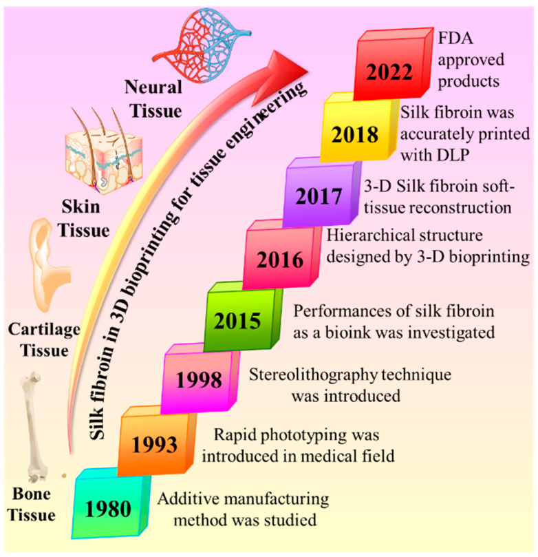
Regenerative medicine has received potential attention around the globe, with improving cell performances, one of the necessary ideas for the advancements of regenerative medicine. It is crucial to enhance cell performances in the physiological system for drug release studies because the variation in cell environments between in vitro and in vivo develops a loop in drug estimation. On the other hand, tissue engineering is a potential path to integrate cells with scaffold biomaterials and produce growth factors to regenerate organs. Scaffold biomaterials are a prototype for tissue production and perform vital functions in tissue engineering. Silk fibroin is a natural fibrous polymer with significant usage in regenerative medicine because of the growing interest in leftovers for silk biomaterials in tissue engineering. Among various natural biopolymer-based biomaterials, silk fibroin-based biomaterials have attracted significant attention due to their outstanding mechanical properties, biocompatibility, hemocompatibility, and biodegradability for regenerative medicine and scaffold applications. This review article focused on highlighting the recent advancements of 3D printing in silk fibroin scaffold technologies for regenerative medicine and tissue engineering.
Keywords: 3D printing; biomaterials; fibroin; regenerative medicine; scaffolds; tissue engineering.
Conflict of interest statement
The authors declare no conflict of interest.
Figures


Similar articles
-
Bioactive Silk Fibroin-Based Hybrid Biomaterials for Musculoskeletal Engineering: Recent Progress and Perspectives.ACS Appl Bio Mater. 2021 Sep 20;4(9):6630-6646. doi: 10.1021/acsabm.1c00654. Epub 2021 Aug 9. ACS Appl Bio Mater. 2021. PMID: 35006966
-
Nonmulberry silk proteins: multipurpose ingredient in bio-functional assembly.Biomed Mater. 2021 Sep 22;16(6). doi: 10.1088/1748-605X/ac20a0. Biomed Mater. 2021. PMID: 34428758 Review.
-
Analysis of the Adherence of Dental Pulp Stem Cells on Two-Dimensional and Three-Dimensional Silk Fibroin-Based Biomaterials.J Craniofac Surg. 2017 Jun;28(4):939-943. doi: 10.1097/SCS.0000000000003596. J Craniofac Surg. 2017. PMID: 28230598
-
Silk fibroin as biomaterial for bone tissue engineering.Acta Biomater. 2016 Feb;31:1-16. doi: 10.1016/j.actbio.2015.09.005. Epub 2015 Sep 7. Acta Biomater. 2016. PMID: 26360593 Review.
-
Silk Fibroin-Based Biomaterials for Tissue Engineering Applications.Molecules. 2022 Apr 25;27(9):2757. doi: 10.3390/molecules27092757. Molecules. 2022. PMID: 35566110 Free PMC article. Review.
Cited by
-
Advancements and challenges in stem cell transplantation for regenerative medicine.Heliyon. 2024 Aug 10;10(16):e35836. doi: 10.1016/j.heliyon.2024.e35836. eCollection 2024 Aug 30. Heliyon. 2024. PMID: 39247380 Free PMC article. Review.
-
Advancements and Limitations in 3D Printing Materials and Technologies: A Critical Review.Polymers (Basel). 2023 May 30;15(11):2519. doi: 10.3390/polym15112519. Polymers (Basel). 2023. PMID: 37299318 Free PMC article. Review.
-
Optimizations of Placenta Extracellular Matrix-Loaded Silk Fibroin/Alginate 3D-Printed Scaffolds Structurally and Functionally for Bone Tissue Engineering.Eng Life Sci. 2025 Jan 9;25(1):e202400085. doi: 10.1002/elsc.202400085. eCollection 2025 Jan. Eng Life Sci. 2025. PMID: 39801563 Free PMC article.
-
3D Printing Technology as a Promising Tool to Design Nanomedicine-Based Solid Dosage Forms: Contemporary Research and Future Scope.Pharmaceutics. 2023 May 10;15(5):1448. doi: 10.3390/pharmaceutics15051448. Pharmaceutics. 2023. PMID: 37242690 Free PMC article. Review.
-
Emerging Multiscale Biofabrication Approaches for Bacteriotherapy.Molecules. 2024 Jan 22;29(2):533. doi: 10.3390/molecules29020533. Molecules. 2024. PMID: 38276612 Free PMC article. Review.
References
-
- Paul K., Darzi S., McPhee G., Del Borgo M.P., Werkmeister J.A., Gargett C.E., Mukherjee S. 3D bioprinted endometrial stem cells on melt electrospun poly ε-caprolactone mesh for pelvic floor application promote anti-inflammatory responses in mice. Acta Biomater. 2019;97:162–176. doi: 10.1016/j.actbio.2019.08.003. - DOI - PubMed
Publication types
LinkOut - more resources
Full Text Sources
